Patients › Wrist
کارپل ٹنل ریلیز
Carpal tunnel release surgery relieves median nerve compression—when it’s needed and what to expect.
یہ آپریشن کیوں تجویز کیا گیا ہے
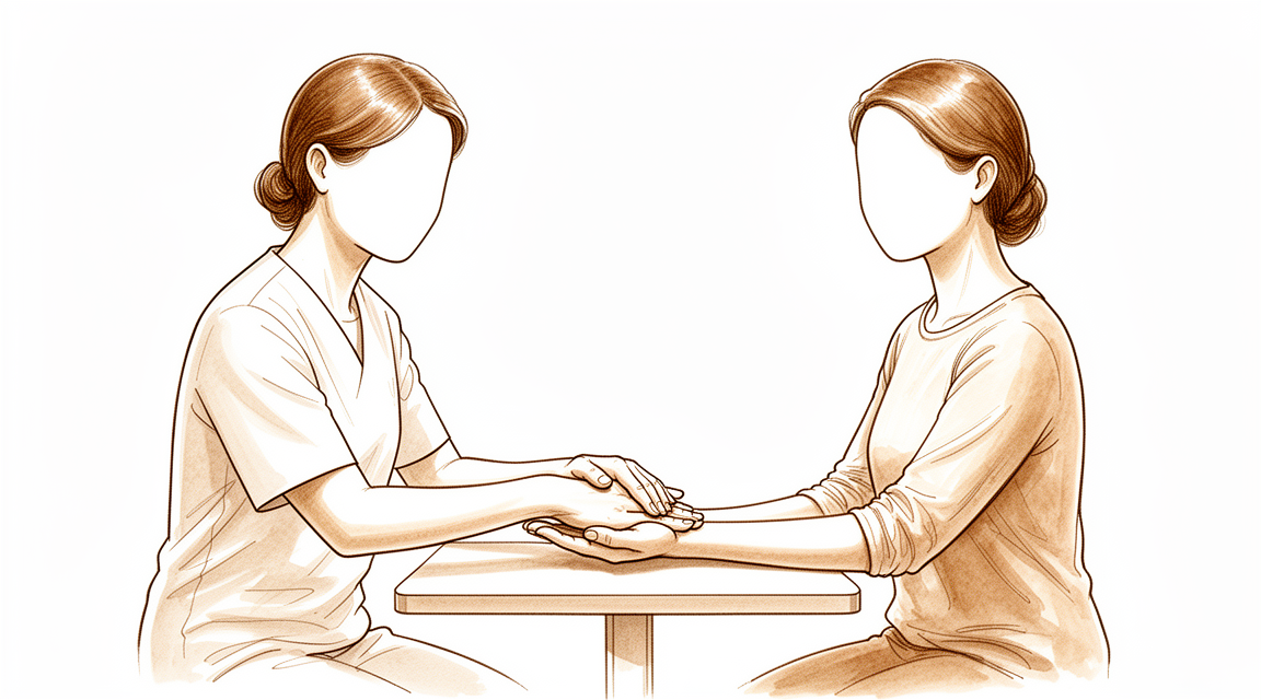
ڈاکٹر کیرن ہِرپرا، جو Mater Private Hospital Rockhampton میں بازو اور ہاتھ کے سرجن (upper-limb surgeon) ہیں، سب سے پہلے وہ طریقے آزماتے ہیں جن میں کم سے کم چیر پھاڑ ہو اور جو آپ کی حالت کے مطابق ہوں۔ مریض عموماً اپنے فیملی ڈاکٹر (GP) کے ریفرل (referral) پر ہمارے کلینک آتے ہیں؛ اگر کسی فزیوتھراپسٹ نے آپ کو ہمیں دکھانے کا مشورہ دیا ہے تو بھی میڈیکیئر (Medicare) کی رقم کی واپسی کا حق دار ہونے کے لیے آپ کو اپنے فیملی ڈاکٹر سے ریفرل لینا ہو گا۔ آپ کی ملاقات پر ہم آپ کی بیماری کی تفصیل لیتے ہیں، آپ کے ہاتھ کا معائنہ کرتے ہیں، اور ضرورت ہو تو اسکین یا اعصاب کے ٹیسٹ کرواتے ہیں۔ کارپل ٹنل سنڈروم (کلائی میں عصب کا دبنا) اس وقت ہوتا ہے جب آپ کی کلائی میں ایک عصب دب جاتا ہے، جس سے ہاتھ میں جھنجھناہٹ، سُن پن اور درد ہوتا ہے۔ ہم عموماً آپریشن کے بغیر علاج سے شروع کرتے ہیں، جیسے اسپلنٹ، ہینڈ تھراپی (ہاتھ کی بحالی کے ماہر سے علاج) یا انجکشن۔ سرجری پر اس وقت غور کیا جاتا ہے جب ان علاجوں سے کافی بہتری نہ آئی ہو۔ اس آپریشن کو کارپل ٹنل ریلیز (carpal tunnel release، یعنی عصب پر دباؤ ختم کرنے کا آپریشن) کہتے ہیں۔ یہ کلائی کے آر پار ایک سخت پٹی کو کاٹ کر عصب پر دباؤ کم کرتا ہے۔ اس آپریشن کے بعد علامات کی شدت اور ہاتھ کی کارکردگی میں نمایاں بہتری کی توقع کی جا سکتی ہے، چاہے آپ کی عمر کچھ بھی ہو، آپ کو دوسری بیماریاں ہوں، یا آپ کا کیس کام کے دوران چوٹ کے معاوضے (workers' compensation) کا ہو۔ ہم آپ سے مختلف راستوں پر بات کریں گے اور آپ کے ساتھ مل کر فیصلہ کریں گے کہ آیا سرجری آپ کے لیے مناسب ہے۔
آپریشن سے پہلے
آپ کے سرجن آپ کو واضح ہدایات دیں گے، لیکن یہاں بتایا گیا ہے کہ کیا توقع رکھیں۔ آپ کو سرجری سے سات گھنٹے پہلے کھانا پینا بند کرنا ہو گا۔ ہم چھ کے بجائے سات گھنٹے اس لیے کہتے ہیں تاکہ اگر آپریشن کی فہرست وقت سے پہلے چل رہی ہو تو آپ کو پہلے بلایا جا سکے۔ اپنی موجودہ دواؤں کی فہرست ساتھ لائیں، اور اپنے سرجن کو خون پتلا کرنے والی کسی بھی دوا کے بارے میں بتائیں، کیونکہ ان میں سے کچھ کو پہلے سے روکنا پڑ سکتا ہے۔ اس بات کا انتظام کریں کہ کوئی آپ کو بعد میں گاڑی میں گھر لے جائے۔ ڈھیلے، آرام دہ کپڑے پہنیں جن کی آستینیں آسانی سے اتر سکیں۔ آپریشن کی منصوبہ بندی کے لیے کچھ اسکین، جیسے ایکسرے، ایم آر آئی یا الٹراساؤنڈ، کی ضرورت پڑ سکتی ہے۔ اگر آپ کو دوسری بیماریاں ہیں تو آپ کو خون کے ٹیسٹ یا بے ہوشی کے ڈاکٹر (anaesthetist) سے معائنے کی ضرورت پڑ سکتی ہے، لیکن زیادہ تر لوگوں کو اس کی ضرورت نہیں ہوتی۔
آپریشن کے دن
سرجری کے دن آپ ہسپتال کے سرجیکل داخلے کے شعبے میں آتے ہیں۔ وہاں کا عملہ آپ کا اندراج کرتا ہے اور آپ کو آپریشن تھیٹر کے لیے تیار کرتا ہے۔ پھر آپ بے ہوشی کے ڈاکٹر سے ملتے ہیں، یعنی وہ ڈاکٹر جو اینستھیزیا دیتے ہیں اور آپریشن کے دوران آپ کی دیکھ بھال کرتے ہیں۔ یہ آپریشن لوکل اینستھیزیا (ایک انجکشن جو صرف آپریشن والی جگہ کو سُن کرتا ہے، اور آپ جاگ رہے ہوتے ہیں) یا جنرل اینستھیزیا (مکمل بے ہوشی) میں کیا جا سکتا ہے۔ زیادہ تر لوگ لوکل اینستھیزیا کا انتخاب کرتے ہیں: صحت یابی زیادہ تیز ہوتی ہے اور آپ جلد ہی گھر جا سکتے ہیں۔ اگر آپ بے ہوش ہونا پسند کریں تو یہ بھی ایک معقول انتخاب ہے؛ اس بارے میں اپنے سرجن اور بے ہوشی کے ڈاکٹر سے بات کریں۔ پھر آپ کو آپریشن تھیٹر میں لے جایا جاتا ہے، جہاں آپریشن کیا جاتا ہے۔ بعد میں آپ ریکوری روم میں ہوش میں آتے ہیں، جہاں اینستھیزیا کا اثر ختم ہونے تک نرسیں آپ کی نگرانی کرتی ہیں۔ جب آپ کی حالت مستحکم ہو جائے تو آپ یا تو گھر چلے جاتے ہیں یا تھوڑی دیر وارڈ میں رہتے ہیں، یہ اس پر منحصر ہے کہ کون سا طریقہ کیا گیا اور آپ کیسے صحت یاب ہو رہے ہیں۔
آپریشن میں کیا کیا جاتا ہے
اس آپریشن کو کارپل ٹنل ریلیز کہتے ہیں۔ آپ کے سرجن آپریشن والی جگہ پر، کلائی اور ہتھیلی پر، ایک ہی چیرا لگاتے ہیں۔ اس چیرے کے ذریعے سرجن ٹشو کی اس سخت پٹی تک پہنچتے ہیں جو کارپل ٹنل کی چھت بناتی ہے، یعنی آپ کی کلائی میں وہ تنگ راستہ جس سے عصب گزرتا ہے۔ اس پٹی کو کاٹنے سے سرنگ کھل جاتی ہے اور عصب پر سے دباؤ ہٹ جاتا ہے۔ خود پٹی کی مرمت کی ضرورت نہیں ہوتی؛ یہ اس طرح بھرتی ہے کہ اندر عصب کے لیے زیادہ جگہ بن جاتی ہے۔
چیرے کو ٹانکوں سے بند کیا جاتا ہے۔ پھر آپ کے ہاتھ اور کلائی کے گرد ایک موٹی گدے دار پٹی لپیٹی جاتی ہے۔ آپ یہ پٹی تقریباً 10 دن تک لگائے رکھیں گے، جیسا کہ اگلے حصے میں بتایا گیا ہے۔
آپریشن خود مختصر ہوتا ہے، اور آپ اسی دن گھر چلے جاتے ہیں۔ رضامندی کے فارم پر دستخط کرنے سے پہلے آپ کے سرجن آپ کو یہ مراحل دوبارہ سمجھائیں گے، اور آپ کسی بھی وقت سوال پوچھ سکتے ہیں۔
آپریشن کے بعد
آپ ریکوری روم میں ہوش میں آئیں گے، جہاں اینستھیزیا کا اثر ختم ہونے تک نرسیں آپ کی نگرانی کرتی ہیں۔ آپ کے ہاتھ پر ایک موٹی گدے دار پٹی ہو گی، اور آپ کو آرام میں رکھنے کے لیے ہم آپ کو درد کش دوا دیں گے۔ یہ ڈے کیس ہے، یعنی آپ اسی دن گھر چلے جائیں گے۔ براہ کرم انتظام کریں کہ پہلے 24 گھنٹے کوئی آپ کے ساتھ رہے۔ ہم پٹی تقریباً 10 دن تک لگی رہنے دیتے ہیں؛ براہ کرم اس سے پہلے اسے نہ اتاریں جب تک ہم آپ کو نہ کہیں۔ جب ہم آپ کو دیکھتے ہیں تو ہم اسے بدلتے یا اتارتے ہیں۔ آرام کرتے وقت اپنا ہاتھ اونچا رکھیں، اور جیسے ہی آپ خود کو اس قابل محسوس کریں، ہلکے کاموں کے لیے اسے آہستگی سے استعمال کریں۔ زیادہ تر لوگ ایک سے دو ہفتوں کے اندر دوبارہ گاڑی چلانے لگتے ہیں، جب زخم آرام دہ ہو جائے اور وہ آپریشن والے ہاتھ کو بچائے بغیر اسٹیئرنگ کو پکڑ اور گھما سکیں۔ دیکھیں بازو اور ہاتھ کی سرجری کے بعد گاڑی چلانا۔
صحت یابی
پہلے چند دن آپ کے ہاتھ میں درد اور سوجن ہو گی، اور گدے دار پٹی بھاری محسوس ہو گی۔ آرام کرتے وقت اپنا ہاتھ اونچا رکھیں، کیونکہ اس سے سوجن اور تکلیف کم ہوتی ہے۔ عموماً سادہ درد کش دوا کافی ہوتی ہے۔ اس آپریشن کے بعد بہت سے لوگوں کو تیز درد کش دوا کی بالکل ضرورت نہیں پڑتی۔
جیسے ہی آپ خود کو اس قابل محسوس کریں، آپ ہلکے کاموں کے لیے اپنا ہاتھ آہستگی سے استعمال کریں گے۔ پٹی اس وقت تک لگی رہتی ہے جب تک ہم آپ کو نہ دیکھ لیں، اور اترنے کے بعد آپ معمول کے مطابق ہاتھ دھو سکتے ہیں اور نہا سکتے ہیں۔ آپ کے ٹانکے فالو اپ ملاقات پر نکالے جاتے ہیں۔ سرجری کے بعد ہینڈ تھراپی Extend Rehabilitation میں Ruby Doolan کے ساتھ ہوتی ہے۔ روبی آپ کی ورزشوں میں رہنمائی کریں گی اور اگر آرام کے لیے ضرورت ہو تو آپ کے لیے اسپلنٹ بنائیں گی۔ ہلکی حرکت عصب کو پرسکون ہونے میں مدد دیتی ہے، اس لیے جیسے جیسے ہفتے گزریں گے آپ اپنا ہاتھ زیادہ استعمال کرنے لگیں گے۔
جیسے جیسے سوجن کم ہو گی، آپ دیکھیں گے کہ جھنجھناہٹ اور سُن پن کم ہو رہا ہے اور آپ کی گرفت واپس آ رہی ہے۔ جب آپ بغیر درد کے پکڑ اور گھما سکیں تو روزمرہ کے کام دوبارہ معمول کے مطابق محسوس ہوتے ہیں۔ عصب پر سے دباؤ بروقت ختم کرنے سے ہاتھ میں احساس اور کارکردگی واپس آ جاتی ہے۔ اگر آپ کی علامات دونوں ہاتھوں میں تھیں تو ہم ایک ہی بار میں دونوں کا آپریشن کرنے پر بات کر سکتے ہیں۔
صحت یابی ہر شخص میں مختلف ہوتی ہے۔ آپ کا وقت مختلف ہو سکتا ہے، اور آپ کے سرجن اور تھراپسٹ اس سارے عرصے میں آپ کی رہنمائی کریں گے۔
کیا مسائل پیدا ہو سکتے ہیں
زیادہ تر مریض ٹھیک رہتے ہیں، لیکن کبھی کبھار مسائل پیدا ہو سکتے ہیں۔ آپ کے سرجن اور ٹیم آپ کی قریب سے نگرانی کرتے ہیں تاکہ کوئی بھی مسئلہ جلد پکڑا جا سکے۔
انفیکشن۔ زخم کے انفیکشن میں چیرے سے باہر کی طرف پھیلتی سرخی، گرمی، یا رطوبت کا رسنا نظر آ سکتا ہے۔ آپ کو گہرا، دھڑکتا ہوا درد محسوس ہو سکتا ہے جو سادہ درد کش دوا سے کم نہ ہو، یا آپ کو بخار ہو سکتا ہے۔ اگر آپ ان میں سے کوئی بھی بات دیکھیں تو اسی دن کلینک کو فون کریں۔ اگر آپ کی طبیعت خراب محسوس ہو یا سرخی تیزی سے پھیل رہی ہو تو ہسپتال کی ایمرجنسی جائیں۔ کچھ چیزیں انفیکشن کا امکان بڑھا سکتی ہیں، جیسے سرجری سے کچھ ہی پہلے کلائی میں اسٹیرائیڈ کا انجکشن یا بعض بیماریاں۔ آپریشن سے پہلے ہم آپ سے ہر اس بات پر بات کریں گے جو آپ سے متعلق ہو۔
زخم کے مسائل۔ بعض اوقات زخم کے کنارے بھرنے میں زیادہ وقت لیتے ہیں یا تھوڑے سے کھل جاتے ہیں، یا جلد کے نیچے خون جمع ہو کر سوجن پیدا کرتا ہے۔ آپ دیکھ سکتے ہیں کہ ٹانکے کھنچ کر الگ ہو رہے ہیں یا چیرے کے قریب ایک سخت، چھونے پر درد کرنے والی گانٹھ محسوس کر سکتے ہیں۔ فالو اپ ملاقات پر اس کا ذکر کریں، یا اگر یہ زیادہ خراب لگے تو اس سے پہلے کلینک کو فون کریں۔
عصب میں جلن (irritation)۔ جس عصب کو آزاد کیا جا رہا ہے وہ آپریشن والی جگہ کے قریب ہوتا ہے، اس لیے سرجری کے دوران اسے چوٹ لگ سکتی ہے یا اس میں جلن پیدا ہو سکتی ہے۔ یہ عموماً ہاتھ میں نئی جھنجھناہٹ، سُن پن یا سوئیاں چبھنے جیسے احساس کی صورت میں، یا جلد کے کسی ایسے حصے کی صورت میں محسوس ہوتا ہے جو پہلے سے مختلف محسوس ہو۔ عصب کی زیادہ تر جلن وقت کے ساتھ ٹھیک ہو جاتی ہے، لیکن کچھ تبدیلیاں باقی رہ سکتی ہیں۔ اگر آپ کو نیا سُن پن محسوس ہو جو بہتر نہ ہو رہا ہو تو اگلے معائنے پر اپنے سرجن کو بتائیں۔
علامات جو واپس آ جائیں یا پوری طرح ٹھیک نہ ہوں۔ کبھی کبھار عصب پر دباؤ پوری طرح ختم نہیں ہوتا، یا مہینوں یا سالوں میں سرنگ دوبارہ تنگ ہو جاتی ہے۔ آپ دیکھ سکتے ہیں کہ پہلے والی جھنجھناہٹ، سُن پن یا رات کا درد واپس آ رہا ہے۔ اگر ایسا ہو تو اپنے معائنے پر اس کا ذکر کریں۔ بعض اوقات مزید سرجری کی ضرورت پڑتی ہے، اور آپ کے سرجن اس کی وجہ جاننے کے لیے ٹیسٹ کروائیں گے۔
اگر آپ تفصیل جاننا چاہیں تو اس صفحے پر پیچیدگیوں کی جدول میں عام شرحیں دی گئی ہیں۔
ہمیں کب فون کریں
زیادہ تر لوگ بغیر کسی مسئلے کے صحت یاب ہو جاتے ہیں، لیکن کچھ علامات پر فوری توجہ کی ضرورت ہوتی ہے۔ اگر آپ کو بخار ہو، زخم سے سرخی یا رطوبت کا اخراج بڑھ رہا ہو، یا درد مسلسل بڑھتا جا رہا ہو تو ہمیں فون کریں۔ اگر آپ کو ہاتھ میں نیا سُن پن محسوس ہو جو بہتر نہ ہو رہا ہو، یا آپ اپنی انگلیاں نہ ہلا سکیں تو ہمیں فون کریں۔ اگر آپ کو اچانک شدید درد ہو، پنڈلی میں سوجن یا درد ہو، یا سانس پھول رہی ہو تو ہسپتال کی ایمرجنسی جائیں۔ جب شک ہو تو کلینک کو فون کریں اور ہم آپ کی رہنمائی کریں گے۔
اس مسئلے کے بارے میں مزید کہاں پڑھیں
یہ صفحہ خود آپریشن کے بارے میں ہے۔ جس بیماری کا یہ علاج کرتا ہے، بشمول اس کے کہ شواہد کیا بتاتے ہیں کہ سرجری کب مدد کرتی ہے اور کب نہیں، اس کی مزید تفصیل کارپل ٹنل سنڈروم کے صفحے پر ہے۔
Evidence & references
This is the clinical evidence summary written for health professionals. It is technical, and it lists the research this page was built from. You do not need to read it to understand your treatment or to make a decision about it.
Overview
- Symptoms of pain, numbness, and tingling in the hands are common in the general population [1].
Anatomy & Pathophysiology
Epidemiology & Symptoms
- Carpal tunnel syndrome with compression of the median nerve at the wrist is the most commonly diagnosed site of nerve compression in the upper extremity [10].
- Symptoms include paresthesia or numbness (or both) in the median nerve distribution, specifically the thumb, index finger, middle finger, and radial side of the ring finger [10].
- Nocturnal paresthesias in the radial three digits of the hand is nearly pathognomonic for carpal tunnel syndrome [10].
- Paresthesias occur characteristically in "fixed wrist activities" such as reading a book or a newspaper, driving, or use of a computer keyboard or mouse [10].
- Patients rarely describe aching in the thenar eminence [10].
- With advanced nerve compression, weakness and atrophy of the abductor pollicis brevis and opponens pollicis muscles occur [10].
- Because of the slow onset of thenar weakness, patients typically adapt to this loss without functional impairments [10].
Pathogenesis
- Bony abnormalities of the carpal bones can decrease the size of the carpal tunnel [5].
- Acromegaly can decrease the size of the carpal tunnel [5].
- Flexion or extension of the wrist can decrease the size of the carpal tunnel [5].
- Forearm and wrist fractures, including Colles fracture and scaphoid fracture, can increase the contents of the carpal canal [5].
- Dislocations and subluxations, including scaphoid rotary subluxation and lunate volar dislocation, can increase the contents of the carpal canal [5].
- Posttraumatic arthritis with osteophytes can increase the contents of the carpal canal [5].
- Musculotendinous variants can increase the contents of the carpal canal [5].
- Aberrant muscles, including lumbrical, palmaris longus, and palmaris profundus, can increase the contents of the carpal canal [5].
- Local tumors, including neuroma, lipoma, multiple myeloma, and ganglion cysts, can increase the contents of the carpal canal [5].
- A persistent medial artery, whether thrombosed or patent, can increase the contents of the carpal canal [5].
- Hypertrophic synovium can increase the contents of the carpal canal [5].
- Hematoma resulting from hemophilia, anticoagulation therapy, or trauma can increase the contents of the carpal canal [5].
- Diabetes mellitus is a neuropathic condition involved in the pathogenesis of carpal tunnel syndrome [5].
- Alcoholism is a neuropathic condition involved in the pathogenesis of carpal tunnel syndrome [5].
- Double-crush syndrome is a neuropathic condition involved in the pathogenesis of carpal tunnel syndrome [5].
- Exposure to industrial solvents is a neuropathic condition involved in the pathogenesis of carpal tunnel syndrome [5].
- Rheumatoid arthritis is an inflammatory condition involved in the pathogenesis of carpal tunnel syndrome [5].
- Gout is an inflammatory condition involved in the pathogenesis of carpal tunnel syndrome [5].
- Nonspecific tenosynovitis is an inflammatory condition involved in the pathogenesis of carpal tunnel syndrome [5].
- Infection is an inflammatory condition involved in the pathogenesis of carpal tunnel syndrome [5].
- Pregnancy involves alterations of fluid balance in the pathogenesis of carpal tunnel syndrome [5].
- Menopause involves alterations of fluid balance in the pathogenesis of carpal tunnel syndrome [5].
- Eclampsia involves alterations of fluid balance in the pathogenesis of carpal tunnel syndrome [5].
- Thyroid disorders, especially hypothyroidism, involve alterations of fluid balance in the pathogenesis of carpal tunnel syndrome [5].
- Renal failure involves alterations of fluid balance in the pathogenesis of carpal tunnel syndrome [5].
- Long-term hemodialysis involves alterations of fluid balance in the pathogenesis of carpal tunnel syndrome [5].
- Raynaud disease involves alterations of fluid balance in the pathogenesis of carpal tunnel syndrome [5].
- Obesity involves alterations of fluid balance in the pathogenesis of carpal tunnel syndrome [5].
- Lupus erythematosus involves alterations of fluid balance in the pathogenesis of carpal tunnel syndrome [5].
- Scleroderma involves alterations of fluid balance in the pathogenesis of carpal tunnel syndrome [5].
- Amyloidosis involves alterations of fluid balance in the pathogenesis of carpal tunnel syndrome [5].
- Paget disease involves alterations of fluid balance in the pathogenesis of carpal tunnel syndrome [5].
- Vibration is an external force involved in the pathogenesis of carpal tunnel syndrome [5].
- Direct pressure is an external force involved in the pathogenesis of carpal tunnel syndrome [5].
Anatomical Structures & Surgical Landmarks
- The palmar cutaneous sensory branch of the median nerve lies in the interval between the palmaris longus and the flexor carpi radialis tendons [4].
- The palmar cutaneous branch of the median nerve is located deep to the thenar crease and radial to the palmaris longus [11].
- The superficial palmar arterial arch is 5 to 8 mm distal to the distal margin of the transverse carpal ligament [4].
- The flexor retinaculum includes the distal deep fascia of the forearm proximally, the transverse carpal ligament, and the aponeurosis between the thenar and hypothenar muscles [4].
- Fibers of the transverse carpal ligament can extend distally farther than expected [4].
- The recurrent branch of the median nerve may perforate the transverse carpal ligament and leave the median nerve on the volar side [4].
- Potential anatomical anomalies include connections between the flexor pollicis longus and the index flexor digitorum profundus tendons [4].
- Potential anatomical anomalies include anomalous flexor digitorum superficialis [4].
- Potential anatomical anomalies include palmaris longus, hypothenar, and lumbrical muscle bellies [4].
- Potential anatomical anomalies include median and ulnar nerve branches and interconnections [4].
- The thenar crease takes a variable course [4].
- A crossing cutaneous branch from the ulnar nerve is identified in a distal portion of the incision about 15% of the time [11].
Diagnostic & Prognostic Factors
- Carpal tunnel syndrome is a clinical diagnosis based on a combination of symptoms and characteristic physical findings [10].
- Electrodiagnostic studies are useful to stage the degree of nerve compression and assist in anticipating the time needed for recovery of nerve function [10].
- Patients with long-standing symptoms, severe atrophy of the thenar musculature, and dense sensory loss should be cautioned that release may not lead to complete recovery of sensation or thenar strength [10].
- Ultrasound sensitivity for carpal tunnel syndrome has been reported to be over 97% when the median nerve diameter is greater than 10 mm² at the level of the pisiform [5].
- In patients with negative electrodiagnostic studies but a clinical diagnosis of carpal tunnel syndrome, high-resolution ultrasonography has a sensitivity of 73% if the cutoff of 9.4 mm² at the inlet of the carpal tunnel is used [5].
- The diagnosis of carpal tunnel syndrome should be based on clinical acumen and physical examination in the vast majority of patients [5].
- Ancillary tests should be reserved for patients without clear presentations [5].
- MRI is not routinely used for diagnosis of carpal tunnel syndrome [5].
- A major advantage of MRI is its high soft-tissue contrast, which gives detailed images of bones and soft tissues [5].
- Reported false-negative rates of electrodiagnostic testing are 10% [5].
- Postoperative electrodiagnostic testing may be helpful in assessing recurrent symptoms [5].
Clinical Presentation
Epidemiology and Risk Factors
- Carpal tunnel syndrome is the most common compression neuropathy of the upper extremity [9].
- The mean age at diagnosis for carpal tunnel syndrome is 50 years [9].
- Carpal tunnel syndrome is more common in women than men by nearly four times [9].
- By the age of 65 years, the prevalence of carpal tunnel syndrome is approximately 5.1% for women and 1.3% for men [9].
- Risk factors for carpal tunnel syndrome include obesity, pregnancy, hypothyroidism, diabetes mellitus, and menopause [9].
- The American Academy of Orthopaedic Surgeons guidelines list body mass index (BMI) and high hand repetition rate as factors with strong evidence of increased risk for development of carpal tunnel syndrome [9].
Clinical Symptoms and Signs
- Classically, carpal tunnel syndrome presents with nocturnal paresthesias in a median nerve distribution that gradually worsen as nerve injury progresses [9].
- Late in the disease course, carpal tunnel syndrome leads to sensory loss and thenar muscle atrophy [9].
- Many patients report pain in the hand and may report symptoms that are not directly referable to the median nerve [9].
- A positive Tinel sign at the wrist can aid in the diagnosis of carpal tunnel syndrome [9].
- Development of symptoms after a provocative Phalen maneuver can aid in the diagnosis of carpal tunnel syndrome [9].
- The reported specificity of the Tinel sign at the wrist varies from 55% to 100% [9].
- The reported specificity of the Phalen test varies from 54% to 98% [9].
- Tinel sign and Phalen maneuver are most reliable as adjuncts to other diagnostic tests [9].
Diagnostic Testing
- Nerve conduction studies are a useful diagnostic tool for carpal tunnel syndrome, assessing focal demyelination by delayed conduction velocities of the median nerve at the wrist [9].
- Needle electromyography is considered an optional adjunct to nerve conduction studies and is mostly used to differentiate carpal tunnel syndrome from other possible causes [9].
- Documenting muscle atrophy and fibrillations on needle EMG can assist with identifying severity of the disease and help with prognostication [9].
- Thenar atrophy and abductor pollicis brevis weakness can often be detected on physical examination [9].
- Ultrasonography allows rapid diagnosis of carpal tunnel syndrome by identification of enlarged, hypoechoic median nerve fascicles proximal to the carpal tunnel [9].
- Controversy remains as to whether ultrasonography evaluation could replace electrophysiology in the diagnosis of carpal tunnel syndrome [9].
- MRI and/or ultrasonography imaging should be considered in patients who have new, persistent, or recurrent symptoms after surgery to delineate the etiology of the symptoms [9].
- Ancillary tests for carpal tunnel syndrome should be reserved for patients without clear presentations [5].
- Reports of MRI in carpal tunnel syndrome are promising, especially with newer techniques such as diffusion tensor imaging [5].
Pathogenesis Factors
- Bony abnormalities of the carpal bones are a factor involved in the pathogenesis of carpal tunnel syndrome [5].
- Acromegaly is a factor involved in the pathogenesis of carpal tunnel syndrome [5].
- Flexion or extension of the wrist is a factor involved in the pathogenesis of carpal tunnel syndrome [5].
- Forearm and wrist fractures, such as Colles fracture and scaphoid fracture, are factors involved in the pathogenesis of carpal tunnel syndrome [5].
- Dislocations and subluxations, such as scaphoid rotary subluxation and lunate volar dislocation, are factors involved in the pathogenesis of carpal tunnel syndrome [5].
- Posttraumatic arthritis with osteophytes is a factor involved in the pathogenesis of carpal tunnel syndrome [5].
- Musculotendinous variants are a factor involved in the pathogenesis of carpal tunnel syndrome [5].
- Aberrant muscles, including lumbrical, palmaris longus, and palmaris profundus, are factors involved in the pathogenesis of carpal tunnel syndrome [5].
- Local tumors, including neuroma, lipoma, multiple myeloma, and ganglion cysts, are factors involved in the pathogenesis of carpal tunnel syndrome [5].
- A persistent medial artery, whether thrombosed or patent, is a factor involved in the pathogenesis of carpal tunnel syndrome [5].
- Hypertrophic synovium is a factor involved in the pathogenesis of carpal tunnel syndrome [5].
- Hematoma from hemophilia, anticoagulation therapy, or trauma is a factor involved in the pathogenesis of carpal tunnel syndrome [5].
- Pregnancy is an alteration of fluid balance involved in the pathogenesis of carpal tunnel syndrome [5].
- Menopause is an alteration of fluid balance involved in the pathogenesis of carpal tunnel syndrome [5].
- Eclampsia is an alteration of fluid balance involved in the pathogenesis of carpal tunnel syndrome [5].
- Thyroid disorders, especially hypothyroidism, are alterations of fluid balance involved in the pathogenesis of carpal tunnel syndrome [5].
- Renal failure is an alteration of fluid balance involved in the pathogenesis of carpal tunnel syndrome [5].
- Long-term hemodialysis is an alteration of fluid balance involved in the pathogenesis of carpal tunnel syndrome [5].
- Raynaud disease is an alteration of fluid balance involved in the pathogenesis of carpal tunnel syndrome [5].
- Obesity is an alteration of fluid balance involved in the pathogenesis of carpal tunnel syndrome [5].
- Lupus erythematosus is an alteration of fluid balance involved in the pathogenesis of carpal tunnel syndrome [5].
- Scleroderma is an alteration of fluid balance involved in the pathogenesis of carpal tunnel syndrome [5].
- Amyloidosis is an alteration of fluid balance involved in the pathogenesis of carpal tunnel syndrome [5].
- Paget disease is an alteration of fluid balance involved in the pathogenesis of carpal tunnel syndrome [5].
Investigations
Clinical Presentation and Prevalence
- The American Academy of Orthopaedic Surgeons (AAOS) guidelines list body mass index (BMI) and high hand repetition rate as factors with strong evidence of increased risk for development of carpal tunnel syndrome [9].
Physical Examination
Electrodiagnostic Testing
- Nerve conduction studies remain a useful diagnostic tool in carpal tunnel syndrome [9].
- Focal demyelination in carpal tunnel syndrome can be assessed by delayed conduction velocities of the median nerve at the wrist [9].
- Needle electromyography is currently considered an optional adjunct to nerve conduction studies [9].
- Needle electromyography is mostly used to differentiate carpal tunnel syndrome from other possible causes [9].
- Pre-operative electrodiagnostic testing predicts time to resolution of symptoms after carpal tunnel release [3].
- Reported false-negative rates of 10% limit the usefulness of electrodiagnostic testing to determine treatment [5].
- Electrodiagnostic testing does not increase the diagnostic value of the four commonly used clinical tests (abnormal hand diagram, abnormal Semmes-Weinstein, positive Durkan compression, and night pain) [5].
- Electrodiagnostic testing does not predict functional recovery or reemployment after carpal tunnel release [5].
Imaging
- MRI is not routinely used for the diagnosis of carpal tunnel syndrome [5].
- Advances in ultrasonography technology have allowed rapid diagnosis of carpal tunnel syndrome by identification of enlarged, hypoechoic median nerve fascicles proximal to the carpal tunnel [9].
- Imaging can help identify causes of postoperative symptoms such as incomplete ligament division, iatrogenic injury, or other causes [9].
Diagnostic Principles
- The diagnosis of carpal tunnel syndrome is made by clinical history, physical examination, and supportive diagnostic testing with exclusion of other possible disorders [9].
Treatment
Operative Technique
- Palmar incisions for carpal tunnel release should be placed well ulnar to the thenar crease to avoid injury to the median nerve palmar cutaneous branch [4].
- A curved incision ulnar and parallel to the thenar crease is not advisable because the palmar cutaneous branch of the median nerve may be at higher risk of injury proximally [4].
- The incision should be extended proximally to the wrist flexion crease and angled toward the ulnar side of the wrist to avoid crossing flexor creases at a right angle [4].
- The palmar cutaneous sensory branch lies in the interval between the palmaris longus and the flexor carpi radialis tendons [4].
- If the palmar sensory branch is severed, it frequently causes a painful neuroma that may later require excision from the scar [4].
- When the palmar sensory branch is severed, repair is not attempted; instead, the nerve is sectioned more proximally to be covered by the middle finger sublimis muscle [4].
- The transverse carpal ligament (TCL) must be carefully divided while avoiding damage to the median nerve and its recurrent branch, which may perforate the ligament and leave the median nerve on the volar side [4].
- Fibers of the TCL can extend distally farther than expected [4].
- The flexor retinaculum includes the distal deep fascia of the forearm proximally, the TCL, and the aponeurosis between the thenar and hypothenar muscles [4].
- A successful carpal tunnel release usually requires division of all components of the flexor retinaculum [4].
- Potential anatomical anomalies to be aware of include connections between the flexor pollicis longus and index flexor digitorum profundus tendons, anomalous flexor digitorum superficialis, palmaris longus/hypothenar/lumbrical muscle bellies, and median/ulnar nerve branches and interconnections [4].
- The superficial palmar arterial arch is located 5 to 8 mm distal to the distal margin of the TCL and must be avoided [4].
- Tenosynovectomy is occasionally indicated, especially in patients with rheumatoid arthritis [4].
- Closure involves only the skin, with wound drainage as needed [4].
Postoperative Care
- A light compression dressing and a volar splint may be applied postoperatively [4].
- The hand is actively used as soon as possible after surgery, but the dependent position is avoided [4].
- The dressing can usually be removed by the patient at home 2 or 3 days after surgery [4].
- Gentle washing and showering of the hand are permitted after dressing removal [4].
- Gradual resumption of normal hand use is encouraged [4].
- Sutures are removed after 10 to 14 days [4].
- A splint may be continued for comfort as needed for 14 to 21 days [4].
Complications
- Transection of the motor branch of the ulnar nerve is a complication of two-portal endoscopic carpal tunnel release [2].
- Scar discomfort can occur after carpal tunnel surgery [2].
- Recurrent carpal tunnel syndrome is a recognized clinical entity following carpal tunnel release [2].
- Poor outcome is associated with neural surgery (epineurotomy or neurolysis) for carpal tunnel syndrome compared with carpal tunnel release alone [2].
- Symptoms may return after carpal tunnel surgery [2].
- Complications related to carpal tunnel release are a documented clinical concern [2].
- Carpal tunnel release in patients with diabetes results in poor outcomes in long-term study [3].
Key Evidence
- [L4] Symptoms of pain, numbness, and tingling in the hands are common in the general population. [1] (10.1001/jama.282.2.153)
References
[1] Prevalence of Carpal Tunnel Syndrome in a General Population. JAMA. 1999. DOI: 10.1001/jama.282.2.153
[2] Campbell S Operative Orthopaedics 4 Volume Set. COMPRESSIVE NEUROPATHIES OF THE HAND, FOREARM, AND ELBOW > CARPAL TUNNEL SYNDROME.
[3] Campbell S Operative Orthopaedics 4 Volume Set. COMPRESSIVE NEUROPATHIES OF THE HAND, FOREARM, AND ELBOW > REFERENCES > CARPAL TUNNEL SYNDROME.
[4] Campbell S Operative Orthopaedics 4 Volume Set. COMPRESSIVE NEUROPATHIES OF THE HAND, FOREARM, AND ELBOW > EXTENDED OPEN CARPAL TUNNEL RELEASE.
[5] Campbell S Operative Orthopaedics 4 Volume Set. COMPRESSIVE NEUROPATHIES OF THE HAND, FOREARM, AND ELBOW > CARPAL TUNNEL SYNDROME > BOX 77.1.
[9] Orthopaedic Knowledge Update 13 Ebook Without Multimedia. Neuropathies, Vascular Conditions: Buerger’s, Raynaud’s; Degenerative Conditions > Upper Extremity Neuropathies > Carpal Tunnel Syndrome.
[10] Green S Operative Hand Surgery. Median Nerve Compression at the Elbow and Forearm > COMPRESSION OF THE MEDIAN NERVE > Median Nerve Compression at the Wrist: Carpal Tunnel Syndrome.
[11] Green S Operative Hand Surgery. Median Nerve Compression at the Elbow and Forearm > AUTHORS’ PREFERRED METHOD OF TREATMENT: OPEN CARPAL TUNNEL RELEASE.