Patients › Elbow
کہنی کی آرتھروسکوپی (Elbow Arthroscopy)
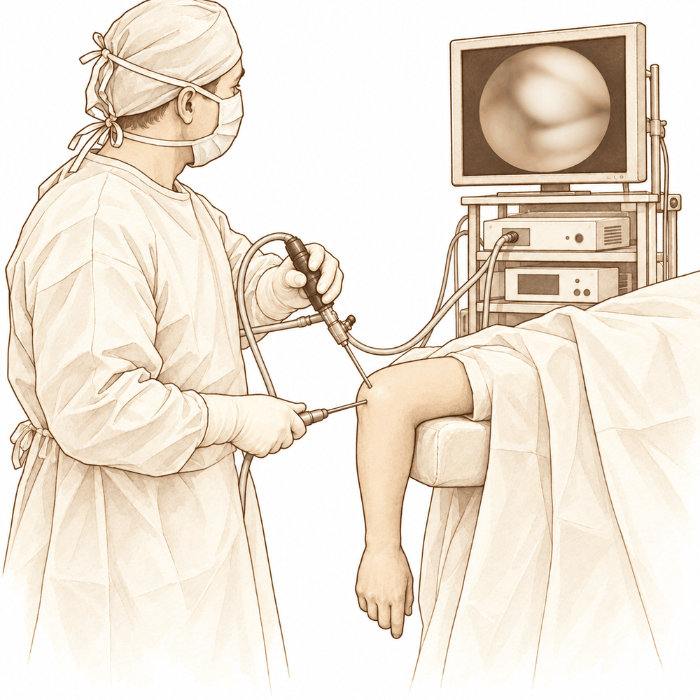
یہ آپریشن کیوں تجویز کیا گیا ہے
ڈاکٹر کیرن ہِرپرا، جو Mater Private Hospital Rockhampton میں بازو اور ہاتھ کے سرجن (upper-limb surgeon) ہیں، علاج کا آغاز ان طریقوں سے کرتے ہیں جن میں جسم پر کم سے کم مداخلت ہو اور جو آپ کے مسئلے کے لیے موزوں ہوں۔ عام طور پر مریضوں کو ان کا فیملی ڈاکٹر (GP) ریفرل (referral) دے کر ہمارے کلینک بھیجتا ہے؛ اگر کسی فزیوتھراپسٹ نے آپ کو ہمیں دکھانے کا مشورہ دیا ہے، تب بھی میڈیکیئر (Medicare) کی طرف سے رقم کی واپسی (rebate) کا حق دار ہونے کے لیے آپ کو اپنے فیملی ڈاکٹر سے ریفرل لینا ہوگا۔ ملاقات پر ہم آپ سے آپ کی تکلیف کی تفصیل پوچھتے ہیں، آپ کی کہنی کا معائنہ کرتے ہیں، اور جہاں ضرورت ہو اسکین کرواتے ہیں۔ پرانے مسائل میں ہم عام طور پر پہلے آپریشن کے بغیر علاج آزماتے ہیں، جیسے سرگرمیوں میں تبدیلی، فزیوتھراپی یا ہینڈ تھراپی (ہاتھ کی بحالی کا علاج)، اسپلنٹ اور انجکشن۔ سرجری کی بات تب ہوتی ہے جب ان اقدامات سے آپ کو کافی بہتری نہ ملی ہو۔
کہنی کی آرتھروسکوپی (دوربین سے جوڑ کے اندر دیکھ کر علاج) دوربین کے ذریعے آپریشن (keyhole) ہے۔ سرجن چھوٹے چیروں کے ذریعے جوڑ کے اندر کام کرتے ہیں، اور ایک پتلے کیمرے کی مدد سے پوری کہنی کو صاف دیکھتے ہیں۔ ہم یہ آپریشن اس وقت تجویز کرتے ہیں جب جوڑ کے اندر کوئی واضح مسئلہ ہو جس کا علاج اس طرح ہو سکتا ہو، جیسے ہڈی یا کارٹلیج (cartilage)، یعنی جوڑ میں ہڈیوں کے سروں پر چکنی، لچکدار تہہ، کے ڈھیلے ٹکڑے جو اٹکتے ہیں اور جوڑ کو جکڑ دیتے ہیں، اکڑی ہوئی کہنی، یا چوٹ کے بعد کا نقصان۔ یہ کہنی کے ہر مسئلے کے لیے موزوں نہیں، اس لیے ہم اسے صرف تب پیش کرتے ہیں جب یہ آپ کی تشخیص کے مطابق ہو۔ مقصد روزمرہ زندگی میں کم درد، زیادہ آزاد حرکت، اور زیادہ مستحکم کہنی ہے۔
آپریشن سے پہلے
سرجری سے پہلے کے ہفتوں میں ہم اسکین کے ذریعے منصوبے کی تصدیق کرتے ہیں۔ زیادہ تر لوگوں کا پہلے ہی ایکسرے ہو چکا ہوتا ہے، جو ہڈیوں کی بنیادی تصویر ہے۔ کچھ لوگوں کو ایم آر آئی اسکین کی بھی ضرورت ہوتی ہے، جو نرم بافتیں دکھاتا ہے جیسے لیگامنٹ (ligament)، یعنی وہ مضبوط بندھن جو ایک ہڈی کو دوسری ہڈی سے جوڑتا ہے، ٹینڈن (tendon)، یعنی وہ مضبوط ڈوری جو پٹھے کو ہڈی سے جوڑتی ہے، اور کارٹلیج، یا پھر الٹراساؤنڈ کی۔ اگر آپ کو کوئی اور بیماری ہے تو ہو سکتا ہے آپ کو خون کے ٹیسٹ یا بے ہوشی کے ڈاکٹر (anaesthetist)، یعنی وہ ڈاکٹر جو آپ کو بے ہوشی کی دوا دیتے ہیں، سے معائنے کی ضرورت ہو۔ زیادہ تر لوگوں کو ان میں سے کسی کی ضرورت نہیں ہوتی۔
آپریشن کے دن، سات گھنٹے پہلے کھانا پینا بند کر دیں۔ ہم سات گھنٹے اس لیے کہتے ہیں تاکہ اگر آپریشن کی فہرست وقت سے پہلے چلنے لگے تو ہم آپ کا آپریشن پہلے کر سکیں۔ آپ کے سرجن آپ کو بتائیں گے کہ آپ اپنی معمول کی دواؤں میں سے کون سی نہ لیں۔ جو کچھ آپ لیتے ہیں اس سب کی لکھی ہوئی فہرست ساتھ لائیں۔ کسی کو ساتھ رکھیں جو بعد میں آپ کو گاڑی میں گھر لے جائے، اور ڈھیلے، آرام دہ کپڑے پہنیں جن کی آستینیں آسانی سے کہنی پر سے گزر جائیں۔
آپریشن کے دن
آپ ہسپتال کے سرجیکل داخلے کے شعبے میں پہنچتے ہیں، جہاں آپ کا اندراج ہوتا ہے اور آپ کو آپریشن تھیٹر کے لیے تیار کیا جاتا ہے۔ پھر آپ کی ملاقات بے ہوشی کے ڈاکٹر سے ہوتی ہے، یعنی وہ ڈاکٹر جو آپ کو بے ہوشی کی دوا دیتے ہیں۔ یہ آپریشن جنرل اینستھیزیا (مکمل بے ہوشی) میں کیا جاتا ہے۔ آپریشن کے بعد درد سے آرام کے لیے کبھی کبھی اعصابی بلاک (nerve block)، یعنی ایک انجکشن جو جسم کے ایک حصے کو سُن کر دیتا ہے، بھی دیا جاتا ہے؛ بے ہوشی کے ڈاکٹر اسی دن آپ سے اس بارے میں بات کریں گے۔
پھر آپ کو آپریشن تھیٹر لے جایا جاتا ہے، جہاں آپریشن کیا جاتا ہے۔ اس کے بعد آپ ریکوری روم میں ہوش میں آتے ہیں، جہاں نرسیں آپ کی نگرانی کرتی ہیں جب تک بے ہوشی کی دوا کا اثر ختم ہوتا ہے۔ جب آپ کی حالت مستحکم ہو جائے تو طریقۂ کار اور آپ کی صحت یابی کی رفتار کے مطابق آپ یا تو وارڈ میں منتقل ہو جاتے ہیں یا اسی دن گھر چلے جاتے ہیں۔
آپریشن میں کیا کیا جاتا ہے
کہنی کی آرتھروسکوپی دوربین کے ذریعے آپریشن ہے۔ آپ کے سرجن کہنی کے گرد دو یا تین چھوٹے چیرے لگاتے ہیں، ہر ایک تقریباً ایک سینٹی میٹر لمبا۔ ایک چیرے سے پتلا کیمرا ڈالا جاتا ہے، اور باقی چیروں سے چھوٹے اوزار جوڑ تک پہنچتے ہیں۔ اوزار ڈالنے سے پہلے جوڑ کے اندر تقریباً 25 سے 30 ملی لیٹر (mL) مائع ڈالا جاتا ہے۔ یہ مائع کام کرنے کے لیے جگہ کھول دیتا ہے تاکہ کیمرا صاف دیکھ سکے۔ چیرے ان بڑے اعصاب اور خون کی نالیوں سے دور لگائے جاتے ہیں جو کہنی کے پاس سے گزرتی ہیں، اور آپ کے سرجن ہر چیرا محفوظ جگہ پر لگانے کے لیے پہلے آپ کی جلد پر ہڈیوں کے نشانات بنا لیتے ہیں۔
اندر کیا کیا جاتا ہے، یہ آپ کی تشخیص پر منحصر ہے۔ ہڈی یا کارٹلیج کے ڈھیلے ٹکڑے جو اٹکتے ہیں اور جوڑ کو جکڑ دیتے ہیں، نکالے جا سکتے ہیں۔ اگر کہنی اکڑی ہوئی ہو تو جوڑ کے گرد کی تنگ بافتوں کو ڈھیلا کیا جا سکتا ہے تاکہ حرکت آزاد ہو۔ خراب کارٹلیج کو ہموار کیا جا سکتا ہے، اور بعض صورتوں میں ہڈی یا کارٹلیج کا ایک چھوٹا ٹکڑا ایسی پنوں کے ذریعے واپس اپنی جگہ پر لگایا جا سکتا ہے جو وقت کے ساتھ جسم میں گھل جاتی ہیں۔ چونکہ کیمرا پورے جوڑ کا صاف منظر دکھاتا ہے، اس لیے آپ کے سرجن ان مسائل کا بھی علاج کر سکتے ہیں جو اسکین میں نظر نہیں آئے تھے۔
چھوٹے چیروں کو ٹانکوں سے بند کر کے ان پر زخم کی پٹی لگا دی جاتی ہے۔ آپریشن ایک لمبے چیرے کے بجائے چھوٹے چیروں کے ذریعے کیا جاتا ہے، اس لیے کھلی سرجری کے مقابلے میں کہنی کے گرد کی نرم بافتوں کو کم چھیڑا جاتا ہے۔
آپریشن کے بعد
آپ ریکوری روم میں ہوش میں آتے ہیں، جہاں نرسیں آپ پر نظر رکھتی ہیں جب تک بے ہوشی کی دوا کا اثر ختم ہوتا ہے۔ آپ کا بازو سلنگ (sling)، یعنی گلے سے لٹکی وہ پٹی جو بازو کو سہارا دیتی ہے، یا ایک سادہ پٹی میں ہوگا، اور جیسے ہی آپ خود کو سنبھلا ہوا محسوس کریں، آپ چل پھر سکتے ہیں۔ درد کی دوا کا منصوبہ آپریشن تھیٹر سے نکلنے سے پہلے بنا لیا جاتا ہے، اس لیے اگر آپ کی کہنی میں درد ہو تو نرسوں کو بتائیں، وہ اسے بدل سکتی ہیں۔ گھر پہنچنے کے بعد پہلے 24 گھنٹے کسی کو آپ کے ساتھ رہنا چاہیے۔ آپ کی ٹیم آپ کو بتائے گی کہ آپ اسی دن گھر جائیں گے یا ایک رات ہسپتال میں رہیں گے۔ ہم زخم کی پٹی تقریباً 10 دن تک لگی رہنے دیتے ہیں؛ براہِ کرم اس سے پہلے اسے نہ اتاریں، جب تک ہم آپ کو ایسا کرنے کو نہ کہیں۔ جب ہم آپ کو دیکھیں گے تو اسے بدل دیں گے یا اتار دیں گے۔
صحت یابی
پہلے چند دن آپ کی کہنی میں درد اور سوجن رہے گی۔ یہ آہستہ آہستہ ٹھیک ہو جاتی ہے۔ آرام، جب ممکن ہو اپنا ہاتھ اونچا رکھنا، اور آپ کے لیے تجویز کی گئی درد کی دوا، سب مدد کرتے ہیں۔ بہت سے لوگوں کو لگتا ہے کہ تکلیف شروع میں سب سے زیادہ ہوتی ہے اور سوجن کم ہونے کے ساتھ مسلسل کم ہوتی جاتی ہے۔
آپ گھر اس حالت میں جائیں گے کہ آپ کا بازو سلنگ یا ایک سادہ پٹی میں ہوگا۔ جیسے ہی آرام محسوس ہو، آپ ہلکے کاموں کے لیے اپنا ہاتھ استعمال کر سکتے ہیں، جیسے کھانا کھانا، ٹائپ کرنا یا بٹن بند کرنا۔ سرجری کے بعد ہینڈ تھراپی روبی ڈولن (Ruby Doolan) کے ساتھ Extend Rehabilitation میں ہوتی ہے۔ روبی ایک ہینڈ تھراپسٹ (ہاتھ کی بحالی کی ماہر) ہیں: وہ آپ کی ورزشوں میں رہنمائی کریں گی اور آپ کو جس اسپلنٹ کی ضرورت ہو وہ بنائیں گی۔ یہ ورزشیں زخم بھرنے کے دوران آپ کی کہنی اور کلائی کو حرکت میں رکھتی ہیں، اور یہ خود آپریشن جتنی ہی اہم ہیں۔ انہیں ہدایت کے مطابق کریں، اور اگر کسی حرکت سے تیز درد ہو تو اپنے تھراپسٹ کو بتائیں۔
روزمرہ سرگرمیاں مرحلہ وار واپس آتی ہیں۔ جب سوجن کم ہو جائے گی تو آپ محسوس کریں گے کہ آپ کی گرفت مضبوط ہو رہی ہے اور چیزوں تک ہاتھ بڑھانا آسان ہو رہا ہے۔ جب آپ کے سرجن کہنی کی حرکت اور بھرنے سے مطمئن ہوں گے تو آپ کو گاڑی چلانے کی اجازت مل جائے گی؛ ہماری رہنما تحریر بازو اور ہاتھ کی سرجری کے بعد گاڑی چلانا لاگو ہونے والے اصول بتاتی ہے، جن میں یہ بھی شامل ہے کہ جب تک آپ کا بازو سلنگ میں ہو گاڑی نہ چلائیں اور تیز درد کش دوائیں چھوڑ چکے ہوں۔ کام اور کھیل تب دوبارہ شروع ہوتے ہیں جب آپ انہیں بغیر درد کے کر سکیں، اور آپ کے سرجن یا تھراپسٹ آپ کو بتائیں گے کہ وہ وقت کب آ گیا ہے۔
صحت یابی ہر شخص میں مختلف ہوتی ہے۔ آپ کا وقت مختلف ہو سکتا ہے، اور آپ کے سرجن اور ہینڈ تھراپسٹ پورے راستے آپ کی رہنمائی کریں گے۔
کیا مسائل پیدا ہو سکتے ہیں
زیادہ تر مریض ٹھیک رہتے ہیں، لیکن کبھی کبھار مسائل پیدا ہو سکتے ہیں۔ آپ کے سرجن اور ٹیم آپ کی قریب سے نگرانی کرتے ہیں تاکہ کسی بھی مسئلے کو جلد پکڑا جا سکے۔
اعصاب میں جلن (irritation) سب سے زیادہ دیکھا جانے والا مسئلہ ہے۔ آپ کو جھنجھناہٹ، سوئیاں چبھنے جیسا احساس، سُن پن، یا جلد کا کوئی ایسا حصہ محسوس ہو سکتا ہے جو مختلف لگے۔ یہ عام طور پر عارضی ہوتا ہے اور خود ہی ٹھیک ہو جاتا ہے۔ اگر ایسا ہو تو اگلے معائنے پر اپنے سرجن کو بتائیں۔
سرجری کے بعد کے دنوں میں انفیکشن ہو سکتا ہے۔ گہرے، دھڑکتے ہوئے درد پر نظر رکھیں جو عام درد کش دوا سے کم نہ ہو، زخم سے باہر کی طرف پھیلتی ہوئی سرخی، یا پٹی میں سے رسنے والا مائع۔ اگر آپ ان میں سے کچھ بھی دیکھیں تو فوراً کلینک کو فون کریں۔ گہرے انفیکشن کا جلد علاج ضروری ہے۔
کچھ چیزیں انفیکشن کا امکان بڑھا دیتی ہیں۔ اگر آپ کو ذیابیطس (شوگر) ہے، اگر آپ تمباکو نوشی کرتے ہیں، یا اگر آپ کا وزن زیادہ ہے، تو آپ کا خطرہ زیادہ ہے۔ جوڑ میں اسٹیرائیڈ (کورٹیسون) کا انجکشن، چاہے سرجری سے کچھ پہلے لگایا جائے یا آپریشن کے آخر میں، بھی خطرہ بڑھاتا ہے۔ آپ کے سرجن پہلے سے آپ کے ساتھ مل کر ان سب باتوں پر غور کریں گے۔
بہت کم صورتوں میں عصب کی چوٹ مستقل ہو سکتی ہے۔ ایسا کم ہوتا ہے، لیکن جب ہوتا ہے تو اس کے اثرات عمر بھر رہتے ہیں۔ سرجری کے لیے آپ کی رضامندی سے پہلے آپ کے سرجن آپ سے اس خطرے پر بات کریں گے۔
کبھی کبھی جوڑ کے اندر یا اس کے گرد ایسی جگہ ہڈی بن جاتی ہے جہاں اسے نہیں بننا چاہیے۔ اسے ہیٹروٹوپک اوسیفیکیشن (heterotopic ossification) کہتے ہیں۔ آپ کو کہنی کے قریب ایک سخت گانٹھ محسوس ہو سکتی ہے، یا آپ دیکھ سکتے ہیں کہ جوڑ پہلے جتنا مڑ یا سیدھا نہیں ہو رہا۔ اپنے معائنے پر اس کا ذکر کریں تاکہ اسے چیک کیا جا سکے۔
کچھ اور مسائل کم ہوتے ہیں لیکن معلوم ہیں۔ کیمرا یا کوئی اوزار جوڑ کی چکنی سطح کو چھو کر رگڑ سکتا ہے۔ جوڑ سے باہر جلد تک مائع نکلنے کا ایک چھوٹا سا راستہ بن سکتا ہے۔ کوئی اوزار جوڑ کے اندر ٹوٹ سکتا ہے۔ سرجری کے دوران بازو پر باندھا جانے والا کف (cuff) بازو پر دباؤ ڈال سکتا ہے اور اس کے نیچے کی جلد میں تھوڑی دیر کے لیے جلن پیدا کر سکتا ہے۔
اگر سرجری کے بعد کے دنوں میں آپ کی طبیعت خراب ہو، یا آپ کی کہنی کے بارے میں کچھ ٹھیک نہ لگے، تو اگلی ملاقات کا انتظار کرنے کے بجائے کلینک کو فون کریں۔ اگر آپ تفصیل جاننا چاہیں تو اس صفحے پر پیچیدگیوں کی جدول میں ان کی عام شرحیں دی گئی ہیں۔
ہمیں کب فون کریں
زیادہ تر مسائل جلد ظاہر ہو جاتے ہیں، اور ہم چاہیں گے کہ آپ انتظار کرنے کے بجائے ہمیں اسی دن بتائیں۔ اگر آپ کی کہنی گرم، زیادہ سوجی ہوئی یا زیادہ دردناک ہو جائے، اگر زخم سے سرخی پھیلے، یا پٹی میں سے مائع رسے تو ہمیں فون کریں۔ اگر آپ کو بخار جیسا محسوس ہو، اگر جھنجھناہٹ یا سُن پن کم ہونے کے بجائے بڑھ جائے، یا آپ اپنا ہاتھ یا انگلیاں پہلے کی طرح حرکت نہ دے سکیں تو ہمیں فون کریں۔ اگر آپ کو اچانک شدید درد ہو، پنڈلی کے پچھلے حصے میں نئی سوجن ہو، یا سانس پھولے، تو ہسپتال کی ایمرجنسی جائیں۔ یہ خون کے لوتھڑے کی علامت ہو سکتی ہیں، اور ان کا فوراً معائنہ ضروری ہے۔ اگر بس کچھ ٹھیک محسوس نہ ہو تو ہمیں فون کریں۔
مزید تفصیل
Advanced reading: the deeper science (optional)
یہ حصہ اس سے آگے جاتا ہے جتنا آپ کو اپنے علاج کے فیصلوں کے لیے جاننے کی ضرورت ہے۔ کہنی کی آرتھروسکوپی کے بارے میں یہ اضافی مطالعہ اس لیے مفید ہے کہ اس کی شائع شدہ پیچیدگیوں کی شرح کوئی ایک عدد نہیں بلکہ اتنی وسیع حد ہے کہ غلطی لگتی ہے، اور یہ سمجھنا کہ ایسا کیوں ہے، اس آپریشن کی زیادہ تر اہم باتیں واضح کر دیتا ہے۔
ایک ہی آپریشن، دو بالکل مختلف جواب
ایک منظم جائزہ (systematic review)، جس میں کہنی کی 16,815 آرتھروسکوپیوں کو یکجا کیا گیا، نے پیچیدگیوں کی درمیانی (median) شرح 3 فیصد بتائی، جبکہ مختلف تحقیقات میں یہ شرح 0 فیصد سے 71 فیصد تک تھی [1]۔ دوبارہ آپریشن میں بھی یہی صورت تھی: درمیانی شرح 2 فیصد، حد 0 فیصد سے 59 فیصد [1]۔ 14,289 آپریشنوں کے ایک الگ میٹا اینالیسس (meta-analysis) نے معقول طور پر یہ نتیجہ نکالا کہ کہنی کی آرتھروسکوپی نسبتاً محفوظ آپریشن ہے جس میں پیچیدگیوں کی شرح کم ہے [2]۔
دونوں درست ہیں۔ ان میں مطابقت پہلے جائزے کے اپنے نتیجے میں ہے: زیادہ پیچیدہ سرجری کے بعد پیچیدگیوں کی شرح زیادہ ہوتی ہے [1]۔ "کہنی کی آرتھروسکوپی" کوئی ایک آپریشن نہیں ہے۔ ایک کشادہ جوڑ سے ڈھیلا ٹکڑا (loose body) نکالنا، اور النر اور ریڈیل اعصاب سے چند ملی میٹر کے فاصلے پر سکڑی ہوئی جوڑ کی جھلی (capsule) کو ڈھیلا کرنا، دونوں کا نام اور اوزار ایک ہے، اور اس کے علاوہ تقریباً کچھ بھی مشترک نہیں۔ خطرے کا بتایا گیا کوئی عدد تقریباً بے معنی ہے جب تک اسے اس مخصوص طریقۂ کار سے نہ جوڑا جائے جو کیا جا رہا ہے۔
کہنی گھٹنے جیسی کیوں نہیں
جن جوڑوں میں معمول کے طور پر دوربین سے آپریشن کیا جاتا ہے، ان میں کہنی وہ جوڑ ہے جس میں جوڑ اور اعصاب کا فاصلہ سب سے کم ہے۔ النر، ریڈیل اور میڈین اعصاب سب دوربین ڈالنے کی معیاری جگہوں (portal sites) سے چند ملی میٹر کے اندر ہوتے ہیں، اور کام کرنے کی جگہ چھوٹی ہے، اور بالکل انہی اکڑی ہوئی، سکڑی ہوئی کہنیوں میں اور بھی چھوٹی ہوتی ہے جنہیں اس آپریشن کی سب سے زیادہ ضرورت ہے۔ یہی بناوٹ وجہ ہے کہ پیچیدگیوں کی جدولوں میں عارضی اعصابی علامات سب سے زیادہ ہیں، اور یہ کہ خطرہ آپریشن میں لگنے والے وقت کے بجائے ڈھیلا کرنے (release) کی وسعت کے ساتھ بڑھتا ہے۔
جہاں دوربین والے طریقے کا کھلی سرجری سے موازنہ کیا گیا ہے
آرتھرائٹس (جوڑ کی خرابی اور سوزش) والی کہنیوں کے لیے، 1,097 مریضوں کے ایک منظم جائزے میں جوڑ کی صفائی والے آپریشن (debridement arthroplasty) کے بعد درمیانی مدت میں اچھی کارکردگی پائی گئی، اور کھلی سرجری کے مقابلے میں دوربین والے طریقے سے پیچیدگیوں میں کوئی اضافہ نہیں ہوا [3]۔ یہ برتری کا ثبوت نہیں بلکہ حقیقی برابری کا نتیجہ ہے۔
ٹینس ایلبو (کہنی کے باہر کی طرف درد) کے لیے، 1,604 مریضوں کے ایک جائزے میں کھلی، دوربین والی اور جلد میں چھوٹے سوراخ کے ذریعے کی جانے والی ریلیز (percutaneous release) کا موازنہ کیا گیا، اور پایا گیا کہ کھلی اور دوربین والی ریلیز سے چھوٹے سوراخ والے طریقے کے مقابلے میں بہتر کارکردگی مل سکتی ہے، جبکہ دوربین والے اور چھوٹے سوراخ والے طریقوں میں درد کم تھا۔ طریقہ کوئی بھی ہو، پیچیدگیوں کا خطرہ ملتا جلتا تھا [4]۔
دونوں میں ایک ہی رجحان ہے: آرتھروسکوپی اپنے آپ میں زیادہ محفوظ یا زیادہ مؤثر نہیں ہے۔ جہاں یہ بہتر ہے، وہاں آپریشن کے نتیجے میں نہیں بلکہ آپریشن کے آس پاس کی صحت یابی میں بہتر ہے۔
ایک دیانت دارانہ منفی نتیجہ
کسی دوسرے آپریشن کے ساتھ "ایک نظر دیکھنے کے لیے" آرتھروسکوپی شامل کرنے کا فائدہ نہیں ہوتا۔ 2,118 مریضوں کے ایک میٹا اینالیسس میں، کہنی کے اندرونی لیگامنٹ کی دوبارہ تعمیر (ulnar collateral ligament reconstruction) سے پہلے معمول کے طور پر کی جانے والی تشخیصی آرتھروسکوپی نے ویلگس ایکسٹینشن اوورلوڈ (valgus extension overload) کے دوسرے مسائل کے لیے بعد میں ہونے والی سرجری کو شماریاتی طور پر نمایاں (statistically significant) حد تک کم نہیں کیا [5]۔ دیکھنا اس بات کے برابر نہیں کہ علاج کے قابل کچھ مل بھی جائے، اور ہر اضافی سوراخ کے ساتھ اوپر بیان کیا گیا اعصابی خطرہ جڑا ہوتا ہے۔
حوالہ جات
[1] de Klerk HH, Verweij LP, Sierevelt IN, Priester-Vink S, Hilgersom NF, Eygendaal D, et al. Wide range in complication rates following elbow arthroscopy in adult and pediatric patients: a systematic review. Arthroscopy. 2023;39(11):2363-87. https://doi.org/10.1016/j.arthro.2023.04.015
[2] Ahmed AF, Alzobi OZ, Hantouly AT, Toubasi A, Farsakoury R, Alkhelaifi K, et al. Complications of elbow arthroscopic surgery: a systematic review and meta-analysis. Orthop J Sports Med. 2022;10(11). https://doi.org/10.1177/23259671221137863
[3] White CHR, Ravi V, Watson J, Badhrinarayanan S, Phadnis J. A systematic review of arthroscopic versus open debridement of the arthritic elbow. Arthroscopy. 2020;37(2):747. https://doi.org/10.1016/j.arthro.2020.09.005
[4] Pierce TP, Issa K, Gilbert BT, Hanly B, Festa A, McInerney VK, et al. A systematic review of tennis elbow surgery: open versus arthroscopic versus percutaneous release of the common extensor origin. Arthroscopy. 2017;33(6):1260. https://doi.org/10.1016/j.arthro.2017.01.042
[5] Looney AM, Bovill JD, Huffman SS, Najarian RG. Routine diagnostic arthroscopy with elbow ulnar collateral ligament reconstruction does not reduce the need for future valgus extension overload-related surgeries: a systematic review and meta-analysis. J Shoulder Elbow Surg. 2022;31(1):e22-e36. https://doi.org/10.1016/j.jse.2021.08.004
Evidence & references
This is the clinical evidence summary written for health professionals. It is technical, and it lists the research this page was built from. You do not need to read it to understand your treatment or to make a decision about it.
Overview
- Elbow arthroscopy is not without complications and morbidity despite being a minimally invasive procedure and advances made in surgical technique [1].
- The available evidence supports the use of elbow arthroscopy in the management of the majority of conditions where it is currently used [2].
- Patients treated arthroscopically benefit from additional diagnostic techniques, improved visualization of the elbow joint, the ability to address coexisting intra-articular pathologic conditions, and minimal soft tissue injury with no clinical consequences in outcomes [3].
- Elbow arthroscopy has become a safer and more effective treatment modality for several elbow pathologies due to advances in equipment and surgical technique [6].
- Elbow arthroscopy has evolved from a diagnostic tool to a therapeutic procedure with expanded indications, though it requires careful attention to surgical anatomy and patient selection to avoid neurovascular complications [7].
- Elbow arthroscopy has expanded indications for diagnosing and treating acute trauma, including radial head fractures and instability, provided patients are carefully selected and neurovascular risks are managed with proper technique [12].
- Predominantly low-level evidence studies demonstrate varying complication rates (median 3%, range 0%-71%) and reoperation rates (median 2%, range 0%-59%) after elbow arthroscopy [5].
- Elbow arthroscopic surgery is a relatively safe procedure with low complication rates [15].
- Elbow arthroscopic debridement for primary degenerative osteoarthritis results in statistically significant and clinically relevant improvement in elbow range of motion and clinical outcomes with low complication and reoperation rates [14].
- Pediatric elbow arthroscopy performed by an experienced surgeon using a standardized technique for a wide variety of elbow conditions has an acceptable complication rate that is similar to rates in the previously published literature on elbow arthroscopy in the pediatric and adult populations [17].
- A significant proportion of pediatric patients who underwent elbow arthroscopy needed subsequent surgery in the following years [17].
Anatomy & Pathophysiology
Bony Anatomy
- The elbow is a trocho-ginglymoid joint consisting of medial and lateral articulations that provide bony stability [39].
- The ulnohumeral joint is formed by the articulation of the trochlea with the ulna within the greater sigmoid notch [39].
- The ulnohumeral articulation provides highly congruent anatomy through almost 180° of articular contact, with the exception of a bare area on the greater sigmoid notch devoid of cartilage [39].
- The coronoid process has medial and lateral facets that buttress the trochlea anteriorly [39].
- The sublime tubercle is located just distal and medial to the coronoid and serves as the attachment site for the anterior bundle of the medial ulnar collateral ligament [39].
- The radiocapitellar joint is formed by the articulation of the capitellum and radial head [39].
- The radial head is a concave elliptical structure covered with articular cartilage along the radiocapitellar joint and approximately 270° of the articular margin [39].
- The radius is held in close approximation to the ulna at the proximal radioulnar joint by the annular ligament [39].
- The area of the ulna that articulates with the margin of the radial head at the proximal radioulnar joint is known as the lesser sigmoid notch [39].
- The distal humeral articulation is angled 30° from the longitudinal axis [39].
- The axis of rotation is angulated 5° to 7° in the coronal plane relative to the epicondylar axis, with the medial side more distal than the lateral side [39].
- The ulna medially bends approximately 8° at 8 cm from the tip of the olecranon [39].
- The articulation to the tip of the coronoid is approximately 30° from the long axis of the ulna in the sagittal plane [39].
- The articular surface of the distal humerus is angled 30 degrees anterior to the humeral shaft axis [42, 43].
- The trochlea has a 300-degree arc of cartilage [44].
- The medial column diverges from the humeral shaft at a 45-degree angle [44].
- The lateral column diverges from the humeral shaft at a 20-degree angle [44].
- The olecranon fossa is an oval structure that is wider in the medial to lateral dimension [34].
Ligamentous Anatomy
- Elbow stability is determined by primary and secondary stabilizers [20].
- The three primary stabilizers of the elbow are the ulnohumeral articulation, the medial ulnar collateral ligament (MUCL), and the lateral ulnar collateral ligament (LUCL) complex [20].
- Secondary stabilizers of the elbow include the radiocapitellar articulation, the common flexor tendon, the common extensor tendon, and the joint capsule [20].
- The medial ulnar collateral ligament complex is comprised of the anterior bundle, posterior bundle, and transverse ligament [38].
- The anterior bundle of the medial ulnar collateral ligament is the strongest component and the primary restraint to valgus stress [38].
- The anterior bundle of the medial ulnar collateral ligament is further subdivided into anterior and posterior bands that provide reciprocal function, with the anterior band tight in extension and the posterior band tight in flexion [38].
- The medial collateral ligament originates on the posterior medial epicondyle and inserts on the sublime tubercle of the medial coronoid process [42, 43].
- The posterior bundle of the medial collateral ligament is the primary restraint to valgus stress with the elbow in maximal flexion [42, 43].
- Stability in full extension is provided by the medial collateral ligament, joint capsule, and ulnohumeral articulation [42, 43].
- The lateral ulnar collateral ligament origin center is located 10.7 mm from the lateral epicondyle [38].
- The lateral ulnar collateral ligament insertion is located 3.3 mm from the apex of the supinator crest [38].
Range of Motion & Biomechanics
- The normal elbow has a range of motion from 0° to 140° from extension to flexion [20].
- The normal elbow has a range of motion of 75° in pronation and 85° in supination [20].
- A functional arc of motion for the elbow is 100° for flexion and extension [20].
- A functional arc of motion for forearm rotation is 100° [20].
- The normal range of elbow flexion/extension is 0 to 150 degrees [42, 43].
- The normal forearm pronosupination is 80 to 85 degrees in each direction [42, 43].
- The functional range of motion for elbow flexion/extension is 30 to 130 degrees [42, 43].
- The functional range of motion for forearm pronosupination is 50 degrees [42, 43].
- The normal valgus carrying angle of the elbow is 5 to 10 degrees for men and 10 to 15 degrees for women [42, 43].
- In full extension, 60% of axial load is transmitted through the radiocapitellar joint [42, 43].
Pathophysiology
- Valgus extension overload syndrome is caused by the olecranon being repeatedly and forcefully driven into the olecranon fossa during throwing, exerting shear forces on the medial aspect of the olecranon tip and the olecranon fossa [21].
- The pathoanatomy of valgus extension overload syndrome includes chondrosis, osteophyte development on the posteromedial olecranon and humerus, and loose bodies [21].
- Medial ligamentous laxity commonly exacerbates valgus extension overload syndrome [21].
- Osteoarthritis of the elbow is characterized by osteophyte formation, capsular contracture, and loose bodies, often with relative preservation of the joint space [34].
- Periarticular hypertrophic osteophytes in elbow osteoarthritis act as a mechanical block at the end ranges of flexion and extension [34].
- Elbow osteoarthritis typically involves the radiocapitellar joint articular cartilage preferentially, with relative preservation of the ulnohumeral articular surfaces [34].
- Stiffness of the elbow may be due to intrinsic (intra-articular) or extrinsic (extra-articular) causes [54].
- Intrinsic causes of elbow stiffness include articular damage or malunion, intraarticular hardware, and loose bodies [54].
- Extrinsic causes of elbow stiffness include a contracted joint capsule and ligaments, heterotopic ossification, prominent hardware, and skin contracture [54].
- Posttraumatic or postsurgical elbow contractures often involve a damaged, abnormally thickened joint capsule and potential for altered anatomy [54].
- In posttraumatic or postsurgical elbow contractures, the joint space is usually contracted and the capsule is scarred down to the bones [54].
- In posttraumatic or postsurgical elbow contractures, the locations of the median and radial nerves may be altered due to contracted anterior soft tissues [54].
- Lateral epicondylitis is an angiofibroblastic hyperplasia of the extensor carpi radialis brevis tendon, a noninflammatory, dysvascular degenerative process caused by repetitive microtrauma [56].
- Osteochondritis dissecans (OCD) of the elbow is caused by repetitive microtrauma to the vulnerable epiphysis, which has a tenuous blood supply [56].
- Repetitive loading of the lateral compartment of the elbow results in subchondral bone degeneration causing cartilage fragmentation in OCD [56].
- During arthroscopic surgery, joint distention moves the capsule away from bone, but the distance between the neurovascular structures and the capsule remains unchanged [34].
- The brachialis muscle protects the median nerve and brachial artery during capsular procedures [34].
Classification
- The Kwak classification categorizes primary elbow osteoarthritis into Stage I, Stage II, and Stage III [71].
- In a retrospective cohort study of 43 patients undergoing arthroscopic osteocapsular arthroplasty, 14 patients were classified as Kwak Stage I, 18 as Stage II, and 11 as Stage III [71].
- Radiologically severe osteoarthritis is associated with worse clinical outcomes after arthroscopic osteocapsular arthroplasty in primary elbow osteoarthritis at medium-term follow-up [71].
- The Mayo Elbow Performance Score (MEPS) classification defines excellent outcomes as a score greater than 89 [72].
- The Mayo Elbow Performance Score (MEPS) classification defines good outcomes as a score between 75 and 89 [72].
- The Mayo Elbow Performance Score (MEPS) classification defines fair outcomes as a score between 60 and 74 [72].
- The Mayo Elbow Performance Score (MEPS) classification defines poor outcomes as a score less than 60 [72].
Clinical Presentation
General Evaluation Principles
- The evaluation of the elbow requires an intimate understanding of anatomy, biomechanics, and diagnostic tests to understand functional biomechanics, perform physical examination maneuvers, and order appropriate imaging studies [20].
- A thorough history and physical examination are invaluable for understanding the type of disease process and the degree to which the condition affects the patient [57].
- Understanding whether a patient has pain throughout the arc of motion or only at terminal limits, as well as any associated mechanical symptoms or instability, is of paramount importance [57].
- Associated conditions such as cubital tunnel syndrome must be considered and evaluated to provide optimal recommendations on management [57].
- The physical examination is directed by the history and the location of the patient's pain in the anterior, posterior, medial, or lateral aspect of the elbow [20].
- The location, quality or type, context, duration, and severity of elbow pain are important for understanding pathology and focusing the physical examination [65].
- Determining the symptom trajectory—whether pain is getting better, worse, or remaining constant over a period of time—is extremely helpful when considering intervention [65].
- A working knowledge of pathologic conditions affecting different locations around the elbow is paramount to making a correct diagnosis [65].
Range of Motion and Stability
- The normal elbow has 75° of pronation and 85° of supination [20].
- A functional arc in each plane is 100° for flexion and extension and forearm rotation [20].
- Elbow stability is determined by primary and secondary stabilizers, and injury to these structures causes elbow instability [20].
Imaging
- Plain radiographs remain the hallmark and the best screening test for elbow evaluation [20].
- Plain radiographs should be obtained during the initial workup to evaluate the articular surface and bony anatomy [57].
- CT scans with 3D reconstructions may be useful for evaluating the extent and location of disease and for surgical planning [57].
- MRI may be useful to evaluate the status of soft tissues, including the medial and lateral collateral ligamentous complexes [57].
- Electromyography and nerve conduction studies (EMG/NCS) may be useful to evaluate the degree of nerve compression and its contribution to elbow pain or dysfunction [57].
Osteoarthritis
- Patients with primary osteoarthritis of the elbow typically present with loss of terminal extension and flexion and painful catching, clicking, or locking of the elbow [34].
- Pain in primary osteoarthritis is typically noted at the end ranges of motion and not through the midrange [34].
- Night pain is not typical for primary osteoarthritis; if present, an inflammatory cause of the arthritis should be considered [34].
- On physical examination for osteoarthritis, pain is usually felt at the end ranges of flexion and extension rather than throughout the arc [34].
- Forearm rotation is relatively preserved until later in the disease process of osteoarthritis [34].
- Ulnar neuropathy is present in up to 50% of patients with osteoarthritis of the elbow [34].
- Radiographs for osteoarthritis typically show osteophyte formation at the coronoid process, coronoid fossa, radial fossa, radial head, olecranon tip, and olecranon fossa [34].
- Radiographs typically underestimate the number of loose bodies present in osteoarthritis [34].
Valgus Extension Overload Syndrome
- Patients with valgus extension overload syndrome report posteromedial elbow pain that occurs during the deceleration phase of throwing as the elbow reaches terminal extension [21].
- Pain in valgus extension overload syndrome may also occur during the acceleration phase of throwing [21].
- Loss of terminal elbow extension may occur in patients with valgus extension overload syndrome [21].
- Crepitus and tenderness over the posteromedial olecranon may be noted on physical examination for valgus extension overload syndrome [21].
- Pain in valgus extension overload syndrome is reproduced when the elbow is forced into extension [21].
- Elbow flexion contracture may be seen in patients with valgus extension overload syndrome [21].
- CT with two-dimensional reconstruction and three-dimensional surface rendering best visualizes the pathology of valgus extension overload syndrome [21].
- MRI may be most helpful in evaluating associated injuries, including partial or complete tears of the medial collateral ligament (MCL), in valgus extension overload syndrome [21].
Medial Collateral Ligament Injuries
- Patients with medial collateral ligament (MCL) injuries report medial elbow pain during the acceleration phase of throwing [62].
- Pain in MCL injuries may occur only when throwing at more than 50% to 75% of maximal effort [62].
- Acute MCL injuries may present suddenly with a pop, sharp pain, and inability to continue throwing [62].
- Point tenderness can be noted at the MCL or toward its insertion sites during physical examination [62].
- Valgus instability is tested with the patient’s elbow flexed between 20° and 30° to unlock the olecranon from its fossa as valgus stress is applied [62].
- The milking maneuver is performed by pulling on the patient’s thumb to create valgus stress while the forearm is supinated and the elbow is flexed beyond 90° [62].
- A subjective feeling of apprehension, instability, or localized pain at the MCL during the milking maneuver indicates injury [62].
- The moving valgus stress test involves applying valgus stress while the elbow is moved through an arc of flexion or extension [62].
- A subjective feeling of apprehension, instability, or localized pain at the MCL during the moving valgus stress test indicates injury [62].
- Valgus stress radiographs with the elbow in 20° to 30° of flexion and the forearm in full supination may be used to measure medial joint line opening [62].
- Medial joint line opening greater than 3 mm on valgus stress radiographs has been considered diagnostic for valgus instability [62].
- Conventional MRI can help identify thickening within the MCL from chronic injury or more obvious full-thickness tears [62].
- Magnetic resonance arthrography enhanced with intra-articular gadolinium improves the diagnosis of partial undersurface tears of the MCL [62].
- Dynamic ultrasonography can help detect increased laxity with valgus stress, though diagnostic quality is operator dependent [62].
Lateral Elbow Pathology
- Patients with lateral elbow tendinopathy report pain when lifting things from a bag with a pronated hand, turning doorknobs, taking milk from the fridge, shaking hands, taking a laptop out of a bag, or bumping the lateral elbow [65].
- Direct palpation of the extensor carpi radialis brevis (ECRB) origin is a physical examination maneuver for lateral elbow tendinopathy [65].
- The tennis elbow shear test is a physical examination maneuver for lateral elbow tendinopathy [65].
- Pain along with resisted wrist or long finger extension is a physical examination finding for lateral elbow tendinopathy [65].
- The laptop test is a physical examination maneuver for lateral elbow tendinopathy [65].
- Patients with radial tunnel syndrome report extensor musculature "forearm aching" [65].
- Wrist flexion and forearm pronation are physical examination maneuvers to elicit radial tunnel syndrome [65].
- The rule of nines test is a physical examination maneuver for radial tunnel syndrome [65].
- Weakness and pain with resisted long finger extension are physical examination findings for radial tunnel syndrome [65].
- Patients with cutaneous neuritis (posterior antebrachial cutaneous nerve or lateral antebrachial cutaneous nerve) report burning or radiating pain, often described as "I want to cut my arm off" [65].
- Direct palpation or percussion, such as the Tinel test, are physical examination maneuvers for cutaneous neuritis [65].
- Patients with posterolateral rotatory instability report not trusting the elbow or feeling of giving way or instability when pushing out of a chair with arms [65].
- The posterolateral rotatory (PLR) drawer test is a physical examination maneuver for posterolateral rotatory instability [65].
- The PLR pivot shift test is a physical examination maneuver for posterolateral rotatory instability [65].
- The supinated push-up test is a physical examination maneuver for posterolateral rotatory instability [65].
- Patients with plica report a pop with associated pain that then "feels better" [65].
- Direct palpation of a "click" with flexion and pronation (anterior) or extension and supination (posterior) are physical examination maneuvers for plica [65].
- Patients with radiocapitellar arthrosis report distant trauma or surgery [65].
- The radiocapitellar (RC) load test, characterized by pain with pronation and resisted extension, is a physical examination maneuver for radiocapitellar arthrosis [65].
- Patients with osteochondritis dissecans (OCD) or osteonecrosis report gradual loss of motion with or without pain, and catching and locking if loose bodies are present [65].
- The RC load test is a physical examination maneuver for OCD or osteonecrosis [65].
- Patients with partial biceps tendon tear report pain in the lateral arm with resisted supination [65].
- Direct palpation of the radial tuberosity with the arm in pronation elicits crepitus and pain in partial biceps tendon tear [65].
Medial Elbow Pathology
- Patients with medial elbow tendiopathy or tendon tear report pain when washing the face or carrying objects with the arm in a supinated position [65].
- Direct palpation of the flexor or pronator tendon origin is a physical examination maneuver for medial elbow tendiopathy [65].
- The face press examination is a physical examination maneuver for medial elbow tendiopathy [65].
- The server tray examination is a physical examination maneuver for medial elbow tendiopathy [65].
- Resisted flexion test is a physical examination maneuver for medial elbow tendiopathy [65].
- The moving valgus test, characterized by pain between 30° and 60° flexion, is a physical examination maneuver for medial elbow tendiopathy [65].
- Patients with snapping triceps report pain with flexion accompanied by a pop or snap and often tingling into the fingers if the ulnar nerve is involved [65].
- Palpation with flexion is a physical examination maneuver for snapping triceps [65].
- Patients with ulnar neuritis or neuropathy report that the ring and small finger go to sleep when the elbow is flexed, such as while reading in bed or waking them up at night [65].
- Direct palpation or Tinel test are physical examination maneuvers for ulnar neuritis or neuropathy [65].
- Patients with medial ulnar collateral ligament (MUCL) strain, tear, or instability report decreased control and velocity while pitching or a history of trauma and dislocation [65].
- The milking maneuver is a physical examination maneuver for MUCL strain, tear, or instability [65].
- The moving valgus stress test is a physical examination maneuver for MUCL strain, tear, or instability [65].
- Patients with valgus extension overload report decreased range of motion and pain with deceleration and follow-through [65].
- The valgus extension overload examination is a physical examination maneuver for valgus extension overload [65].
- The arm bar examination is a physical examination maneuver for valgus extension overload [65].
- Patients with varus posteromedial rotatory instability report decreased range of motion after traumatic dislocation with continued varus deformity and pain with activities with the arm away from the body [65].
- The gravity-assisted varus grind test is a physical examination maneuver for varus posteromedial rotatory instability [65].
- Patients with ulnohumeral arthritis report a history of inflammatory conditions or trauma [65].
- Painful range of motion through the midarc with or without a load is a physical examination finding for ulnohumeral arthritis [65].
- Patients with medial epicondyle or condyle fracture report a history of trauma [65].
- Direct palpation, valgus stress, and moving valgus stress test are physical examination maneuvers for medial epicondyle or condyle fracture [65].
- Patients with medial antebrachial cutaneous nerve (MABCN) neuroma or neuritis report localized pain or burning with an area of hypersensitivity over an area of injury or prior surgery [65].
- Palpation or Tinel test are physical examination maneuvers for MABCN neuroma or neuritis [65].
- Patients with median nerve compression report vague forearm pain that may radiate from hand to forearm [65].
- Palpation or Tinel test are physical examination maneuvers for median nerve compression [65].
Investigations
Physical Examination
- The physical examination of the elbow is directed by the history and the location of the patient's pain in the anterior, posterior, medial, or lateral aspect [20].
- Pathologic entities associated with specific compartments aid the examiner in detecting pathologic conditions [20].
- Active and passive flexion, extension, supination, and pronation should be evaluated using a goniometer for accurate measurement [45].
- The contralateral elbow should be examined for comparison during range of motion assessment [45].
- If the elbow has less than 90° to 100° of flexion, the posterior bundle of the medial collateral ligament is contracted and must be released to restore flexion [45].
- Pain during the mid-arc of motion is more common with intrinsic disease and may not improve with contracture release alone [45].
- The ulnar nerve is of utmost importance in the neurovascular examination due to its anatomic proximity to the elbow [45].
- The posterior bundle of the medial collateral ligament forms the floor of the cubital tunnel along the course of the ulnar nerve [45].
- Electromyography and nerve conduction velocity studies should be performed if there is any question about neurologic dysfunction [45].
- An assessment for ulnar nerve subluxation should be performed [45].
- Ulnar nerve subluxation is a relative contraindication for an arthroscopic procedure secondary to possible iatrogenic nerve injury [45].
- The soft tissue surrounding the elbow should be examined for previous skin incisions, grafts, eschar, or infection [45].
- Crepitus and tenderness over the posteromedial olecranon may be noted in valgus extension overload syndrome [21].
- Pain is reproduced when the elbow is forced into extension in valgus extension overload syndrome [21].
- Elbow flexion contracture may be seen in valgus extension overload syndrome [21].
- Neurovascular status should be documented both before and after elbow reduction in acute dislocation [48].
- Open injuries and compartment syndrome should be ruled out during the physical examination of acute elbow dislocation [48].
Imaging
- AP, lateral, and oblique radiographs are standard for evaluating elbow stiffness [45].
- Serial radiography is used as follow-up when heterotopic ossification is present [45].
- CT is helpful when assessing for malunion architecture and the location and pattern of osteophytes and/or loose bodies [45].
- Three-dimensional CT is used to check for heterotopic ossification [45].
- CT is not necessary when elbow stiffness is entirely soft-tissue related [45].
- CT is beneficial if any joint incongruity or abnormal bony anatomy is present [45].
- MRI can be used to evaluate ligaments and tendons but is rarely indicated for elbow stiffness [45].
- AP and lateral radiographs of the elbow are necessary to document congruent reduction in acute dislocation [48].
- Oblique views may be useful to identify periarticular fractures in acute dislocation [48].
- CT is useful to identify associated osseous injury in acute dislocation [48].
- CT or MRI should be considered to identify potential incarcerated osteocartilaginous fragments if the reduction is incongruous [48].
- AP, lateral, oblique, and axillary views of the elbow may reveal posteromedial olecranon osteophytes and/or loose bodies in valgus extension overload syndrome [21].
- MRI may be most helpful in evaluating associated injuries including partial or complete tears of the medial collateral ligament in valgus extension overload syndrome [21].
- Radiographs, CT, ultrasonography, and MRI each have a role in elbow imaging [46].
- CT can be helpful in identifying mineralized intra-articular loose bodies or delineating the anatomy of a complex intra-articular fracture [46].
- Ultrasonographic soft-tissue evaluation in the elbow is most useful in evaluating the distal biceps and the common flexor and extensor tendons [46].
- Ultrasonography allows dynamic imaging, which may be useful in evaluating for ulnar nerve subluxation or a snapping triceps [46].
- MRI is the imaging modality best suited for evaluating soft-tissue structures in the elbow including ligaments, tendons, cartilage, and nerves [46].
- Conventional MRI sequences should be obtained in all three planes using T1-weighted and fluid-sensitive sequences [46].
- Magnetic resonance arthrography is particularly beneficial in the evaluation of osteochondral lesions, loose bodies, and ulnar collateral ligament injury in a throwing athlete [46].
- Coronal MRI studies should be obtained along a line connecting the medial and lateral epicondyles [46].
- Sagittal MRI studies should be perpendicular to the coronal studies [46].
- MRI units with a 3-Tesla magnetic field strength can generate high signal-to-noise ratios and are more able to show normal anatomy than a 1.5-Tesla unit [46].
- 3-Tesla imaging can show mild signal alterations of tendons, ligaments, and nerves of the elbow that may not be symptomatic [46].
- Ligaments and tendons appear anechoic on all MRI imaging sequences [46].
- Tears are diagnosed on MRI by identifying signal in the tissue that brightens to the level of simple fluid, representing focal discontinuity of tendon or ligament fibers [46].
Treatment
General Indications and Safety
- Arthroscopy offers advantages such as less scarring, decreased risk of infection, and less postoperative pain [23].
- The majority of the top 50 cited articles in elbow arthroscopy comprised case series exhibiting Level IV or V evidence [10].
Specific Pathologies
- Patients treated arthroscopically for chronic elbow instability benefit from additional diagnostic techniques, improved visualization of the elbow joint, the ability to address coexisting intra-articular pathologic conditions, and minimal soft tissue injury with no clinical consequences in outcomes [3].
- Arthroscopic capsular release of the elbow is effective for restoring a functional arc of motion in the short term in most patients with extrinsic contractures [19].
- Patients with stiff elbows who underwent arthroscopic arthrolysis achieved satisfactory clinical outcomes very early postoperatively [11].
- Overall, patients saw improvement in elbow ROM, but many still had residual symptoms from their underlying disease after arthroscopic elbow capsular release [13].
- Recent advances in arthroscopic instrumentation and techniques have led to growing interest in arthroscopic treatment of elbow osteoarthritis [16].
Pediatric Population
- Pediatric patients who underwent elbow arthroscopy had an 86% return-to-sport rate, a 12% reoperation rate, and a 3.7% complication rate [17].
Postoperative Care and Complications
- A significant proportion of patients from a large cohort of elbow arthroscopy patients visited the emergency department at least once in the 90 days following surgery [8].
- Additional peripheral nerve block combined with a postoperative nerve block catheter in arthroscopic arthrolysis in cases of elbow stiffness may be an opportunity to enhance postoperative outcomes by achieving better functional ROM, perhaps through reduced postoperative pain [68].
Surgical Technique and Positioning
- Patient positioning for elbow arthroscopy is based primarily on surgeon preference [63].
- The modern concept of arm suspension from the supine position, keeping the arm in 90° of shoulder abduction and 90° of elbow flexion, was introduced in 1985 [63].
- The modified supine position suspends the arm over the chest with the elbow in 90° of flexion while the forearm, wrist, and hand are secured in a commercially available mechanical holder [63].
- The modified supine position decreases the risk of injury to the anterior neurovascular structures by allowing them to drop away from the anterior capsule [63].
- In the lateral decubitus position, the patient is positioned laterally on a beanbag with the surgical arm flexed to 90° and suspended over a well-padded post, with joint distraction provided by a weight attached to the hand [63].
- In the prone position, the arm is suspended off the table in an arm holder with the arm in 90° of shoulder abduction and the elbow in 90° of flexion [63].
- It is crucial to assess the elbow for access to each compartment and to portal sites before starting the procedure [63].
- Pressure on the antecubital fossa should be avoided to decrease the risk of injury to anterior neurovascular structures [63].
- A tourniquet is placed as proximal on the arm as possible and can be insufflated as needed [63].
- Arthroscopic elbow arthrolysis via posterior and posterolateral approaches with humeral fenestration according to the Outerbridge-Kashiwagi method was performed for patients with elbow stiffness [69].
- The posterolateral portal for arthroscopic arthrolysis is located 2 cm proximal to the tip of the olecranon, adjacent to the lateral border of the triceps tendon [69].
- The posterior portal for arthroscopic arthrolysis is midway between the epicondyles, between 2 and 4 cm from the tip of the olecranon to the middle of the triceps [69].
- The olecranon fossa is hollowed out with radiofrequency energy until bone contact, avoiding damage to the cartilage border of the trochlea [69].
- The perforation and fenestration of the bottom of the olecranon fossa uses a 5.5-mm oval bur until the olecranon fossa is in communication with the coronoid fossa [69].
- The diameter of the humeral fenestration averaged 10 mm, allowing adequate visualization to perform anterior capsulectomy [69].
Complications
Overall Safety and Rates
- Predominantly low-level evidence studies demonstrate varying complication rates (median 3%, range 0%-71%) after elbow arthroscopy [5].
- Predominantly low-level evidence studies demonstrate varying reoperation rates (median 2%, range 0%-59%) after elbow arthroscopy [5].
- Elbow arthroscopy has historically been associated with complication rates as high as 20% [61].
- Overall rates of complication were lower following arthroscopic approaches in a cohort of surgeons undergoing arthroscopic versus open elbow debridements [37].
- Elbow arthroscopy is mostly safe and effective, but life-changing complications such as deep infection and permanent nerve injury do occur [77].
Neurovascular and Nerve Injuries
- The most common complication of elbow arthroscopy is neurovascular injury resulting from surgeon inexperience, poor technique, and lack of knowledge of elbow anatomy [61].
- Compression from cannulas, fluid extravasation into surrounding soft tissues, local anesthesia, and laceration with the scalpel or cannula are the most commonly cited insults for neurovascular injury [61].
- Most neurovascular injuries from elbow arthroscopy are transient and resolve without residual deficit [61].
- Nerve injuries are exceedingly rare, with reported transient nerve injury rates ranging from 1.7% to 2.0% [61].
Infection and Risk Factors
- An increased risk of postoperative infection was noted in patients receiving an intra-articular steroid injection at the end of the procedure [61].
- Intra-articular steroid injection at the end of the procedure is not recommended due to increased infection risk [61].
- Significant increases in postoperative infection risk in patients who underwent elbow arthroscopy were noted with intraoperative intra-articular corticosteroid injection administration or preoperative corticosteroid injection administration within 4 weeks of surgery [78].
- Significant increases in postoperative infection risk in patients who underwent elbow arthroscopy were noted with BMI >30 [78].
- Significant increases in postoperative infection risk in patients who underwent elbow arthroscopy were noted with diabetes [78].
- Significant increases in postoperative infection risk in patients who underwent elbow arthroscopy were noted with smoking tobacco [78].
- The average mention rate for risk factors across studies reporting complication rates after elbow arthroscopy was 31% [67].
- Non-modifiable risk factors were mentioned more often than modifiable ones in studies reporting complication rates after elbow arthroscopy [67].
Other Specific Complications
- Other known complications of elbow arthroscopy include articular cartilage injury, synovial fistula formation, instrument breakage, and tissue injury secondary to use of a tourniquet [61].
- Heterotopic ossification was a minor complication of elbow arthroscopy with a prevalence rate of 6.3% among 205 patients [36].
- Heterotopic ossification after elbow arthroscopy was usually located on the medial compartment of the elbow [36].
- Pediatric elbow arthroscopy has an acceptable complication rate that is similar to rates in the previously published literature on elbow arthroscopy in the pediatric and adult populations [17].
- Elbow arthroscopic debridement for primary degenerative osteoarthritis results in low complication and reoperation rates [14].
Recovery
- Elbow arthroscopy is associated with complications and morbidity despite being a minimally invasive procedure [1].
- A significant proportion of patients visited the emergency department at least once in the 90 days following elbow arthroscopy [8].
- Predominantly low-level evidence studies demonstrate varying complication rates with a median of 3% and a range of 0%-71% after elbow arthroscopy [5].
- Predominantly low-level evidence studies demonstrate varying reoperation rates with a median of 2% and a range of 0%-59% after elbow arthroscopy [5].
- Overall rates of complication were lower following arthroscopic approaches compared to open approaches in a cohort of surgeons [37].
- Patients saw improvement in elbow range of motion after arthroscopic elbow capsular release, but many still had residual symptoms from their underlying disease [13].
- Arthroscopic elbow contracture release can improve function and range of motion, though outcomes may vary based on preoperative patient characteristics [26].
- Elbow arthroscopic debridement for primary degenerative osteoarthritis results in statistically significant and clinically relevant improvement in elbow range of motion and clinical outcomes [14].
- Elbow arthroscopic debridement for primary degenerative osteoarthritis is associated with low complication and reoperation rates [14].
- Clinical outcomes for patients with primary elbow osteoarthritis who underwent arthroscopic osteocapsular arthroplasty improved from preoperative assessment to short- and medium-term follow-up [66].
- Range of motion decreased between short- and medium-term follow-up in patients with primary elbow osteoarthritis who underwent arthroscopic osteocapsular arthroplasty [66].
- Professional baseball players saw an improvement in several performance metrics after elbow arthroscopy [76].
Key Evidence
- [L3] Elbow arthroscopy is not without complications and morbidity despite being a minimally invasive procedure and advances made in surgical technique. [1] (10.1177/17585732241249393)
- [L4] The available evidence supports the use of elbow arthroscopy in the management of the majority of conditions where it is currently used. [2] (10.1016/j.arthro.2011.10.007)
- [L4] Patients treated arthroscopically benefit from additional diagnostic techniques, improved visualization of the elbow joint, the ability to address coexisting intra-articular pathologic conditions, and minimal soft tissue injury with no clinical consequences in outcomes. [3] (10.1016/j.arthro.2013.08.016)
- [L4] Predominantly low-level evidence studies demonstrate varying complication rates (median 3%, range 0%-71%) and reoperation rates (median 2%, range 0%-59%) after elbow arthroscopy. [5] (10.1016/j.arthro.2023.04.015)
- [L5] Elbow arthroscopy has become a safer and more effective treatment modality for several elbow pathologies due to advances in equipment and surgical technique. [6] (10.5435/00124635-200810000-00003)
- [L5] Elbow arthroscopy has evolved from a diagnostic tool to a therapeutic procedure with expanded indications, though it requires careful attention to surgical anatomy and patient selection to avoid neurovascular complications. [7] (10.1177/03635465990270022401)
- [L3] A significant proportion of patients from a large cohort of elbow arthroscopy patients visited the ED at least once in the 90 days following surgery. [8] (10.1016/j.jseint.2024.03.015)
- [L4] The majority of the top 50 cited articles in elbow arthroscopy comprised case series exhibiting Level IV or V evidence. [10] (10.1016/j.jisako.2024.04.011)
- [L1] Patients with stiff elbows who underwent arthroscopic arthrolysis achieved satisfactory clinical outcomes very early postoperatively. [11] (10.1016/j.jse.2024.06.009)
- [L5] Elbow arthroscopy has expanded indications for diagnosing and treating acute trauma, including radial head fractures and instability, provided patients are carefully selected and neurovascular risks are managed with proper technique. [12] (10.1016/j.hcl.2004.07.003)
- [L4] Overall, patients saw improvement in elbow ROM, but many still had residual symptoms from their underlying disease after arthroscopic elbow capsular release. [13] (10.1177/23259671231190381)
- [L1] Elbow arthroscopic debridement for primary degenerative osteoarthritis results in statistically significant and clinically relevant improvement in elbow range of motion and clinical outcomes with low complication and reoperation rates. [14] (10.1016/j.arthro.2017.08.247)
- [L1] The results of this study showed that elbow arthroscopic surgery is a relatively safe procedure with low complication rates. [15] (10.1177/23259671221137863)
- [L5] Recent advances in arthroscopic instrumentation and techniques have led to growing interest in arthroscopic treatment of elbow osteoarthritis. [16] (10.1016/j.jhsa.2017.05.023)
- [L4] Pediatric elbow arthroscopy performed by an experienced surgeon using a standardized technique for a wide variety of elbow conditions has an acceptable complication rate that is similar to rates in the previously published literature on elbow arthroscopy in the pediatric and adult populations; however, a significant proportion of patients needed subsequent surgery in the following years. [17] (10.1016/j.asmr.2024.100952)
- [L5] Arthroscopic capsular release of the elbow is effective for restoring a functional arc of motion in the short term in most patients with extrinsic contractures. [19] (10.5435/00124635-201105000-00004)
- [L5] Arthroscopy is emerging as an invaluable tool for diagnosing and treating elbow pathology, offering advantages such as less scarring, decreased risk of infection, and less postoperative pain. [23] (10.1016/j.hcl.2009.05.009)
- [L4] Arthroscopic elbow contracture release can improve function and range of motion; however, outcomes may vary based on preoperative patient characteristics. [26] (10.1016/j.jseint.2026.101621)
- [L4] Among 205 patients who underwent elbow arthroscopy, HO was a minor complication of elbow arthroscopy, with a prevalence rate of 6.3%, and was usually located on the medial compartment of the elbow. [36] (10.1177/03635465231198862)
- [L3] Overall rates of complication were lower following arthroscopic approaches in this cohort of surgeons. [37] (10.1177/23259671261425647)
- [L4] Serial assessment of patients with primary elbow OA who underwent arthroscopic OCA showed that the clinical outcomes improved from preoperative assessment to short- and medium-term follow-up, although ROM decreased between short- and medium-term follow-up. [66] (10.1177/23259671231162398)
- [L2] The average mention rate for risk factors across studies reporting complication rates after elbow arthroscopy was 31%, with non-modifiable risk factors mentioned more often than modifiable ones. [67] (10.1016/j.arthro.2024.02.004)
- [L3] Additional peripheral nerve block combined with a postoperative nerve block catheter in arthroscopic arthrolysis in cases of elbow stiffness may be an opportunity to enhance postoperative outcomes by achieving better functional ROM, perhaps through reduced postoperative pain. [68] (10.1016/j.jseint.2024.10.009)
- [L4] [69] (10.1016/j.asmr.2024.101029)
- [L3] [71] (10.1016/j.jse.2023.05.041)
- [L4] [72] (10.1177/23259671251365973)
- [L3] After elbow arthroscopy, professional baseball players saw an improvement in several performance metrics. [76] (10.1177/03635465261424876)
- [L5] Elbow arthroscopy is mostly safe and effective, but life-changing complications such as deep infection and permanent nerve injury do occur; surgeons must strive to minimize patient exposure to these events by mastering indications, anatomy, and technique. [77] (10.1016/j.arthro.2023.06.011)
- [L3] Significant increases in postoperative infection risk in patients who underwent arthroscopy of the ankle, knee, hip, shoulder, and elbow were noted with intraoperative intra-articular CSI administration or preoperative CSI administration within 4 weeks of surgery, BMI >30, diabetes, and smoking tobacco. [78] (10.1177/03635465261429468)
References
[1] 30-Day complications, operative time, and overnight admission following elective elbow arthroscopy. Shoulder & Elbow. 2024. DOI: 10.1177/17585732241249393
[2] Evidence‐Based Indications for Elbow Arthroscopy. Arthroscopy. 2012. DOI: 10.1016/j.arthro.2011.10.007
[3] The Role of Arthroscopy in Chronic Elbow Instability. Arthroscopy. 2013. DOI: 10.1016/j.arthro.2013.08.016
[5] Wide Range in Complication Rates Following Elbow Arthroscopy in Adult and Pediatric Patients: A Systematic Review. Arthroscopy. 2023. DOI: 10.1016/j.arthro.2023.04.015
[6] Elbow Arthroscopy. Journal of the American Academy of Orthopaedic Surgeons. 2008. DOI: 10.5435/00124635-200810000-00003
[7] Arthroscopy of the Elbow. The American Journal of Sports Medicine. 1999. DOI: 10.1177/03635465990270022401
[8] Emergency department utilization after elbow arthroscopy. JSES International. 2024. DOI: 10.1016/j.jseint.2024.03.015
[10] It is time to move forward: a bibliometric analysis of top 50 cited articles in elbow arthroscopy. Journal of ISAKOS. 2024. DOI: 10.1016/j.jisako.2024.04.011
[11] Does tranexamic acid reduce elbow swelling and improve early function following arthroscopic arthrolysis? A double-blind randomized controlled trial. Journal of Shoulder and Elbow Surgery. 2024. DOI: 10.1016/j.jse.2024.06.009
[12] Arthroscopic management of elbow trauma. Hand Clinics. 2004. DOI: 10.1016/j.hcl.2004.07.003
[13] Incidence of Repeat Elbow Capsular Release After Arthroscopic Elbow Capsular Release. Orthopaedic Journal of Sports Medicine. 2023. DOI: 10.1177/23259671231190381
[14] Arthroscopic Debridement for Primary Degenerative Osteoarthritis of the Elbow Leads to Significant Improvement in Range of Motion and Clinical Outcomes: A Systematic Review. Arthroscopy. 2017. DOI: 10.1016/j.arthro.2017.08.247
[15] Complications of Elbow Arthroscopic Surgery: A Systematic Review and Meta-analysis. Orthopaedic Journal of Sports Medicine. 2022. DOI: 10.1177/23259671221137863
[16] Arthroscopic Management of Elbow Osteoarthritis. The Journal of Hand Surgery. 2017. DOI: 10.1016/j.jhsa.2017.05.023
[17] Pediatric Patients Who Underwent Elbow Arthroscopy Had an 86% Return‐to‐Sport Rate, a 12% Reoperation Rate, and a 3.7% Complication Rate. Arthroscopy, Sports Medicine, and Rehabilitation. 2024. DOI: 10.1016/j.asmr.2024.100952
[19] Arthroscopic Management of the Stiff Elbow. American Academy of Orthopaedic Surgeon. 2011. DOI: 10.5435/00124635-201105000-00004
[20] Orthopaedic Knowledge Update 13 Ebook Without Multimedia. Anatomy, Biomechanics, Physical Examination, and Imaging of the Elbow > Summary and Conclusions.
[21] Aaos Comprehensive Orthopaedic Review 3. Elbow Injuries in the Athlete* > III. Valgus Extension Overload Syndrome and Posterior Impingement.
[23] The Emerging Role of Elbow Arthroscopy in Chronic Use Injuries and Fracture Care. Hand Clinics. 2009. DOI: 10.1016/j.hcl.2009.05.009
[26] Preoperative risk factors associated with patient outcomes following arthroscopic elbow contracture release. JSES International. 2026. DOI: 10.1016/j.jseint.2026.101621
[34] Aaos Comprehensive Orthopaedic Review 3. Arthritis and Arthroplasty of the Elbow > I. Osteoarthritis.
[36] Prevalence, Timing, Locational Distribution, and Risk Factors for Heterotopic Ossification After Elbow Arthroscopy. The American Journal of Sports Medicine. 2023. DOI: 10.1177/03635465231198862
[37] Arthroscopic Versus Open Elbow Debridements Among ABOS Part II Candidates: A Decline in Arthroscopic Volume yet Fewer Complications After Arthroscopic Procedures. Orthopaedic Journal of Sports Medicine. 2026. DOI: 10.1177/23259671261425647
[38] Orthopaedic Knowledge Update 13 Ebook Without Multimedia. Anatomy, Biomechanics, Physical Examination, and Imaging of the Elbow > Annotated References.
[39] Orthopaedic Knowledge Update 13 Ebook Without Multimedia. Anatomy, Biomechanics, Physical Examination, and Imaging of the Elbow > Anatomy > Bony Anatomy.
[42] Miller S Review Of Orthopaedics. SECTION 16 PATELLAR TRACKING IN TOTAL KNEE ARTHROPLASTY > ELBOW.
[43] Miller S Review Of Orthopaedics. ELBOW.
[44] A Lange Medical Book Current Diagnosis Treatment In Orthopedics Fifth Edition. 2Musculoskeletal Trauma Surgery > INJURIES AROUND THE ELBOW.
[45] Aaos Comprehensive Orthopaedic Review 3. Elbow Stiffness* > IV. Evaluation.
[46] Orthopaedic Knowledge Update Sports Medicine 6. Magnetic Resonance Imaging of the Elbow > Introduction.
[48] Aaos Comprehensive Orthopaedic Review 3. Acute and Recurrent Elbow Instability* > III. Acute Dislocation.
[54] Green S Operative Hand Surgery. Arthroscopic Release of the Stiff Posttraumatic or Postsurgical Elbow.
[56] Orthopaedic Knowledge Update Sports Medicine 6. Elbow Arthroscopy and the Thrower’s Elbow > Specific Procedures.
[57] Orthopaedic Knowledge Update 13 Ebook Without Multimedia. Elbow Degenerative Conditions and Nerve Disorders > Evaluation.
[61] Orthopaedic Knowledge Update Sports Medicine 6. Elbow Arthroscopy and the Thrower’s Elbow > Complications.
[62] Aaos Comprehensive Orthopaedic Review 3. Elbow Injuries in the Athlete* > II. Medial Collateral Ligament Injuries.
[63] Orthopaedic Knowledge Update Sports Medicine 6. Elbow Arthroscopy and the Thrower’s Elbow > Patient Positioning.
[65] Orthopaedic Knowledge Update 13 Ebook Without Multimedia. Anatomy, Biomechanics, Physical Examination, and Imaging of the Elbow > Biomechanics > Clinical Examination.
[66] Serial Changes in Clinical Outcomes After Arthroscopic Osteocapsular Arthroplasty for Primary Elbow Osteoarthritis: A Medium-term Follow-up Study. Orthopaedic Journal of Sports Medicine. 2023. DOI: 10.1177/23259671231162398
[67] Does Literature on Elbow Arthroscopy Put Complications Into Perspective?. Arthroscopy. 2024. DOI: 10.1016/j.arthro.2024.02.004
[68] A comparative analysis of short-term results in range of motion following arthroscopic arthrolysis with vs. without peripheral nerve block in cases of elbow stiffness. JSES International. 2025. DOI: 10.1016/j.jseint.2024.10.009
[69] Anterior Capsulectomy Through Humeral Fenestration in Arthroscopic Arthrolysis for Elbow Stiffness Is Safe and Effective. Arthroscopy, Sports Medicine, and Rehabilitation. 2024. DOI: 10.1016/j.asmr.2024.101029
[71] Radiologically severe osteoarthritis is related to worse clinical outcomes after arthroscopic osteocapsular arthroplasty in primary elbow osteoarthritis at medium-term follow-up: a retrospective cohort study. Journal of Shoulder and Elbow Surgery. 2023. DOI: 10.1016/j.jse.2023.05.041
[72] Time to Return to Play After Arthroscopic Elbow Osteocapsular Arthroplasty in Professional and Elite Baseball Players. Orthopaedic Journal of Sports Medicine. 2025. DOI: 10.1177/23259671251365973
[76] Elbow Arthroscopy and the Risk of Future Ulnar Collateral Ligament Surgery in Professional Baseball Pitchers. The American Journal of Sports Medicine. 2026. DOI: 10.1177/03635465261424876
[77] Editorial Commentary: Avoiding Complications in Elbow Arthroscopy: Know the Indications, Learn the Anatomy, and Master a Safe Technique. Arthroscopy. 2023. DOI: 10.1016/j.arthro.2023.06.011
[78] Risk Factors for Infection After Ankle, Knee, Hip, Shoulder, or Elbow Arthroscopy: A Systematic Review and Meta-analysis. The American Journal of Sports Medicine. 2026. DOI: 10.1177/03635465261429468