Patients › Hand
دائمی پیرونیکیا کے لیے ایپونیکیل مارسوپیئلائزیشن (Eponychial marsupialisation)
یہ آپریشن کیوں تجویز کیا گیا ہے
ڈاکٹر کیرن ہِرپرا، جو Mater Private Hospital Rockhampton میں بازو اور ہاتھ کے سرجن (upper-limb surgeon) ہیں، علاج کا آغاز ان کم سے کم مداخلت والے طریقوں سے کرتے ہیں جو آپ کے مسئلے کے لیے موزوں ہوں۔ دائمی پیرونیکیا (chronic paronychia، ناخن کے گرد جلد کی پرانی سوزش) میں ناخن کے ارد گرد جلد کی تہیں لمبے عرصے سے سوجی ہوئی اور دکھتی رہتی ہیں۔ یہ عموماً پانی سے بار بار واسطہ پڑنے سے ہوتا ہے، جیسے کئی ہفتوں تک ٹھنڈا پانی۔ پھر نرم پڑی ہوئی جلد میں بیکٹیریا اور خمیر (yeast) پنپ سکتے ہیں۔ ہم اکثر پہلے آپریشن کے بغیر علاج آزماتے ہیں، جیسے ہاتھوں کو خشک رکھنا، مرہم یا گولیاں۔ اگر ان سے کافی بہتری نہ آئی ہو تو اگلا قدم سرجری ہو سکتا ہے۔
اس آپریشن کو ایپونیکیل مارسوپیئلائزیشن (eponychial marsupialisation) کہتے ہیں۔ اس کا مطلب ہے ناخن کی جڑ پر جلد کی تہہ میں ایک چھوٹا سا سوراخ بنانا تاکہ نیچے کا سوجا ہوا ٹشو آزادانہ طور پر خارج ہو سکے۔ بعض اوقات اسی وقت ناخن کی پلیٹ بھی نکال دی جاتی ہے۔ مقصد یہ ہے کہ سوجے ہوئے ٹشو کو بغیر رکاوٹ خارج ہونے دے کر پیرونیکیا کو ٹھیک کیا جائے۔ اس سے آپ کا درد کم ہونا چاہیے اور ناخن کی تہہ بھر جانی چاہیے۔ مریضوں کو عام طور پر ان کا فیملی ڈاکٹر (GP) ہمارے کلینک بھیجتا ہے؛ اگر کسی فزیوتھراپسٹ نے آپ کو ہم سے ملنے کا مشورہ دیا ہے، تب بھی میڈیکیئر (Medicare) کی رقم کی واپسی کا حق دار ہونے کے لیے آپ کو اپنے فیملی ڈاکٹر سے ریفرل (referral) لینا ہو گا۔
آپریشن سے پہلے
آپ کے سرجن آپ کی انگلی کا معائنہ کریں گے اور آپریشن کی منصوبہ بندی کے لیے ایکسرے، ایم آر آئی یا الٹراساؤنڈ جیسی جانچ کا انتظام کر سکتے ہیں۔ زیادہ تر لوگوں کو کسی اور ٹیسٹ کی ضرورت نہیں ہوتی۔ اگر آپ کو کوئی اور بیماری ہے تو ہو سکتا ہے آپ کو خون کے ٹیسٹ یا بے ہوشی کے ڈاکٹر (anaesthetist)، یعنی وہ ڈاکٹر جو آپ کو بے ہوشی کی دوا دیتے ہیں، سے معائنے کی ضرورت ہو۔ اپنی تمام موجودہ دواؤں کی فہرست ساتھ لائیں، جن میں مرہم اور گولیاں بھی شامل ہوں۔ آپ کے سرجن آپ کو بتائیں گے کہ سرجری سے پہلے کون سی دوائیں کب بند کرنی ہیں۔ آپریشن سے سات گھنٹے پہلے تک نہ کچھ کھائیں اور نہ کچھ پئیں۔ ہم چھ کے بجائے سات گھنٹے کا کہتے ہیں تاکہ اگر آپریشن کی فہرست وقت سے پہلے چل رہی ہو تو آپ کی سرجری کا وقت پہلے کیا جا سکے۔ آپریشن کے بعد آپ کو گاڑی میں گھر لے جانے کے لیے کسی کا انتظام کریں۔ اس دن آرام دہ، ڈھیلے کپڑے پہنیں۔
آپریشن کے دن
آپ ہسپتال کے سرجیکل داخلے کے شعبے میں پہنچیں گے، جہاں آپ کا اندراج ہوتا ہے اور آپ کو آپریشن تھیٹر کے لیے تیار کیا جاتا ہے۔ پھر آپ بے ہوشی کے ڈاکٹر سے ملیں گے، یعنی وہ ڈاکٹر جو آپ کو بے ہوشی کی دوا دیتے ہیں۔ یہ آپریشن جنرل اینستھیزیا (مکمل بے ہوشی) میں کیا جاتا ہے۔ آپریشن کے بعد درد میں آرام کے لیے بعض اوقات اعصابی بلاک (nerve block)، یعنی ایک انجکشن جو جسم کے ایک حصے کو سُن کر دیتا ہے، بھی شامل کیا جاتا ہے؛ بے ہوشی کے ڈاکٹر اسی دن آپ سے اس پر بات کریں گے۔ پھر آپ کو آپریشن تھیٹر میں لے جایا جاتا ہے، جہاں آپریشن کیا جاتا ہے۔
آپ ریکوری روم میں جاگیں گے، جہاں بے ہوشی کی دوا کا اثر ختم ہونے تک نرسیں آپ کی نگرانی کرتی ہیں۔ جب آپ کی حالت مستحکم ہو جائے تو طریقہ کار اور آپ کی صحت یابی کے مطابق آپ یا تو وارڈ میں جاتے ہیں یا گھر چلے جاتے ہیں۔
آپریشن میں کیا کیا جاتا ہے
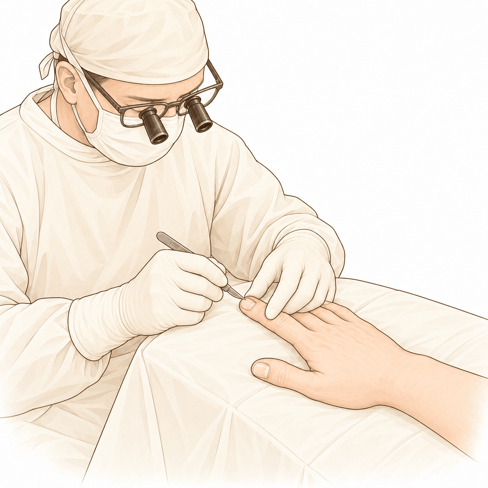
آپ کے سرجن آپ کے ناخن کی جڑ پر جلد کی تہہ میں ایک چھوٹا سا سوراخ بناتے ہیں۔ جلد کی اس تہہ کو ایپونیکیئم (eponychium) کہتے ہیں۔ یہ سوراخ نیچے کے سوجے ہوئے، سوزش والے ٹشو کو آزادانہ طور پر خارج ہونے دیتا ہے۔ یہی اخراج اس آپریشن کا پورا مقصد ہے۔
بعض اوقات ناخن کی جڑ پر جلد کی پوری تہہ ایک ہی ٹکڑے میں نکال دی جاتی ہے، جسے ایک ٹکڑے میں نکالنے کا طریقہ (en bloc excision) کہتے ہیں۔ اگر ناخن کی پلیٹ بے ترتیب یا خراب ہو تو آپ کے سرجن اسے بھی نکال سکتے ہیں، کیونکہ اس سے تہہ کو بھرنے میں مدد مل سکتی ہے۔ آپریشن میں جلد کی موٹی اور سوزش زدہ تہہ کی ایک پتلی ہلال نما پٹی نکالی جاتی ہے، جس سے نیا ناخن بنانے والے ٹشو کو چھیڑے بغیر پھنسی ہوئی سوجن کو نکلنے کا راستہ مل جاتا ہے۔
پھر سوراخ پر پٹی کی جاتی ہے۔ آپ پٹی لگے ہوئے گھر جائیں گے، اور تقریباً 10 دن بعد آپ کے معائنے پر ہماری ٹیم آپ کو انگلی کی دیکھ بھال کا طریقہ بتائے گی۔
آپریشن کے بعد
آپ ریکوری وارڈ میں جاگیں گے، جہاں بے ہوشی کی دوا کا اثر ختم ہونے تک نرسیں آپ پر کڑی نظر رکھتی ہیں۔ آپ کی انگلی پر پٹی ہو گی، اور ہم آپ کو اس کی دیکھ بھال کا طریقہ بتائیں گے۔ اس آپریشن میں درد عموماً ہلکا ہوتا ہے، اور زیادہ تر لوگوں کے لیے عام درد کش دوا کافی ہوتی ہے۔ آپ معمول کے مطابق چل پھر سکتے ہیں اور اپنا ہاتھ آہستگی سے استعمال کر سکتے ہیں۔ گھر پہنچنے کے بعد پہلے 24 گھنٹے کسی کو آپ کے ساتھ رہنا چاہیے۔ آپ کی ٹیم آپ کو بتائے گی کہ آپ اسی دن گھر جائیں گے یا ایک رات ہسپتال میں رہیں گے۔ زیادہ تر لوگوں کو اس آپریشن کے بعد اینٹی بائیوٹکس کی ضرورت نہیں ہوتی۔ ہم پٹی تقریباً 10 دن تک لگی رہنے دیتے ہیں؛ براہ کرم اس سے پہلے اسے نہ اتاریں، جب تک ہم آپ سے نہ کہیں۔ جب ہم آپ کو دیکھیں گے تو پٹی تبدیل کریں گے یا اتار دیں گے۔
صحت یابی
پہلے چند دنوں میں جہاں سوراخ بنایا گیا تھا وہاں آپ کی انگلی کو چھونے پر درد ہو گا اور وہ تھوڑی سوجی ہوئی ہو گی۔ یہ آہستہ آہستہ ٹھیک ہو جاتا ہے۔ عام درد کش دوا عموماً کافی ہوتی ہے، اور آرام کرتے وقت ہاتھ کو تکیے پر اونچا رکھنے سے ٹیس کم ہو سکتی ہے۔ آپ گھر میں اپنا ہاتھ آہستگی سے استعمال کر سکتے ہیں، لیکن اسے پانی میں بھگونے یا ناخن کی تہہ پر دباؤ ڈالنے سے بچیں۔
پٹی تقریباً 10 دن تک لگی رہتی ہے، اس لیے آپ کا زیادہ تر روزمرہ معمول عام طور پر جاری رہتا ہے۔ جب آپ آرام محسوس کریں تو کپڑے پہن سکتے ہیں، کھانا پکا سکتے ہیں اور ہلکے کام کر سکتے ہیں۔ جب تک جلد بھر نہ جائے، زور سے پکڑنے، گیلے کام اور ہر اس چیز سے بچیں جس سے انگلی کو ٹکر لگے۔
جب معائنے پر پٹی اتاری جائے تو ہو سکتا ہے کھلا حصہ ابھی پوری طرح نہ بھرا ہو۔ ناخن کی تہہ وقت کے ساتھ ٹھیک ہوتی ہے، اور کوئی بھی نیا ناخن جڑ سے آہستہ آہستہ اگتا ہے۔ آپ کو پتا چل جائے گا کہ سب ٹھیک جا رہا ہے جب دکھن ختم ہو جائے، سوجن کم ہو جائے اور آپ انگلی کو بغیر درد کے پکڑنے اور استعمال کرنے کے قابل ہو جائیں۔
سرجری کے بعد ہینڈ تھراپی Extend Rehabilitation میں Ruby Doolan کے ساتھ ہوتی ہے۔ وہ آپ کی ورزشوں میں رہنمائی کریں گی اور ضرورت ہو تو اسپلنٹ بنائیں گی۔
صحت یابی ہر شخص میں مختلف ہوتی ہے۔ آپ کا وقت مختلف ہو سکتا ہے، اور آپ کے سرجن اور تھراپسٹ ہر مرحلے پر آپ کی رہنمائی کریں گے۔
کیا مسائل پیدا ہو سکتے ہیں
زیادہ تر مریض ٹھیک رہتے ہیں، لیکن کبھی کبھار مسائل پیدا ہو سکتے ہیں۔ آپ کے سرجن اور ٹیم کسی بھی مسئلے کو جلد پکڑنے کے لیے آپ کی قریب سے نگرانی کرتے ہیں۔
بعض اوقات ناخن کے ارد گرد انفیکشن ابتدائی علاج سے ٹھیک نہیں ہوتا۔ اگر انفیکشن انگلی کے نرم ٹشوز میں مزید گہرائی تک پھیل جائے تو آپ دیکھ سکتے ہیں کہ سوجن اور سرخی ناخن کی تہہ سے باہر کی طرف پھیل رہی ہے، انگلی گرم محسوس ہوتی ہے، یا درد کم ہونے کے بجائے بڑھتا جا رہا ہے۔ ایسا ہو تو کلینک کو فون کریں، یا اگر آپ کی طبیعت خراب محسوس ہو یا سرخی تیزی سے پھیل رہی ہو تو ہسپتال کی ایمرجنسی جائیں۔
انفیکشن انگلی کی ہڈی تک بھی پہنچ سکتا ہے۔ اسے ہڈی کا انفیکشن (osteomyelitis) کہتے ہیں۔ یہ گہرے، دھڑکتے ہوئے درد کی طرح محسوس ہو سکتا ہے جو عام درد کش دواؤں سے کم نہ ہو، اور اکثر ساتھ میں ہڈی کے اوپر مسلسل سوجن اور چھونے پر درد ہوتا ہے۔ انگلی سرخ نظر آ سکتی ہے اور گرم محسوس ہو سکتی ہے۔ اس کا فوری علاج ضروری ہے، اس لیے اسی دن کلینک سے رابطہ کریں یا اگر درد شدید ہو تو ہسپتال کی ایمرجنسی جائیں۔
اگر انفیکشن والی ناخن کی تہہ کا ابتدائی علاج اچھی طرح کام نہ کرے تو مسئلہ لمبے عرصے تک رہ سکتا ہے۔ ہو سکتا ہے آپ دیکھیں کہ ناخن کے ارد گرد سوجن اور دکھن ہفتوں یا مہینوں تک بار بار لوٹ آتی ہے، اور جلد کی تہیں سرخ ہوتی ہیں، چھونے پر درد کرتی ہیں اور پانی لگنے پر بھڑک اٹھتی ہیں۔ اپنے اگلے معائنے پر اس کا ذکر کریں تاکہ علاج کا منصوبہ بدلا جا سکے۔
ناخن کا بستر (nail bed)، یعنی ناخن کی پلیٹ کے نیچے ٹشو کی وہ تہہ جس سے ناخن اگتا ہے، بھی مستقل طور پر خراب ہو سکتا ہے اگر انفیکشن کا بروقت اچھی طرح علاج نہ ہو۔ ہو سکتا ہے آپ دیکھیں کہ ناخن موٹا، دھاری دار یا بگڑی ہوئی شکل میں اگ رہا ہے، یا ٹھیک سے اگ ہی نہیں رہا۔ ایسی کسی بھی تبدیلی کا اپنے معائنے پر ذکر کریں۔
خود آپریشن میں پیچیدگیوں کا خطرہ کم ہے، اور بعد میں سوجن اور دکھن کے لوٹ آنے کا امکان بھی کم ہے۔ اگر صحت یابی کے دوران آپ کو کوئی تشویشناک بات نظر آئے، جیسے بڑھتا ہوا درد، پھیلتی ہوئی سرخی یا سوراخ سے رطوبت کا اخراج، تو اگلی ملاقات کا انتظار کرنے کے بجائے کلینک سے رابطہ کریں۔
اگر آپ تفصیلات جاننا چاہتے ہیں تو اس صفحے پر پیچیدگیوں کی جدول میں عام شرحیں درج ہیں۔
ہمیں کب فون کریں
زیادہ تر لوگ آسانی سے صحت یاب ہو جاتے ہیں، لیکن بعض علامات پر فوری توجہ کی ضرورت ہوتی ہے۔ اگر آپ کو بخار ہو، اگر زخم کی سرخی یا اس سے رطوبت کا اخراج بڑھ رہا ہو، یا اگر درد اچانک شدید ہو جائے تو ہمیں فون کریں۔ اگر آپ کی پنڈلی میں سوجن ہو، سانس پھولے یا سینے میں درد ہو تو ہسپتال کی ایمرجنسی جائیں۔ اگر آپ کی انگلی سُن ہو جائے، ٹھنڈی محسوس ہو، یا آپ اسے ہلا نہ سکیں تو فوراً ہمیں فون کریں۔ شک ہو تو کلینک کو فون کریں۔ ہم چاہیں گے کہ آپ جلد ہم سے رابطہ کریں بجائے اس کے کہ آپ گھر پر انتظار کرتے رہیں۔
Evidence & references
This is the clinical evidence summary written for health professionals. It is technical, and it lists the research this page was built from. You do not need to read it to understand your treatment or to make a decision about it.
Anatomy & Pathophysiology
Cutaneous Anatomy and Functional Units
- The dorsal integument of the distal phalanx is a unique cutaneous unit characterized by the presence of the nail bed and its matrix [3].
- The dorsal covering of the interphalangeal articulations of the digits forms a unique cutaneous unit that exhibits a considerable excess of skin when the digits are in extension [3].
- The fine, tight skin of the dorsal aspect of the middle phalanx constitutes a distinct functional cutaneous unit [3].
- The palmar integument of the digits is subdivided into phalangeal units separated by digital flexion folds, with three folds for the fingers and two for the thumb [3].
- When a digit is completely flexed, the integument of adjacent phalanges comes into contact at the flexion creases, establishing areas of cutaneous contact in the form of a diamond [3].
- The sides of the diamond-shaped cutaneous contact zones do not undergo variations in length during flexion and extension movements [3].
- Incisions made along the lines of the diamond-shaped cutaneous contact zones present a minimal chance of retraction [3].
- The web spaces are formed from the union of two nonsymmetrical cutaneous surfaces [3].
- The dorsal slope of the web space has a gradual incline and its supple skin is not adherent to the subjacent region [3].
- The palmar surface of the web space is flat and precipitously interrupted, with skin that is densely adherent to the commissural skeleton [3].
- The commissural skeleton of the web space is formed by the interdigital palmar (natatory) ligament between the fingers and by the distal transverse ligament at the level of the thumb web [3].
- The distal transverse ligament at the level of the thumb web is the deepest and most mobile component of the commissural skeleton [3].
Vascular Anatomy
- The arteries of the thumb vary in both size and number, making surgical reconstruction delicate [8].
- The palmar aspect of the thumb is divided into three segments defined by the opposition, metacarpophalangeal, and interphalangeal creases [8].
- In the classical layout, the "princeps pollicis" artery crosses the first intermetacarpal space and runs along the ulnar side of the first metacarpal bone [8].
- The "princeps pollicis" artery emerges onto the subcutaneous palmar tissue at the level of the cutaneous flexion crease of the metacarpophalangeal joint [8].
- At the metacarpophalangeal joint level, the "princeps pollicis" artery divides into two terminal rami known as the collateral palmar arteries of the thumb [8].
- The collateral palmar arteries of the thumb run along the digital tunnel symmetrically and are of equal caliber [8].
- The collateral palmar arteries of the thumb head distally to finally unite in the pulp arcade [8].
- During their transit in the digital tunnel, the collateral palmar arteries break off into numerous collateral branches that are cutaneous, articular, or osseous [8].
- An arcade located deep in the flexor tendon joins the two collateral arteries at the level of the distal metaphysis of the first phalanx [8].
- Vessels originating from the subtendinous arcade at the distal metaphysis of the first phalanx enter the "vincula" and irrigate the flexor tendon [8].
- Only 15% of anatomical dissections of the palmar arteries of the thumb fall into the classical "typical" category [8].
- In the second segment of the thumb, the two arteries run alongside the flexor tendon and behind the collateral nerves [8].
- In the second segment of the thumb, the main artery is typically the ulnar collateral artery [8].
- The subtendinous anastomosis situated at the level of the neck of the first phalanx acts as a "moderator" between the two collateral arteries [8].
- In cases where the palmar ulnar collateral artery is absent, the dorsal artery takes its place by means of a branch through the subtendinous arcade [8].
- In the pulp segment of the thumb, the two arteries are of similar size and run through the thick fatty subcutaneous padding [8].
- In the pulp segment of the thumb, the arteries cross over and convert into the ends of the digital nerves at the level of the median axis [8].
- The posterior area of the thumb is vascularized by two arteries which originate from the palmar arteries at the level of the first metacarpal [8].
- These dorsal arteries run laterally along the metacarpophalangeal joint and continue obliquely from volar to dorsal before heading distally on the side of the two distal phalanges [8].
- At the level of the neck of the first phalanx, an anastomosis originating from the palmar arteries can be found on the dorsal aspect [8].
- The dorsal arteries of the thumb are joined by three arcades: one inconstant arcade under the extensor tendon at the neck of the first phalanx, the arcade of the nail matrix, and the arcade of the nailbed [8].
- The ulnar dorso-collateral artery generally stems from the "princeps pollicis" onto the medial border of the neck of the first metacarpal [8].
General Hand Architecture
- The hand consists of 19 bones, 17 articulations, and 19 muscles situated entirely within the hand [2].
- The hand contains about the same number of tendons activated by the forearm muscles as it has intrinsic muscles [2].
- The open hand, with fingers extended and in contact, forms a balanced graceful oval in its longitudinal axis [2].
- The proximal "carpometacarpal" half of the hand is flattened, presenting two faces with unique anatomical and functional significance [2].
- The posterior or dorsal aspect of the hand is convex, while the anterior, palmar, or volar aspect is concave [2].
- The distal half of the hand is separated into five digits which flex toward the palm [2].
- The digits converge in closing by flexing and adducting, and diverge in opening by extending and abducting [2].
- The thumb has a more proximal and lateral position, allowing movement inward and outward from the palm [2].
- The four fingers are the distal extension of the carpometacarpal part of the hand [2].
- The hinges of finger movements are located at the thenar crease and at the transverse distal palmar crease, not at the bases of the digits [2].
- When the fingers are extended and separated, the tips of the fingers lie on the circumference of a circle whose center is the head of the third metacarpal [2].
- The web space of the thumb is the largest and deepest among the digital web spaces [2].
Investigations
- Clinical evaluation of the hand and wrist requires combining patient history with a careful physical examination to pinpoint or narrow the scope of possible pathologic processes [1].
- Diagnostic tests such as imaging and serum laboratory studies are useful in determining pathology but can be expensive, time consuming, and often nonspecific [1].
- A careful physical examination is essential to direct care and future testing if indicated [1].
- An 8-MHz Doppler tone assessment may be used to identify superficially displaced neurovascular bundles when Dupuytren cords lie beneath soft fleshy prominences [11].
- False-negatives are possible with 8-MHz Doppler tone assessment for identifying neurovascular bundles [11].
- Doppler imaging is a promising improvement over handheld Doppler assessment but higher resolution imaging technology is needed [11].
- MR assessment of Dupuytren’s disease is hindered by the resolution of current equipment, orientation issues due to multiplanar deformities of the fingers, and lack of intraoperative availability [11].
- MRI is probably most useful in identifying additional pathology such as flexor tendon bowstringing [11].
- MRI may be helpful in providing a quantitative noninvasive measure of cellularity of affected areas, which is an index of biologic activity [11].
- The potential staging tool of MRI for measuring cellularity has not been investigated yet on a large scale [11].
References
[1] Orthopaedic Knowledge Update 13 Ebook Without Multimedia. Anatomy, Evaluation, Clinical Examination, and Imaging > Evaluation and Clinical Examination: Current Concepts.
[2] Exam Of The Hand Wrist 2Ed. INTRODUCTION.
[3] Exam Of The Hand Wrist 2Ed. Functional cutaneous units.
[8] Exam Of The Hand Wrist 2Ed. Techniques of investigation of the arterial supply by J P Melki > Vascularization of the thumb > Palmar aspect.
[11] Dupuytren S Disease And Related Hyperproliferative Disorders. 54. The Future of Dupuytren’s Research and Treatment > 54.4 Mechanical Measurements and Procedures > 54.4.4 Imaging.