Patients › Hand
انگوٹھے کے جوڑ کی تبدیلی: ٹچ (Touch) آرتھروپلاسٹی
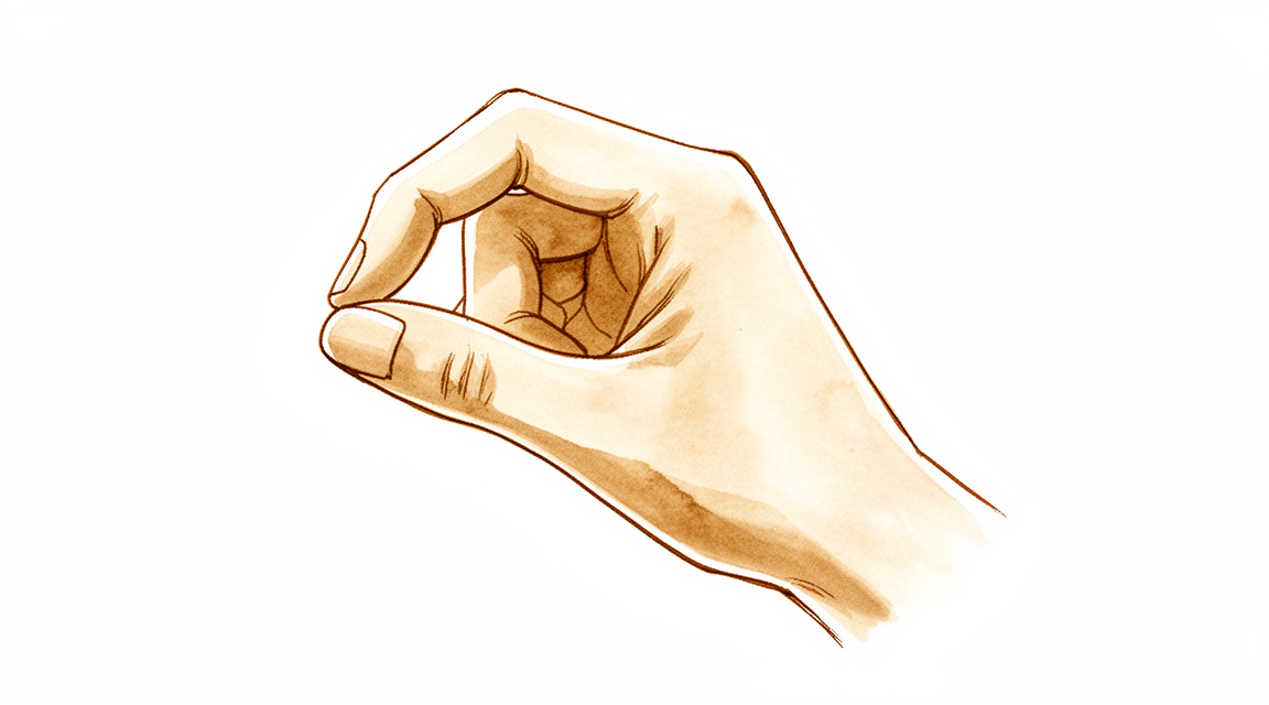
یہ آپریشن کیوں تجویز کیا گیا ہے
ڈاکٹر کیرن ہِرپرا، جو Mater Private Hospital Rockhampton میں بازو اور ہاتھ کے سرجن (upper-limb surgeon) ہیں، علاج کا آغاز ان کم سے کم چیر پھاڑ والے طریقوں سے کرتے ہیں جو آپ کی حالت کے لیے موزوں ہوں۔ مریضوں کو عام طور پر ان کے فیملی ڈاکٹر (GP) ہمارے کلینک کے لیے ریفر کرتے ہیں؛ اگر کسی فزیوتھراپسٹ نے آپ کو ہمیں دکھانے کا مشورہ دیا ہے، تب بھی میڈیکیئر (Medicare) سے رقم کی واپسی (rebate) کا اہل ہونے کے لیے آپ کو اپنے فیملی ڈاکٹر سے ریفرل (referral) لینا ہوگا۔ آپ کی ملاقات پر ہم آپ کی تکلیف کی تفصیل پوچھتے ہیں، آپ کے ہاتھ کا معائنہ کرتے ہیں، اور اگر تشخیص کی تصدیق کے لیے ضرورت ہو تو ایکسرے یا اسکین جیسے تصویری ٹیسٹ (imaging) کا انتظام کرتے ہیں۔
انگوٹھے کی جڑ میں جوڑ کے گھسنے والا آرتھرائٹس (wear-and-tear arthritis) درد اور کمزوری کی ایک عام وجہ ہے۔ اس کی وجہ سے چٹکی سے پکڑنا، گرفت کرنا اور چابی گھمانا مشکل ہو سکتا ہے۔ ہم عام طور پر پہلے بغیر آپریشن کے علاج آزماتے ہیں، جیسے ہاتھ کے استعمال کا طریقہ بدلنا، ہینڈ تھراپی (hand therapy، ہاتھ کی بحالی کا علاج) اور اسپلنٹ۔ جب ان طریقوں سے آپ کو کافی بہتری نہ ملے تو سرجری پر غور کیا جاتا ہے۔
یہ آپریشن، یعنی انگوٹھے کے جوڑ کی تبدیلی (thumb joint replacement)، آپ کے انگوٹھے کی جڑ کے گھسے ہوئے جوڑ کی جگہ ایک مصنوعی امپلانٹ (جسم میں لگایا گیا مصنوعی پرزہ) لگاتا ہے۔ یہ عام طور پر ان لوگوں کو پیش کیا جاتا ہے جن کا آرتھرائٹس صرف اسی جوڑ تک محدود ہو اور جن کا درد بغیر آپریشن کے علاج سے ٹھیک نہ ہوا ہو۔ ہو سکتا ہے ہم نے یہ آپ کے لیے اس لیے تجویز کیا ہو کہ اس کا مقصد درد کم کرنا اور حرکت، طاقت اور استحکام بحال کرنا ہے، تاکہ آپ اپنا انگوٹھا اعتماد کے ساتھ استعمال کر سکیں۔ زیادہ تر ٹچ (Touch) امپلانٹ 2 سال بعد بھی اپنی جگہ پر موجود ہوتے ہیں، اور امپلانٹ کے کام کرتے رہنے کی شرح 96 فیصد بتائی گئی ہے۔ یہ آپریشن آپ کے لیے موزوں ہے یا نہیں، اس کا فیصلہ ہم آپ کے ساتھ مل کر کرتے ہیں۔
آپریشن سے پہلے
سرجری سے پہلے کے ہفتوں میں ہم آپ کے انگوٹھے کے تصویری ٹیسٹ (imaging)، جیسے ایکسرے، ایم آر آئی یا الٹراساؤنڈ، کے ذریعے منصوبے کی تصدیق کرتے ہیں۔ یہ تصویریں گھسا ہوا جوڑ دکھاتی ہیں اور جوڑ کی تبدیلی کی منصوبہ بندی میں ہماری مدد کرتی ہیں۔ تیاری کے بارے میں ہماری ٹیم آپ کو واضح ہدایات دے گی۔ آپ کو آپریشن سے سات گھنٹے پہلے کھانا پینا بند کرنا ہوگا۔ ہم چھ کے بجائے سات گھنٹے کا کہتے ہیں تاکہ اگر آپریشن کی فہرست وقت سے پہلے چل رہی ہو تو آپ کو پہلے بلایا جا سکے۔ کچھ دواؤں کو عارضی طور پر روکنا پڑتا ہے، اور ہم آپ کو بتائیں گے کہ کون سی دوائیں اور کب۔ اپنی موجودہ دواؤں کی فہرست ساتھ لائیں۔ بعد میں آپ کو گاڑی میں گھر لے جانے کے لیے کسی کا انتظام کریں۔ ڈھیلے اور آرام دہ کپڑے پہنیں۔ اگر آپ کو دوسری طبی بیماریاں ہیں تو ہو سکتا ہے آپ کو خون کے ٹیسٹ یا بے ہوشی کے ڈاکٹر (anaesthetist) سے معائنے کی ضرورت ہو۔
آپریشن کے دن
آپریشن کے دن آپ ہسپتال کے سرجری کے داخلے کے شعبے میں آئیں گے۔ وہاں آپ کا اندراج کیا جاتا ہے اور آپ کو آپریشن تھیٹر کے لیے تیار کیا جاتا ہے۔ آپ کی ملاقات بے ہوشی کے ڈاکٹر سے ہوگی، یعنی وہ ڈاکٹر جو سرجری کے دوران آپ کی بے ہوشی اور آرام کا خیال رکھتے ہیں۔ یہ آپریشن جنرل اینستھیزیا (مکمل بے ہوشی) میں کیا جاتا ہے۔ آپریشن کے دوران آپ مکمل طور پر بے ہوش ہوں گے۔ کچھ مریضوں کو آپریشن کے بعد درد میں آرام کے لیے اعصابی بلاک (nerve block)، یعنی ایک انجکشن جو جسم کے ایک حصے کو سُن کر دیتا ہے، بھی لگایا جا سکتا ہے؛ بے ہوشی کے ڈاکٹر آپ کے انفرادی حالات کی بنیاد پر اسی دن اس کا فیصلہ کرتے ہیں۔
پھر آپ کو آپریشن تھیٹر میں لے جایا جاتا ہے، جہاں آپریشن کیا جاتا ہے۔ اس کے بعد آپ ریکوری روم میں جاگتے ہیں، جہاں بے ہوشی کی دوا کا اثر ختم ہونے تک نرسیں آپ کی نگرانی کرتی ہیں۔ جب آپ کی حالت مستحکم ہو جائے تو، طریقۂ علاج اور آپ کی صحت یابی کے مطابق، آپ یا تو وارڈ میں منتقل ہوتے ہیں یا گھر چلے جاتے ہیں۔
آپریشن میں کیا کیا جاتا ہے
گھسے ہوئے جوڑ تک پہنچنے کے لیے آپ کے سرجن آپ کے انگوٹھے کی جڑ پر ایک ہی چیرا لگاتے ہیں۔ جوڑ کی خراب سطحیں نکال دی جاتی ہیں اور ان کی جگہ مصنوعی حصے لگائے جاتے ہیں: انگوٹھے کی ہڈی میں لگایا جانے والا ایک چھوٹا تنا (stem) اور اس کے ساتھ والی چھوٹی ہڈی میں بٹھایا جانے والا ایک کپ (cup)۔ نئے حصے اس طرح بنائے گئے ہیں کہ وہ ایک صحت مند جوڑ کی طرح ایک دوسرے کے ساتھ حرکت کریں، تاکہ آپ دوبارہ چٹکی سے پکڑ اور گرفت کر سکیں۔
مقصد آپ کے انگوٹھے کو اس کی قدرتی لمبائی اور سیدھ میں رکھنا ہے، جس سے انگوٹھا دیکھنے اور کام کرنے میں جتنا ممکن ہو معمول کے قریب رہتا ہے۔ چونکہ جوڑ کو نکالنے کے بجائے تبدیل کیا جاتا ہے، اس لیے آپ کے انگوٹھے کو حرکت دینے والے پٹھوں اور ٹینڈنوں (tendon، یعنی وہ مضبوط ڈوریاں جو پٹھے کو ہڈی سے جوڑتی ہیں) کو چٹکی بھرنے کے لیے اتنا زور نہیں لگانا پڑتا۔
جب نیا جوڑ اپنی جگہ لگ جائے تو آپ کے سرجن چیرے کو ٹانکوں سے بند کرتے ہیں اور اس پر زخم کی پٹی لگاتے ہیں۔ جب آپ جاگیں گے تو آپ کا انگوٹھا پٹی میں لپٹا ہوگا، اور زخم کی پٹی تقریباً 10 دن تک لگی رہتی ہے۔
اس کے بعد ایک ہینڈ تھراپسٹ (ہاتھ کی بحالی کے ماہر) آپ کی صحت یابی میں رہنمائی کرتے ہیں۔ وہ آپ کو دکھاتے ہیں کہ انگوٹھا ٹھیک ہونے کے دوران اسے کیسے حرکت دیں اور استعمال کریں، اور وہ بتاتے ہیں کہ آپ کب اور کتنا وزن اٹھا سکتے ہیں۔ نئے جوڑ پر بھاری بوجھ نہ ڈالنے سے یہ زیادہ دیر تک چلتا ہے۔
آپریشن کے بعد
زیادہ تر مریض اس آپریشن کے بعد ایک رات ہسپتال میں رہتے ہیں، اگرچہ کچھ اسی دن گھر جا سکتے ہیں۔ آپ ریکوری روم میں جاگتے ہیں، پھر وارڈ میں منتقل ہوتے ہیں۔ نرسیں آپ کا درد جانچتی ہیں اور آپ کو آرام میں رکھنے کے لیے دوا دیتی ہیں۔ آپ کا انگوٹھا زخم کی پٹیوں میں لپٹا ہوگا اور اسے ایک اسپلنٹ کا سہارا دیا گیا ہوگا۔ آرام کرتے وقت اپنا ہاتھ تکیوں پر اونچا رکھیں؛ اس سے سوجن کم ہونے میں مدد ملتی ہے۔ آپ اٹھ کر وارڈ میں چل پھر سکتے ہیں، اور زیادہ تر کاموں کے لیے آپ اپنا دوسرا ہاتھ استعمال کریں گے۔ براہ کرم گھر پر پہلے 24 گھنٹے اپنے ساتھ رہنے کے لیے کسی کا انتظام کریں۔ ہم زخم کی پٹی تقریباً 10 دن تک لگی رہنے دیتے ہیں؛ براہ کرم اس سے پہلے اسے نہ اتاریں، جب تک ہم آپ کو ایسا کرنے کو نہ کہیں۔ جب ہم آپ سے ملتے ہیں تو اسے بدل دیتے ہیں یا اتار دیتے ہیں۔
صحت یابی
پہلے چند دنوں تک آپ کے انگوٹھے میں درد اور سوجن رہے گی۔ اپنا ہاتھ تکیوں پر اونچا رکھنے سے سوجن کم ہونے میں مدد ملتی ہے۔ عام درد کش دوا، ہدایت کے مطابق لی جائے تو، سب سے مشکل دن گزرنے تک آپ کو آرام میں رکھتی ہے۔
جب آپ گھر جائیں گے تو آپ کا انگوٹھا اسپلنٹ میں ہوگا، اور زخم کی پٹی تقریباً 10 دن تک لگی رہتی ہے۔ سرجری کے بعد ہینڈ تھراپی Extend Rehabilitation میں Ruby Doolan کے ساتھ ہوتی ہے۔ Ruby ایک ہینڈ تھراپسٹ ہیں: وہ آپ کو ہلکی ورزشیں سکھاتی ہیں جو نئے جوڑ کے ٹھیک ہونے کے دوران آپ کی انگلیوں کو حرکت میں رکھتی ہیں، اور انگوٹھے کی صحت یابی کے دوران آپ کو جس اسپلنٹ کی بھی ضرورت ہو وہ بناتی ہیں۔ وہ یہ بھی بتاتی ہیں کہ آپ کب اور کتنا وزن اٹھا سکتے ہیں، کیونکہ نئے جوڑ پر بھاری بوجھ نہ ڈالنے سے یہ زیادہ دیر تک چلتا ہے۔
روزمرہ زندگی میں شروع میں آپ زیادہ تر کاموں کے لیے اپنا دوسرا ہاتھ استعمال کریں گے۔ جب تک اسپلنٹ لگا ہو، آپ گاڑی نہیں چلا سکتے، کیونکہ اس کی وجہ سے آپ اسٹیئرنگ کو محفوظ طریقے سے نہیں پکڑ سکتے۔ جب اسپلنٹ اتر جائے اور آپ کے سرجن اجازت دے دیں تو گاڑی چلانا شروع کیا جا سکتا ہے؛ ہمارا صفحہ بازو اور ہاتھ کی سرجری کے بعد گاڑی چلانا دیکھیں۔ جیسے جیسے سوجن کم ہوتی ہے اور حرکت واپس آتی ہے، آپ روزمرہ کے ہلکے کاموں کے لیے انگوٹھا استعمال کرنا شروع کریں گے، اور پھر آہستہ آہستہ بغیر درد کے گرفت کرنے اور چٹکی سے پکڑنے تک پہنچیں گے۔
صحت یابی ہر شخص میں مختلف ہوتی ہے۔ آپ کی صحت یابی کا وقت مختلف ہو سکتا ہے، اور آپ کے سرجن اور ہینڈ تھراپسٹ ہر مرحلے پر آپ کی رہنمائی کریں گے۔
کیا مسائل پیدا ہو سکتے ہیں
زیادہ تر مریض ٹھیک رہتے ہیں، لیکن کبھی کبھار مسائل پیدا ہو سکتے ہیں۔ آپ کے سرجن اور ٹیم کسی بھی مسئلے کو جلد پکڑنے کے لیے آپ کی قریبی نگرانی کرتے ہیں۔
نئے جوڑ کے حصے کبھی کبھی ڈھیلے ہو سکتے ہیں۔ یہ عام طور پر انگوٹھے کی جڑ میں گہرے دکھن والے درد کی طرح محسوس ہوتا ہے جو کچھ عرصہ آرام رہنے کے بعد واپس آ جائے یا بڑھ جائے۔ اگر سرجری کے ہفتوں یا مہینوں بعد آپ کے انگوٹھے میں دوبارہ درد ہونے لگے تو کلینک کو فون کریں تاکہ ہم تصویری ٹیسٹ کا انتظام کر سکیں اور دیکھ سکیں کہ کیا ہو رہا ہے۔
جوڑ اپنی جگہ سے نکل (dislocation) بھی سکتا ہے، یعنی نئے حصے اپنی جگہ سے کھسک جاتے ہیں۔ آپ کو اچانک کھٹکے (clunk) جیسا احساس ہوگا، جس کے بعد درد ہوگا اور انگوٹھا معمول کے مطابق حرکت نہیں کرے گا۔ اگر ایسا ہو تو فوراً کلینک کو فون کریں۔
نئے جوڑ کے آس پاس کی چھوٹی ہڈیوں میں سے کسی ایک کا فریکچر (ہڈی کا ٹوٹنا) سرجری کے دوران یا بعد میں ہو سکتا ہے۔ یہ تیز درد اور سوجن کی طرح محسوس ہوتا ہے جو توقع سے کہیں زیادہ ہو۔ کلینک کو فون کریں تاکہ ہم اس کا معائنہ کر سکیں۔
انگوٹھے کے قریب کے اعصاب میں جلن (irritation) پیدا ہو سکتی ہے یا انہیں چوٹ لگ سکتی ہے۔ آپ کو انگوٹھے کی پشت پر جھنجھناہٹ، سوئیاں چبھنے جیسا احساس، یا سُن پن محسوس ہو سکتا ہے۔ اعصابی بلاک کے بعد پہلے 24 گھنٹوں میں سُن پن اور کمزوری متوقع ہیں۔ بلاک کا اثر ختم ہونے کے بعد اگر سُن پن برقرار رہے یا آپ اپنا انگوٹھا یا انگلیاں نہ ہلا سکیں تو کلینک کو فون کریں۔
انفیکشن کم ہوتا ہے لیکن سنگین ہوتا ہے۔ اگر آپ کو بخار ہو، زخم سے باہر کی طرف پھیلتی ہوئی سرخی ہو، یا زخم سے رطوبت یا پیپ رس رہی ہو تو اسی دن کلینک کو فون کریں۔ اگر آپ کا انگوٹھا سفید پڑ جائے، ٹھنڈا ہو جائے، یا اس کا رنگ سفید، نیلا یا گہرا ہو جائے تو ہسپتال کی ایمرجنسی جائیں، کیونکہ یہ خون کی گردش کا مسئلہ ہو سکتا ہے۔
کچھ مریضوں کو سرجری کے بعد ہاتھ میں مسلسل درد اور حساسیت ہو جاتی ہے، بعض اوقات سوجن اور جلد کے رنگ یا درجۂ حرارت میں تبدیلی کے ساتھ۔ اگر درد کی دواؤں کے باوجود درد بڑھتا ہی جائے تو اسی دن کلینک کو فون کریں۔
اگر نیا جوڑ کامیاب نہ ہو تو کبھی کبھی مزید سرجری کی ضرورت پڑتی ہے۔ اس کا مطلب امپلانٹ کو ٹھیک کرنا یا بدلنا ہو سکتا ہے، یا ایک مختلف آپریشن کرنا جس میں گھسا ہوا جوڑ نکال دیا جاتا ہے اور خالی جگہ کو آپ کے اپنے ٹشو (tissue) سے گدی کی طرح بھر دیا جاتا ہے۔ اگر کبھی ایسی صورت پیش آئی تو ہم آپ کے ساتھ تمام راستوں پر تفصیل سے بات کریں گے۔
اگر آپ تفصیلی اعداد و شمار جاننا چاہتے ہیں تو اس صفحے پر پیچیدگیوں کا جدول عام شرحیں دکھاتا ہے۔
ہمیں کب فون کریں
زیادہ تر مسائل جلد ظاہر ہو جاتے ہیں، اور ہم چاہتے ہیں کہ ہمیں ان کے بارے میں جلد پتا چلے۔ اگر آپ کو بخار ہو، زخم سے باہر کی طرف پھیلتی ہوئی سرخی ہو، یا زخم سے رطوبت یا پیپ رس رہی ہو، یا درد کی دواؤں کے باوجود درد بڑھتا ہی جائے تو اسی دن کلینک کو فون کریں۔ اگر اعصابی بلاک کا اثر ختم ہونے کے بعد، یعنی سرجری کے تقریباً 24 گھنٹے بعد، سُن پن برقرار رہے یا آپ اپنا انگوٹھا یا انگلیاں نہ ہلا سکیں تو ہمیں فون کریں۔ اگر آپ کی پنڈلی کے پچھلے حصے (calf) میں سوجن یا درد ہو، سانس پھولے یا سینے میں درد ہو تو ہسپتال کی ایمرجنسی جائیں، کیونکہ یہ خون کے لوتھڑے کی علامات ہو سکتی ہیں۔ اگر آپ کا انگوٹھا سفید پڑ جائے، ٹھنڈا ہو جائے، یا اس کا رنگ سفید، نیلا یا گہرا ہو جائے تو بھی یہی کریں۔ اگر دفتری اوقات کے بعد یا ہفتے کے آخر میں آپ کلینک سے رابطہ نہ کر سکیں تو اپنے قریب ترین ہسپتال کی ایمرجنسی جائیں۔
اس مسئلے کے بارے میں مزید کہاں پڑھیں
یہ صفحہ خود آپریشن کے بارے میں ہے۔ جس بیماری کا یہ علاج کرتا ہے، اور اس بارے میں کہ شواہد کیا دکھاتے ہیں کہ سرجری کب مدد کرتی ہے اور کب نہیں، اس کی تفصیل انگوٹھے کی جڑ کا آرتھرائٹس والے صفحے پر دی گئی ہے۔
Evidence & references
This is the clinical evidence summary written for health professionals. It is technical, and it lists the research this page was built from. You do not need to read it to understand your treatment or to make a decision about it.
Overview
- The Touch® trapeziometacarpal joint arthroplasty demonstrated a 96% implant survival rate at 2 years [4].
- The Touch® prosthesis is recommended as the standard treatment for patients with isolated trapeziometacarpal joint osteoarthritis [4].
- The Touch1 prosthesis is a safe and stable implant providing good satisfaction and very good functional scores [7].
- Patients using the Touch1 prosthesis experience a fast return to work and leisure activity [7].
- Trapeziometacarpal prosthesis enhances function, thumb length, and patient recovery [2].
- Total joint arthroplasty for primary trapeziometacarpal joint arthritis results in low pain levels, excellent mobility, and high patient satisfaction [10].
- The MAÏA trapeziometacarpal prosthesis is a reliable long-term surgical procedure that improves overall function beyond 10 years [23].
- The MAÏA TMC total joint arthroplasty provides very good results for pain relief, strength, mobility, and restoration of thumb length [13].
- The MAÏA TMC total joint arthroplasty provides correction of most thumb z-deformities [13].
- The Elektra prosthesis had a revision rate of 44% after 72 months [1].
- The Elektra prosthesis is not recommended due to poor outcomes observed after only 2 years [8].
- The main complication for the Elektra prosthesis was loosening, with a rate never dropping below 17% at 4 years [9].
- The Moovis prosthesis is recommended for the treatment of advanced trapeziometacarpal osteoarthritis in patients without too many manual constraints [5].
- Dual mobility arthroplasty for thumb CMC joint osteoarthritis is a satisfactory solution with good functional results and absence of early and middle term dislocations [12].
- The Ivory arthroplasty is a reliable option for treating advanced trapeziometacarpal osteoarthritis, providing important improvement in overall function and pain reduction [15].
- Not every complication associated with the Ivory prosthesis requires surgical revision [14].
- Correct implant position leads to reliable medium-term results after trapeziometacarpal joint arthroplasty [11].
- Limiting the magnitude of thumb loads after arthroplasty may contribute positively to the longevity of the procedure [3].
- Scaphometacarpal arthroplasty using a trapeziometacarpal prosthesis is not sufficiently reliable to be a routine solution for surgical revision of failed TMC arthroplasty or trapeziectomy [21].
- Scaphometacarpal arthroplasty using a trapeziometacarpal prosthesis should be used with caution, primarily as a salvage solution if no safer alternative is available [21].
- The Touch® prosthesis is compatible with the Ivory® trapeziometacarpal prosthesis stem during revision surgery [16].
- If the trial neck does not align with the centre of the trapezium, the cup position should be dictated by the neck to represent the new centre of rotation [17].
Anatomy & Pathophysiology
Bony Anatomy
- The skeleton of the hand consists of 27 bones, of which 19 are long bones [30].
- The radial ray or first ray is the shortest and is made up of only three bones: a metacarpal and two phalanges [30].
- The trapezium is clearly angled out in front of the carpal plane so that the first metacarpal makes an angle of about 45 degrees with the second metacarpal in the sagittal plane [30].
- The thumb metacarpal is independent and articulates with the trapezium [36].
- The base of each metacarpal articulates with the distal row of the carpus [30].
- The thumb ray continues the external column of the carpus formed by the scaphoid and trapezium [30].
Joint Mechanics & Kinematics
- The trapeziometacarpal joint has a saddle shape in opposing planes of the articular surfaces [30].
- The first metacarpal makes an angle of about 45 degrees with the second metacarpal in the sagittal plane, which explains the gap between the first ray and the palm [30].
- The thumb ray is more mobile, shorter, and more proximal than the other rays, allowing it to project in front of the plane of the palm to oppose itself to the other four rays [30].
- The wrist has three axes of movement, permitting the hand to be positioned in any spatial configuration [30].
- The combined movements of the wrist and forearm place the hand in a position for grasping [31].
- The thumb has a more proximal and lateral position, allowing movement inward and outward from the palm [31].
Musculature & Tendon Anatomy
- The first (most radial) extensor compartment contains the abductor pollicis longus and the extensor pollicis brevis [29].
- The abductor pollicis longus inserts at the base of the thumb metacarpal and radially abducts the thumb [29].
- The extensor pollicis brevis inserts on the dorsum of the proximal aspect of the proximal phalanx of the thumb and actively extends the metacarpophalangeal joint of the thumb [29].
- The extensor pollicis longus runs longitudinally down the forearm through the third compartment and turns abruptly radialward about Lister tubercle [29].
- The extensor pollicis longus inserts on the distal phalanx and provides forceful extension of the thumb interphalangeal joint [29].
- The flexor pollicis longus originates from two heads: a radial head from the proximal radius and interosseous membrane, and an accessory head from the coronoid process of the ulna and medial epicondyle of the humerus [40].
- The flexor pollicis longus inserts into the proximal base of the thumb distal phalanx and is innervated by the anterior interosseous branch of the median nerve [40].
- The flexor pollicis longus flexes both the interphalangeal and metacarpophalangeal joints of the thumb [40].
- The tenosynovial sheath of the flexor pollicis longus is continuous with the radial bursa [40].
Vascular Anatomy
- The "princeps pollicis" artery, a terminal branch of the radial artery, crosses the first intermetacarpal space and runs along the ulnar side of the first metacarpal bone [37].
- The princeps pollicis artery emerges onto the subcutaneous palmar tissue at the level of the cutaneous flexion crease of the metacarpophalangeal joint [37].
- The princeps pollicis artery divides into two terminal rami, the collateral palmar arteries of the thumb, which run along the digital tunnel symmetrically [37].
- An arcade located deep in the flexor tendon joins together the two palmar arteries at the level of the distal metaphysis of the first phalanx [37].
- The dorsal arteries of the thumb originate from terminal branches of the radial artery at the level of the anatomical snuff-box [37].
- The posterior area of the thumb is vascularized by two arteries which originate from the palmar arteries at the level of the first metacarpal [37].
Soft Tissue & Skin Anatomy
- The superficial palmar fascia covers a triangular area of the central palm, with no central band for the thumb [43].
- The dorsal skin of the hand is thin and possesses loose connections with deeper planes, allowing free gliding [42].
- The palmar integument is subdivided into two separate zones by the oppositional crease of the thumb [32].
- The skin of the radial portion of the palm covers the thenar eminence and is the mobile portion [32].
- The skin of the ulnar and distal portion covers the hypothenar eminence where the skin has poor mobility [32].
- The web spaces are formed from the union of two nonsymmetrical cutaneous surfaces, with the palmar surface being flat and densely adherent to the commissural skeleton [32].
- The distal transverse ligament at the level of the thumb web is the deepest and most mobile commissural skeleton [32].
Prosthesis Design & Biomechanics
- The Touch® prosthesis consists of a mobile prosthetic head in a polyethylene insert that is freely moveable within a metal cup [22].
- The Touch® prosthesis comprises two concentric articulations: a smaller articulation between the head and polyethylene insert, and a larger articulation between the polyethylene insert and metal cup [22].
- The Touch® prosthesis uses metal-on-polyethylene bearings [22].
- The Touch® prosthesis is modular, composed of a titanium alloy stem (Ti–6Al–4 V), an M30 stainless steel intermediate implant, and an M30 stainless steel hemispheric cup [22].
- The Touch® prosthesis head has a 4-mm diameter covered by a pre-assembled high-density 7-mm diameter polyethylene insert [22].
- The Touch® prosthesis cup is available in two sizes: 9 and 10 mm in diameter [22].
- Implant fixation for the Touch® prosthesis is primary by a press-fit effect and secondary by means of a double-layer porous titanium and hydroxyapatite coating [22].
- The total in vitro range of motion of the Touch® prosthesis is 117 degrees [22].
Classification
- The Touch® prosthesis reproduces the dual mobility concept consisting of a mobile prosthetic head in a polyethylene insert that is freely moveable within a metal cup [22].
- The Touch® prosthesis utilizes metal-on-polyethylene bearings [22].
- The Touch® prosthesis stem is available in five sizes [22].
- The Touch® prosthesis intermediate implant has a 4-mm diameter head covered by a pre-assembled high-density 7-mm diameter polyethylene insert [22].
- The Touch® prosthesis polyethylene insert is available in three lengths and two angles [22].
- The Touch® prosthesis hemispheric cup is available in two sizes: 9 and 10 mm in diameter [22].
- The double-layer porous titanium and hydroxyapatite coating promotes and accelerates osteointegration [22].
Clinical Presentation
- A 53-year-old male professional cook presented with persistent clicking during thumb motion 12 months after Touch trapeziometacarpal arthroplasty [18].
- The patient reported no antecedent trauma or pain associated with the clicking noise [18].
- Grip and pinch strength were clinically evaluated and showed no difference compared with the opposite side [18].
- Standard radiographs taken 1 year after surgery did not demonstrate loosening of the implant [18].
- Computed tomography (CT) scan demonstrated a well-aligned implant without evidence of loosening [18].
- CT scan revealed a hint of a very small bone cyst but no loosening of the cup [18].
- Stress-view imaging using a fluoroscopic image intensifier revealed instability of the carpometacarpal I joint during the application of translatory stress forces on the thumb [18].
- Stress-view imaging showed a hint of possible decentralization of the head compared with the cup [18].
- The new diagnosis based on imaging was a fracture of the polyethylene (PE) liner [18].
- Revision surgery confirmed a fractured PE liner with an intact metallic head and stem [18].
- Exchanging only the liner restored stability [18].
- Follow-up radiographs 6 weeks after revision surgery confirmed the implant position remained satisfactory [18].
- Dynamic radiographic imaging with longitudinal traction is important for making a timely diagnosis of polyethylene liner fracture when standard radiographs are normal [20].
Investigations
- Standard radiographs (x-ray) are used to evaluate preoperative osteoarthritis and postoperative implant position, including stem subsidence, periprosthetic radiolucent lines, osteolysis, implant fracture, polyethylene wear, periprosthetic ossifications, and prosthetic dislocation or subluxation [27].
- Subsidence of the metacarpal component is evaluated by comparing the A/B ratio in percentage on strict lateral views immediately after surgery and at the last follow-up [27].
- A difference of more than 25% in the A/B ratio on strict lateral views is indicative of implant subsidence [27].
- Computed tomography (CT) scans can demonstrate a well-aligned implant without evidence of loosening even when clinical symptoms such as clicking are present [18].
- Stress-view imaging (dynamic x-ray) using a fluoroscopic image intensifier can reveal instability of the carpometacarpal I joint and possible decentralization of the head compared with the cup during the application of translatory stress forces [18].
- Preoperative assessment includes evaluation of the scaphotrapeziotrapezoid (STT) joint and confirmation of the absence of other thumb affections [27].
- Osteoarthritis is assessed preoperatively on standard anteroposterior and lateral radiographic views using the Eaton classification [27].
Treatment
Implant Design and Biomechanics
- The Touch® prosthesis head has a 4-mm diameter and is covered by a pre-assembled high-density 7-mm diameter polyethylene insert [22].
Surgical Technique
- A dorsolateral approach is used, protecting the superficial branch of the radial nerve and artery [25].
- The first dorsal compartment is released routinely, and the TMJ capsule is exposed between the extensor pollicis brevis and abductor pollicis longus tendons [25].
- A complete circumferential soft tissue release of the thumb metacarpal base is performed, including resection of the volar lip to facilitate delivery of the metacarpal base out of the wound [25].
- Osteophytes on the base of the metacarpal and the trapezium are resected to reduce the risk of impingement and dislocation [25].
- The distal articular surface of the trapezium is cut to be parallel with the STT joint [25].
- A K-wire is inserted under image guidance, centred in the trapezium, and the trapezium is reamed over the wire as recommended by the manufacturer [25].
- The MAÏA® dual mobility hemispherical cup is inserted with spikes facing the four corners of the trapezium to provide rotational stability [25].
- Attentive reaming of the trapezium and careful cup impaction are crucial steps to avoid trapezial fracture and early cup failure [19].
Clinical Outcomes and Survival
- The Touch® prosthesis has a 96% implant survival rate at 2 years [4].
- The Touch® prosthesis provides promising patient-reported and clinical outcomes [4].
- Surgery with the Touch® prosthesis is recommended as the standard treatment for patients with isolated trapeziometacarpal joint osteoarthritis [4].
- The Touch1 prosthesis allows for a fast return to work and leisure activity [7].
- Total joint arthroplasty in primary trapeziometacarpal joint arthritis results in low pain levels, excellent mobility, and clinical function with high patient satisfaction [10].
- The Elektra prosthesis observed fast pain relief, maintenance of mobility, and a gradual increase in grip strength [1].
- Due to poor outcomes after only 2 years, the Elektra implant cannot be recommended [8].
- The main complication for the Elektra prosthesis was loosening, which occurred frequently before 18 months in early versions [9].
- The loosening rate for the Elektra prosthesis never dropped below 17% at 4 years [9].
- The MAÏA trapeziometacarpal prosthesis represents a long-term solution for surgical treatment of thumb rhizarthrosis [6].
- MAÏA TMC joint prosthesis is a reliable long-term surgical procedure for TMC joint osteoarthritis, improving overall function beyond 10 years [23].
- MAêA TMC total joint arthroplasty provides very good results for pain relief, strength, mobility, and restoration of the thumb length [13].
- MAÏA TMC total joint arthroplasty provides correction of most thumb z-deformities [13].
- Correct implant position can lead to reliable medium-term results after trapeziometacarpal joint arthroplasty with unconstrained cups [11].
- The Moovis prosthesis is recommended for the treatment of advanced trapeziometacarpal osteoarthritis for people having an activity without too many manual constraints [5].
- Dual mobility arthroplasty for the thumb CMC joint osteoarthritis is a satisfactory solution with good functional results and absence of early and middle term dislocations [12].
- The Moovis prosthesis provides very good results at 1 year for rhizarthrosis [26].
- The Ivory arthroplasty provides improvement in overall function and pain reduction, and not every complication requires surgical revision [14].
Complications and Revision
- Polyethylene liner fracture in the Touch prosthesis can present as persistent clicking during thumb motion without antecedent trauma or pain [18].
- Standard radiographs may appear normal in cases of polyethylene liner fracture, while stress-view imaging reveals instability of the carpometacarpal I joint [18].
- Revision surgery for a fractured PE liner involves exchanging only the liner, which restores stability [18].
- Revision surgery for the Ivory® trapeziometacarpal prosthesis allows the stem to be retained due to compatibility with the Touch® prosthesis [16].
- Clinical and radiographic follow-up at 6 months after revision surgery for the Ivory® prosthesis was uneventful [16].
Complications
Implant Failure and Loosening
- Aseptic cup loosening is cited as the most common reason for implant failure in thumb carpometacarpal joint replacement arthroplasty [24].
- Aseptic cup loosening leads to revision rates of 42%–51% in thumb carpometacarpal joint replacement arthroplasty [24].
- The Elektra prosthesis had a loosening rate that never dropped below 17% at 4 years [9].
- Loosening of the Elektra prosthesis occurred frequently before 18 months in early versions [9].
- The Touch® trapeziometacarpal joint arthroplasty reported a 96% implant survival rate at 2 years [4].
Polyethylene Liner Fracture
- Standard radiographs and CT scans may show a well-aligned implant without evidence of loosening in cases of polyethylene liner fracture [18].
- Stress-view imaging using a fluoroscopic image intensifier can reveal instability of the carpometacarpal I joint and possible decentralization of the head compared with the cup in cases of polyethylene liner fracture [18].
- Revision surgery for polyethylene liner fracture in the Touch prosthesis can involve exchanging only the liner while retaining the intact metallic head and stem [18].
Intraoperative and Early Complications
- Attentive reaming of the trapezium and careful cup impaction are crucial steps to avoid trapezial fracture and early cup failure in Moovis trapeziometacarpal joint arthroplasty [19].
- Osteophytes on the base of the metacarpal and the trapezium are resected to reduce the risk of impingement and dislocation during MAÏA® dual mobility CMC joint arthroplasty [25].
Instability and Dislocation
- The Moovis prosthesis showed an absence of early and middle term dislocations in a short series of 19 cases [12].
- The Moovis prosthesis showed an absence of prosthesis instability in a retrospective functional study of 28 prostheses [5].
- The Touch1 prosthesis appeared to be a safe and stable implant [7].
Revision and Salvage Considerations
- The Ivory® trapeziometacarpal prosthesis stem can be retained during revision surgery with the Touch® prosthesis [16].
- Not every complication of the Ivory arthroplasty requires surgical revision [14].
Recovery
- The Elektra prosthesis was associated with fast pain relief, maintenance of mobility, and a gradual increase in grip strength [1].
- The Touch prosthesis demonstrated a 96% implant survival rate at 2 years [4].
- The Touch prosthesis provided promising patient-reported and clinical outcomes [4].
- The authors of the 2-year Touch study recommend surgery with the Touch prosthesis as the standard treatment for patients with isolated trapeziometacarpal joint osteoarthritis [4].
- The Moovis prosthesis was recommended for the treatment of advanced trapeziometacarpal osteoarthritis in people having an activity without too many manual constraints due to the absence of prosthesis instability [5].
- The Touch prosthesis provided good satisfaction and very good functional scores [7].
- The Touch prosthesis allowed for a fast return to work and leisure activity [7].
- The Elektra prosthesis had poor outcomes after only 2 years, leading the authors to not recommend this implant [8].
- Loosening was the main complication for the Elektra prosthesis, occurring frequently before 18 months in early versions [9].
- The rate of loosening for the Elektra prosthesis never dropped below 17% at 4 years [9].
- Correct implant position can lead to reliable medium-term results after trapeziometacarpal joint arthroplasty [11].
- Dual mobility arthroplasty for the thumb CMC joint provided good functional results and an absence of early and middle term dislocations [12].
- MAÏA TMC total joint arthroplasty provided very good results for pain relief, strength, mobility, and restoration of the thumb length [13].
- MAÏA TMC total joint arthroplasty provided correction of most thumb z-deformities [13].
- The Ivory arthroplasty resulted in an improvement in overall function and pain reduction [14].
- Not every complication following Ivory arthroplasty requires surgical revision [14].
- The Ivory arthroplasty is a reliable option for treating advanced trapeziometacarpal osteoarthritis, providing an important improvement in overall function and pain reduction [15].
- A 53-year-old male professional cook developed persistent clicking during thumb motion 12 months after Touch prosthesis implantation without antecedent trauma or pain [18].
- Grip and pinch strength were clinically evaluated and showed no difference compared with the opposite side in the patient with a fractured PE liner [18].
- Standard radiographs and CT scans in the patient with a fractured PE liner demonstrated a well-aligned implant without evidence of loosening [18].
- Exchanging only the liner restored stability in the patient with a fractured PE liner [18].
- Follow-up radiographs 6 weeks after liner exchange confirmed the implant position remained satisfactory [18].
- Dynamic radiographic imaging with longitudinal traction is important to make a timely diagnosis of polyethylene liner fracture when standard radiographs are normal [20].
Key Evidence
- [L4] Although the study observed fast pain relief, maintenance of mobility, and a gradual increase in grip strength, there was an increasing need for revision with a rate of 44% after 72 months. [1] (10.1177/1753193412443501)
- [L3] Trapeziometacarpal prosthesis shows promise for TMC arthrosis, enhancing function, thumb length, and patient recovery, warranting further research and x-ray guidance. [2] (10.1016/j.jhsg.2024.03.004)
- [L5] Limiting the magnitude of thumb loads after arthroplasty may contribute positively to the longevity of this procedure. [3] (10.1177/1753193416659230)
- [L4] The study reports a 96% implant survival rate at 2 years with promising patient-reported and clinical outcomes, leading the authors to recommend surgery with the TouchRV prosthesis as the standard treatment for patients with isolated trapeziometacarpal joint osteoarthritis. [4] (10.1177/17531934231179581)
- [L4] The absence of prosthesis instability encourages us to recommend this technique for the treatment of advanced trapeziometacarpal osteoarthritis for people having an activity without too many manual constraints. [5] (10.1177/1558944718797341)
- [L4] The MAÏA trapeziometacarpal prosthesis represents a long-term solution for surgical treatment of thumb rhizarthrosis. [6] (10.1177/17531934221136442)
- [L4] The Touch1 prosthesis appeared to be a safe and stable implant, providing good satisfaction and very good functional scores and fast return to work and leisure activity. [7] (10.1016/j.hansur.2021.08.005)
- [L3] Because of these poor outcomes after only 2 years, the authors cannot recommend this implant. [8] (10.1177/1753193411414505)
- [L4] The main complication was loosening, which occurred frequently before 18 months in early versions, with a rate never dropping below 17% at 4 years. [9] (10.1016/j.main.2013.10.082)
- [L3] Total joint arthroplasty in primary trapeziometacarpal joint arthritis results in low pain levels, excellent mobility and clinical function with high patient satisfaction. [10] (10.1186/s12891-024-07439-5)
- [L4] This study shows that correct implant position can lead to reliable medium-term results after trapeziometacarpal joint arthroplasty. [11] (10.1177/1753193417741237)
- [L4] Dual mobility arthroplasty for treatment of the thumb CMC joint osteoarthritis had proved, in this short series, to be a satisfactory solution for patients with good functional results and absence of early and middle term dislocations. [12] (10.1016/j.hansur.2017.10.128)
- [L4] MAÏA TMC total joint arthroplasty may be a reliable treatment option for TMC joint osteoarthritis, with very good results for pain relief, strength, mobility, and restoration of the thumb length, providing correction of most thumb z-deformities. [13] (10.1016/j.jhsa.2017.06.008)
- [L3] Nevertheless, there is an improvement in overall function and pain reduction and not every complication requires surgical revision. [14] (10.1016/j.hansur.2017.10.094)
- [L3] These long term results suggest that the Ivory arthroplasty is a reliable option for treating advanced trapeziometacarpal osteoarthritis, since it gives an important improvement in overall function and pain reduction. [15] (10.1016/j.hansur.2017.10.093)
- [L5] Clinical and radiographic follow-up at 6 months after surgery was uneventful. [16] (10.1016/j.hansur.2020.08.003)
- [L5] They agree that if the trial neck does not align with the centre of the trapezium, the cup position should be dictated by the neck to represent the new centre of rotation. [17] (10.1177/1753193418791435)
- [L5] [18] (10.1016/j.jhsg.2026.101045)
- [L2] Attentive reaming of the trapezium and careful cup impaction are crucial steps to avoid trapezial fracture and early cup failure. [19] (10.1177/1753193420921307)
- [L4] This case highlights the importance of dynamic radiographic imaging with longitudinal traction to make a timely diagnosis of polyethylene liner fracture when standard radiographs are normal. [20] (10.1177/17531934241227918)
- [L5] Scaphometacarpal arthroplasty using a trapeziometacarpal prosthesis is not sufficiently reliable to be a routine solution for surgical revision of failed TMC arthroplasty or trapeziectomy; instead, it should be used with caution, primarily as a salvage solution if no safer alternative is available. [21] (10.1177/17531934231201914)
- [L4] [22] (10.1177/17531934211024500)
- [L4] MAÏA TMC joint prosthesis is a reliable long-term surgical procedure for TMC joint osteoarthritis, improving overall function beyond 10 years. [23] (10.1016/j.jhsa.2024.03.019)
- [L3] [24] (10.1177/1753193419873230)
- [L4] [25] (10.1177/17531934251410360)
- [L4] L’arthroplastie trapézométacarpienne par prothèse Moovis semble être une solution adaptée à la rhizarthrose avec de très bons résultats à 1 an. [26] (10.1016/j.main.2015.10.074)
- [L4] [27] (10.1177/1753193420901435)
References
[1] Elektra prosthesis for trapeziometacarpal osteoarthritis: a follow-up of 39 consecutive cases. Journal of Hand Surgery (European Volume). 2012. DOI: 10.1177/1753193412443501
[2] Comparative Analysis of Prosthetic (Touch) and Arthroplastic Surgeries for Trapeziometacarpal Arthrosis: Functional Outcomes and Patient Satisfaction With a 2-Year Follow-Up. Journal of Hand Surgery Global Online. 2024. DOI: 10.1016/j.jhsg.2024.03.004
[3] Total arthroplasty of basal thumb joint with Elektra prothesis: an in vitro analysis. Journal of Hand Surgery (European Volume). 2016. DOI: 10.1177/1753193416659230
[4] Low complication rate and high implant survival at 2 years after Touch® trapeziometacarpal joint arthroplasty. Journal of Hand Surgery (European Volume). 2023. DOI: 10.1177/17531934231179581
[5] Total Thumb Carpometacarpal Joint Arthroplasty: A Retrospective Functional Study of 28 MOOVIS Prostheses. HAND. 2018. DOI: 10.1177/1558944718797341
[6] Long-term survival analysis of 191 MAÏA® prostheses for trapeziometacarpal arthritis. Journal of Hand Surgery (European Volume). 2022. DOI: 10.1177/17531934221136442
[7] Touch® double mobility arthroplasty for trapeziometacarpal osteoarthritis: outcomes for 92 prostheses. Hand Surgery and Rehabilitation. 2021. DOI: 10.1016/j.hansur.2021.08.005
[8] Two-year outcomes of Elektra prosthesis for trapeziometacarpal osteoarthritis: a longitudinal cohort study. Journal of Hand Surgery (European Volume). 2012. DOI: 10.1177/1753193411414505
[9] Complications et évolution de la prothèse trapézo-métacarpienne Elektra dans une série homogène de 1100 implants. Chirurgie de la Main. 2013. DOI: 10.1016/j.main.2013.10.082
[10] Mid- and long-term clinical results of the Elektra and Moovis prosthesis for trapeziometacarpal joint replacement. BMC Musculoskeletal Disorders. 2024. DOI: 10.1186/s12891-024-07439-5
[11] Can surgical guidelines minimize complications after Maïa® trapeziometacarpal joint arthroplasty with unconstrained cups?. Journal of Hand Surgery (European Volume). 2017. DOI: 10.1177/1753193417741237
[12] Arthroplastie par prothèse trapézo-métacarpienne Moovis – une étude fonctionnelle rétrospective de 19 cas. Hand Surgery and Rehabilitation. 2017. DOI: 10.1016/j.hansur.2017.10.128
[13] MAÏA Trapeziometacarpal Joint Arthroplasty: Clinical and Radiological Outcomes of 80 Patients With More than 6 Years of Follow-Up. The Journal of Hand Surgery. 2017. DOI: 10.1016/j.jhsa.2017.06.008
[14] L’arthroplastie à rotule (Ivory[r]) comme traitement de l’arthrose trapezométacarpienne chez l’homme – le taux d’échec est-il plus élevé ?. Hand Surgery and Rehabilitation. 2017. DOI: 10.1016/j.hansur.2017.10.094
[15] Résultats de l’arthroplastie de type Ivory (r) dans le traitement de l’arthrose trapezométacarpienne avec un suivi d’au moins 10 ans – étude prospective cohorte d’un seul centre. Hand Surgery and Rehabilitation. 2017. DOI: 10.1016/j.hansur.2017.10.093
[16] Revision surgery for the Ivory® trapeziometacarpal prosthesis: compatibility with the Touch® prosthesis allows the stem to be retained. Hand Surgery and Rehabilitation. 2020. DOI: 10.1016/j.hansur.2020.08.003
[17] Letter about a Published Paper: Can surgical guidelines minimize complications after Maïa® trapeziometacarpal joint arthroplasty with unconstrained cups?. Journal of Hand Surgery (European Volume). 2018. DOI: 10.1177/1753193418791435
[18] Polyethylene Liner Fracture in Touch Trapeziometacarpal Prosthesis: A Case Report. Journal of Hand Surgery Global Online. 2026. DOI: 10.1016/j.jhsg.2026.101045
[19] Two-year results of the Moovis trapeziometacarpal joint arthroplasty with focus on early complications. Journal of Hand Surgery (European Volume). 2020. DOI: 10.1177/1753193420921307
[20] Polyethylene liner fracture in dual mobility trapeziometacarpal total joint replacement: how to make a timely diagnosis?. Journal of Hand Surgery (European Volume). 2024. DOI: 10.1177/17531934241227918
[21] Scaphometacarpal arthroplasty with a TOUCH® prosthesis: feasibility and biomechanical impact in a cadaver model. Journal of Hand Surgery (European Volume). 2023. DOI: 10.1177/17531934231201914
[22] Dual mobility trapeziometacarpal prosthesis: a prospective study of 107 cases with a follow-up of more than 3 years. Journal of Hand Surgery (European Volume). 2021. DOI: 10.1177/17531934211024500
[23] MAÏA Trapeziometacarpal Joint Arthroplasty: Clinical and Radiological Outcomes of 76 Patients With More Than 10 Years of Follow-Up. The Journal of Hand Surgery. 2024. DOI: 10.1016/j.jhsa.2024.03.019
[24] Elektra prosthesis versus resection-suspension arthroplasty for thumb carpometacarpal osteoarthritis: a long-term cohort study. Journal of Hand Surgery (European Volume). 2019. DOI: 10.1177/1753193419873230
[25] Short-to-medium-term outcomes of MAÏA® dual mobility CMC joint arthroplasty. Journal of Hand Surgery (European Volume). 2026. DOI: 10.1177/17531934251410360
[26] Arthroplastie trapézométacarpienne à double mobilité Moovis – résultats préliminaires à 1 an de recul d’une série prospective. Chirurgie de la Main. 2015. DOI: 10.1016/j.main.2015.10.074
[27] The Moovis® implant for trapeziometacarpal osteoarthritis: results after 2 to 6 years. Journal of Hand Surgery (European Volume). 2020. DOI: 10.1177/1753193420901435
[29] A Lange Medical Book Current Diagnosis Treatment In Orthopedics Fifth Edition. 9Hand Surgery > Image DISORDERS OF THE MUSCULATURE OF THE HAND.
[30] Exam Of The Hand Wrist 2Ed. 1.1 SKELETON OF THE HAND > The osseous skeleton.
[31] Exam Of The Hand Wrist 2Ed. INTRODUCTION.
[32] Exam Of The Hand Wrist 2Ed. Functional cutaneous units.
[36] Exam Of The Hand Wrist 2Ed. The arches of the hand > The metacarpal arch.
[37] Exam Of The Hand Wrist 2Ed. Techniques of investigation of the arterial supply by J P Melki > Vascularization of the thumb > Palmar aspect.
[40] A Lange Medical Book Current Diagnosis Treatment In Orthopedics Fifth Edition. 9Hand Surgery > FLEXOR TENDON INJURY.
[42] Exam Of The Hand Wrist 2Ed. The dorsal skin.
[43] Green S Operative Hand Surgery. PERTINENT ANATOMY.