Patients › Hand
میوکس سسٹ نکالنے کا آپریشن (مقامی جلد کے فلیپ کے ساتھ)
یہ آپریشن کیوں تجویز کیا گیا ہے
ڈاکٹر کیرن ہِرپرا، جو Mater Private Hospital Rockhampton میں بازو اور ہاتھ کے سرجن (upper-limb surgeon) ہیں، آپ کی حالت کے مطابق سب سے کم چیر پھاڑ والے طریقوں سے آغاز کرتے ہیں۔ مریضوں کو عام طور پر ان کا فیملی ڈاکٹر (GP) ہمارے کلینک ریفر کرتا ہے؛ اگر کسی فزیوتھراپسٹ نے آپ کو ہمیں دکھانے کا مشورہ دیا ہے، تب بھی میڈیکیئر (Medicare) کی رقم کی واپسی (rebate) کا حق دار ہونے کے لیے آپ کو اپنے فیملی ڈاکٹر سے ریفرل (referral) لینا ہو گا۔ کلینک میں معائنہ، جس میں آپ کی تکلیف کی تفصیل، جسمانی معائنہ اور ضرورت ہو تو ایکسرے یا اسکین شامل ہیں، تشخیص طے کرتا ہے۔
میوکس سسٹ (mucous cyst) سیال سے بھری ایک چھوٹی گانٹھ ہے جو انگلی کے آخری جوڑ کے قریب بنتی ہے، عموماً اس جلد میں جو اس جوڑ کے گھسنے والے آرتھرائٹس سے متاثر ہو۔ یہ آپریشن سسٹ کو نکالتا ہے اور اس جگہ کو قریب کی جلد کے ایک چھوٹے ٹکڑے (flap) سے ڈھانپ دیتا ہے۔ ہم عموماً پہلے آپریشن کے بغیر علاج آزماتے ہیں، جیسے سرگرمیوں میں تبدیلی، ہینڈ تھراپی یا اسپلنٹ، اور جب اس سے کافی بہتری نہ ہو تو سرجری پر غور کرتے ہیں۔ کچھ لوگوں کے لیے سرجری کا مشورہ شروع ہی میں دیا جا سکتا ہے۔
آپریشن کا مقصد سسٹ کو ہمیشہ کے لیے نکال دینا ہے۔ اس قسم کی سرجری کے بعد سسٹ کے دوبارہ بننے کی شرح کم ہے، یعنی 1.4 فیصد، اور زیادہ تر لوگ زخم کے نشان کی شکل سے خوش ہوتے ہیں اور دوبارہ بھی یہی آپریشن کروانا پسند کریں گے۔ ہم بات کریں گے کہ یہ آپ کے لیے مناسب ہے یا نہیں، اور آپ کے ساتھ مل کر فیصلہ کریں گے۔
آپریشن سے پہلے
آپ کا سرجن ایکسرے، ایم آر آئی یا الٹراساؤنڈ جیسے اسکین کی مدد سے آپریشن کی منصوبہ بندی کرے گا۔ زیادہ تر لوگوں کو کسی اور ٹیسٹ کی ضرورت نہیں ہوتی۔ اگر آپ کو دوسری طبی بیماریاں ہیں تو آپ کو خون کے ٹیسٹ یا بے ہوشی کے ڈاکٹر (anaesthetist) سے معائنے کی ضرورت پڑ سکتی ہے۔ سرجری سے سات گھنٹے پہلے کھانا پینا بند کر دیں؛ ہم معمول سے تھوڑا زیادہ وقت اس لیے مانگتے ہیں تاکہ اگر آپریشن کی فہرست جلدی چل رہی ہو تو آپ کا وقت پہلے کیا جا سکے۔ آپ کا سرجن آپ کو بتائے گا کہ کون سی دوائیں کب بند کرنی ہیں۔ جو کچھ بھی آپ لیتے ہیں اس کی فہرست ساتھ لائیں، گھر واپسی کے لیے سواری کا انتظام کریں، اور ڈھیلی آستین والے آرام دہ کپڑے پہنیں۔
آپریشن کے دن
آپ ہسپتال کے سرجری کے داخلے کے شعبے میں پہنچتے ہیں، جہاں آپ کا اندراج کیا جاتا ہے اور آپ کو آپریشن تھیٹر کے لیے تیار کیا جاتا ہے۔ وہاں آپ کی ملاقات بے ہوشی کے ڈاکٹر سے ہو گی۔ یہ آپریشن جنرل اینستھیزیا (مکمل بے ہوشی) میں، یا لوکل اینستھیزیا (ایک انجکشن جو صرف انگلی کو سُن کر دیتا ہے، اور آپ جاگتے رہتے ہیں) میں کیا جا سکتا ہے۔ آپریشن سے پہلے ہم آپ کے ساتھ بات کریں گے کہ آپ کے لیے کون سا طریقہ مناسب ہے۔
پھر آپ کو آپریشن تھیٹر میں لے جایا جاتا ہے، جہاں آپریشن کیا جاتا ہے۔ اس کے بعد آپ ریکوری روم میں جاتے ہیں، جہاں نرسیں آپ کی نگرانی کرتی ہیں (اگر آپ کو جنرل اینستھیزیا دیا گیا تھا تو اس کا اثر ختم ہونے تک)۔ جب آپ کی حالت مستحکم ہو جائے تو آپریشن اور آپ کی صحت یابی کے مطابق آپ یا تو وارڈ میں جاتے ہیں یا گھر۔
آپریشن میں کیا کیا جاتا ہے
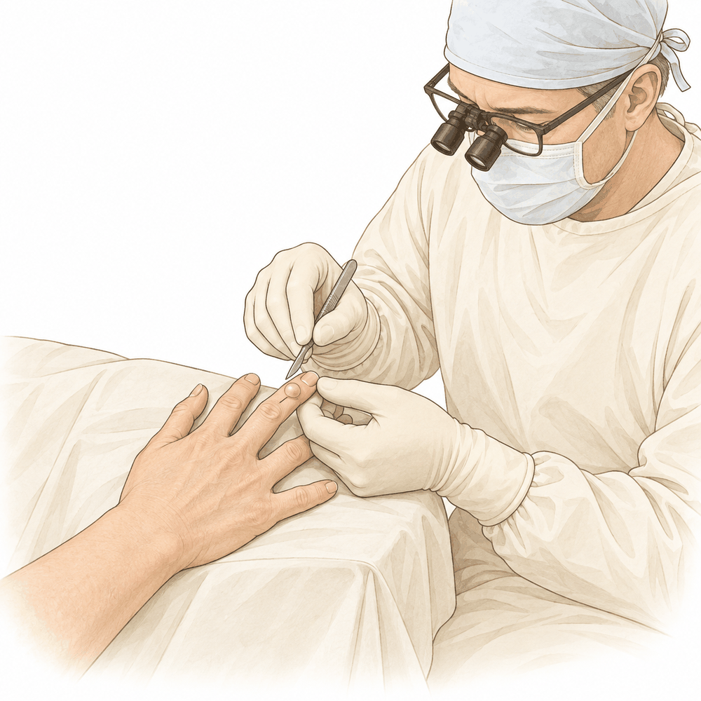
آپ کا سرجن انگلی کے آخری جوڑ کے قریب گانٹھ کے اوپر ایک چھوٹا چیرا لگاتا ہے۔ اس چیرے کے ذریعے وہ سسٹ کو نکالتا ہے اور ہڈی کے اس چھوٹے نوکیلے ابھار (bone spur) کو صاف کرتا ہے جو اکثر اس کے ساتھ ہوتا ہے۔ یہ ابھار جوڑ کے گھسنے والے آرتھرائٹس سے بنتا ہے اور یہ ایک عام وجہ ہے کہ یہ سسٹ بار بار واپس آتے ہیں، اس لیے اسے نکالنا آپریشن کا ایک اہم حصہ ہے۔
جب سسٹ اور ہڈی کا ابھار نکل جائیں تو آپ کا سرجن اس جگہ کو قریب کی جلد کے ایک چھوٹے ٹکڑے (flap) سے ڈھانپ دیتا ہے۔ اس ٹکڑے کو اٹھا کر سسٹ کی چھوڑی ہوئی خالی جگہ پر کھسکایا جاتا ہے اور ٹانکوں سے اپنی جگہ پر سی دیا جاتا ہے۔ اس سے جوڑ کے اوپر صحت مند جلد آ جاتی ہے اور یہ جگہ ایک ہی بار میں بھرنے میں مدد ملتی ہے۔ ٹانکے خود گھل جانے والے ہو سکتے ہیں یا نکالے جانے والے، اور آپ کا سرجن آپ کو بتائے گا کہ کون سی قسم استعمال کی گئی ہے۔
پورا آپریشن انگلی کے سرے پر اسی ایک چھوٹی جگہ کے ذریعے کیا جاتا ہے۔ گہرے جوڑوں یا ٹینڈنوں (tendons، یعنی وہ مضبوط ڈوریاں جو پٹھوں کو ہڈی سے جوڑتی ہیں) کے ساتھ کچھ نہیں کیا جاتا، سوائے اس کے کہ سرجری کے دوران سامنے آنے والی صورتِ حال اس کا تقاضا کرے۔
آپریشن کے بعد
آپ ریکوری روم میں جاتے ہیں، جہاں نرسیں آپ پر نظر رکھتی ہیں (اگر آپ کو جنرل اینستھیزیا دیا گیا تھا تو اس کا اثر ختم ہونے تک)۔ آپ کی انگلی پر زخم کی پٹی ہو گی اور یہ کچھ دیر تک سُن محسوس ہو سکتی ہے۔ سُن پن ختم ہونے سے پہلے ہی آپ کے لیے درد کش دوا کا انتظام کر دیا جاتا ہے؛ اگر آپ کو تکلیف ہو تو نرسوں کو بتائیں، کیونکہ وہ اسے تبدیل کر سکتی ہیں۔ جیسے ہی آپ خود کو سنبھلا ہوا محسوس کریں آپ چل پھر سکتے ہیں، اور آپ کے ہاتھ کو ساکن رکھنے کی ضرورت نہیں۔ پہلے 24 گھنٹے کسی کو آپ کے ساتھ رہنا چاہیے۔ آپ کی ٹیم آپ کو بتائے گی کہ آپ اسی دن گھر جائیں گے یا ایک رات ہسپتال میں رہیں گے۔ ہم زخم کی پٹی تقریباً 10 دن تک لگی رہنے دیتے ہیں؛ براہِ کرم اس سے پہلے اسے نہ اتاریں جب تک ہم آپ سے نہ کہیں۔ جب ہم آپ کو دیکھتے ہیں تو اسے بدلتے یا اتارتے ہیں۔
صحت یابی
پہلے چند دنوں تک آپ کی انگلی میں درد ہو گا اور تھوڑی سوجن ہو گی۔ یہ بتدریج کم ہو جاتی ہے۔ ہاتھ کو تکیے پر اونچا رکھنے سے، خاص طور پر رات کو، ٹیسیں کم ہو جاتی ہیں۔ ٹیم کی ہدایت کے مطابق عام درد کش دوا عموماً کافی ہوتی ہے۔
آپ زخم کی پٹی کے ساتھ گھر جاتے ہیں، اور یہ تقریباً 10 دن تک لگی رہتی ہے، جب تک ہم آپ کو دیکھ کر اسے بدل نہ دیں۔ آپ کے ہاتھ کو ساکن رکھنے کی ضرورت نہیں، اس لیے جیسے ہی آپ خود کو سنبھلا ہوا محسوس کریں، آپ اسے گھر کے ہلکے پھلکے کاموں کے لیے استعمال کر سکتے ہیں۔ زور سے پکڑنے سے پرہیز کریں، اور زخم کی پٹی کو خشک رکھیں۔
پٹی اترنے تک جوڑ کے اوپر جلد کا ٹکڑا عموماً اچھی طرح بھر رہا ہوتا ہے۔ زخم کا نشان وقت کے ساتھ نرم اور ہلکا پڑ جاتا ہے، اور شائع شدہ تحقیقات کے مطابق اس کی شکل اچھی طرح بیٹھ جاتی ہے۔ سوجن کم ہونے کے ساتھ انگلی کے آخری جوڑ کی حرکت واپس آ جاتی ہے۔ سرجری کے بعد ہینڈ تھراپی Extend Rehabilitation میں Ruby Doolan کے ساتھ ہوتی ہے؛ وہ آپ کی ورزشوں میں رہنمائی کریں گی اور ضرورت ہو تو اسپلنٹ بنائیں گی۔
صحت یابی ہر شخص میں مختلف ہوتی ہے، اور آپ کا وقت مختلف ہو سکتا ہے۔ آپ کا سرجن اور آپ کا ہینڈ تھراپسٹ (ہاتھ کی بحالی کے ماہر) ہر معائنے پر آپ کی رہنمائی کریں گے، اور آپ کو پتا چل جائے گا کہ سب ٹھیک چل رہا ہے جب سوجن کم ہو، زخم بند ہو جائے، اور آپ بغیر درد کے انگلی سے پکڑ سکیں اور اسے موڑ سکیں۔
کیا مسائل پیدا ہو سکتے ہیں
زیادہ تر مریض ٹھیک رہتے ہیں، لیکن کبھی کبھار مسائل پیدا ہو سکتے ہیں۔ آپ کا سرجن اور ٹیم آپ کی قریب سے نگرانی کرتے ہیں تاکہ کسی بھی مسئلے کو جلد پہچان سکیں۔
کبھی کبھی سسٹ اسی جگہ دوبارہ بن جاتا ہے۔ آپ کو انگلی کے آخری جوڑ کے قریب دوبارہ ایک چھوٹی، سخت گانٹھ نظر آئے گی، بالکل پہلے والی جیسی۔ اگر آپ کو ایسی گانٹھ بنتی نظر آئے تو اگلے معائنے پر اس کا ذکر کریں۔ جوڑ کے ساتھ موجود ہڈی کا چھوٹا ابھار اسی لیے نکالا جاتا ہے تاکہ ایسا نہ ہو۔
اس جگہ کو ڈھانپنے کے لیے استعمال ہونے والے جلد کے ٹکڑے کا اچھی طرح بھرنا ضروری ہے۔ اگر ایسا نہ ہو تو آپ دیکھ سکتے ہیں کہ زخم کے کنارے الگ ہو رہے ہیں، جلد کا ٹکڑا سفید پڑ گیا ہے یا گہرا (سیاہی مائل) ہو گیا ہے، یا وہ جگہ بند ہونے کے بجائے کچی اور رِستی ہوئی رہتی ہے۔ زخم توقع سے زیادہ آہستہ بھی بھر سکتا ہے۔ اگر آپ کو ان میں سے کوئی بھی نشانی نظر آئے تو کلینک سے رابطہ کریں تاکہ ہم زخم کا معائنہ کر سکیں اور آپ کے علاج میں تبدیلی کر سکیں۔
زخم کا انفیکشن ایک اور چیز ہے جس پر نظر رکھنی چاہیے۔ یہ عموماً ایسی سرخی کی صورت میں نظر آتا ہے جو زخم سے باہر کی طرف پھیلتی ہے، اور ساتھ میں گرمی، سوجن یا چھونے پر درد ہوتا ہے جو کم ہونے کے بجائے بڑھتا جاتا ہے۔ آپ کو زخم سے سیال یا پیپ رِستی نظر آ سکتی ہے، یا گہرا، ٹیسیں مارتا درد محسوس ہو سکتا ہے جو عام درد کش دواؤں سے کم نہ ہو۔ آپ کو بخار جیسی گرمی اور عمومی طور پر طبیعت خراب محسوس ہو سکتی ہے۔ اگر آپ کو یہ نشانیاں نظر آئیں تو فوراً کلینک کو فون کریں۔ اگر آپ ہم سے رابطہ نہ کر سکیں اور آپ کی طبیعت خراب ہو تو اپنے قریب ترین ہسپتال کی ایمرجنسی جائیں۔
اچھی خبر یہ ہے کہ جب یہ آپریشن جلد کے ٹکڑے کے ساتھ کیا جاتا ہے تو رپورٹ شدہ کیسوں میں اس طرح کے زخم کے مسائل کم ہوتے ہیں، اور سسٹ شاذ و نادر ہی دوبارہ بنتا ہے [2, 3]۔
اگر معائنوں کے درمیان آپ کو اپنی انگلی کے بارے میں کوئی بھی بات پریشان کرے تو انتظار نہ کریں۔ ہمیں فون کریں اور بتائیں کہ آپ کیا دیکھ اور محسوس کر رہے ہیں۔ جلد توجہ دینے سے زیادہ تر مسائل کو حل کرنا آسان ہو جاتا ہے۔
اگر آپ تفصیلات جاننا چاہیں تو اس صفحے پر پیچیدگیوں کی جدول میں عام شرحیں درج ہیں۔
ہمیں کب فون کریں
اگر آپ کو بخار محسوس ہو، زخم کے ارد گرد سرخی پھیلتی نظر آئے، یا زخم سے سیال یا پیپ رِستی نظر آئے تو ہمیں فون کریں۔ اگر درد اچانک بڑھ جائے اور عام درد کش دوائیں اس پر کوئی اثر نہ کریں، اگر آپ کی انگلی سُن محسوس ہو، یا اگر آپ اسے موڑ یا سیدھا نہ کر سکیں تو ہمیں فون کریں۔ اگر آپ کی پنڈلی میں سوجن ہو یا سانس پھولے، یا اگر آپ کو بخار جیسی گرمی اور عمومی طور پر طبیعت خراب محسوس ہو اور آپ ہم سے رابطہ نہ کر سکیں، تو اپنے قریب ترین ہسپتال کی ایمرجنسی جائیں۔ اگر معائنوں کے درمیان انگلی کے بارے میں کوئی بھی بات آپ کو پریشان کرے تو انتظار نہ کریں۔
مزید تفصیل
Advanced reading: the deeper science (optional)
یہ حصہ اس سے زیادہ تفصیل میں جاتا ہے جتنی آپ کو اپنے علاج کے فیصلوں کے لیے ضرورت ہے۔ میوکس سسٹ کی سرجری کے بارے میں اضافی مطالعہ اس لیے فائدہ مند ہے کہ یہ آپریشن دراصل سسٹ کے بارے میں ہے ہی نہیں، اور جب آپ اس کی وجہ سمجھ لیتے ہیں تو اس کے کرنے کے طریقے کی ہر بات سمجھ میں آ جاتی ہے۔
سسٹ ایک علامت ہے، اصل بیماری آسٹیوفائٹ ہے
میوکس سسٹ ناخن کے سب سے قریب والے جوڑ کا ایک چھوٹا گینگلیئن سسٹ (ganglion) ہے، اور یہ تقریباً ہمیشہ اس جوڑ کے گھسنے والے آرتھرائٹس کے اوپر ہوتا ہے۔ گھسے ہوئے جوڑ میں اضافی ہڈی کے چھوٹے نوکیلے ابھار (osteophytes) بن جاتے ہیں، جوڑ کا سیال ان کے پاس سے باہر نکلنے کا راستہ ڈھونڈ لیتا ہے، اور سسٹ وہ جگہ ہے جہاں یہ انگلی کی پشت کی پتلی جلد کے نیچے جمع ہو جاتا ہے۔
اسی لیے صرف سسٹ نکالنے کا ریکارڈ خراب ہے: آسٹیوفائٹ چھوڑ دیں تو رساؤ اسے دوبارہ بھر دیتا ہے۔ اس کا سب سے واضح ثبوت ایک ایسی تحقیق سے ملتا ہے جس میں اس کا الٹ کیا گیا، یعنی صرف آسٹیوفائٹ نکالے گئے اور سسٹ کو چھوڑ دیا گیا، اور زیادہ تر صورتوں میں مسئلہ مکمل طور پر ختم ہو گیا [1]۔ اس آپریشن کو یوں سمجھنا بہتر ہے کہ یہ جوڑ کی صفائی (joint debridement) ہے جس کے ساتھ ساتھ سسٹ کا بھی علاج ہو جاتا ہے، نہ کہ سسٹ نکالنے کا آپریشن جس میں ہڈی بعد کا خیال ہو۔
جلد کا ٹکڑا (flap) کیوں، اور یہ زیادہ اہم کیوں نہیں کہ کون سا
پرانے سسٹ کے اوپر کی جلد اکثر کھنچ کر کاغذ کی طرح پتلی ہو جاتی ہے، اور جب سسٹ اور پتلی جلد نکال دی جائیں تو براہِ راست بند کرنے کے لیے کافی صحت مند جلد باقی نہیں رہتی۔ یہی کام مقامی فلیپ (local flap) کرتا ہے: ساتھ کی جلد کا ایک چھوٹا زبان نما ٹکڑا گھما کر خالی جگہ پر لایا جاتا ہے، اور یہ اپنی خون کی فراہمی ساتھ لاتا ہے۔
ڈاکٹر ہِرپرا جو طریقہ سب سے زیادہ استعمال کرتے ہیں وہ جانسن (Johnson) اور ان کے ساتھیوں کے بیان کردہ طریقے پر مبنی ہے [2]: ایک ہی دن میں گھر جانے والا آپریشن، انگلی کی جڑ پر سُن کرنے والے انجکشن (local anaesthetic ring block) کے تحت؛ سسٹ کو پتلی جلد سمیت مکمل طور پر بیضوی چیرے سے نکالنا، سسٹ کی گردن کا پیچھا کرتے ہوئے جوڑ تک جانا اور اسے ساتھ جڑی جوڑ کی جھلی سمیت کاٹ کر نکالنا؛ انگلی کو سیدھا کرنے والے ٹینڈن (extensor tendon) کو بچاتے ہوئے انگلی کی پشت پر قابلِ رسائی آسٹیوفائٹ نکالنا؛ اور خالی جگہ کو انگلی کی اسی طرف سے اٹھائے گئے جلد کی پوری موٹائی والے مقامی ایڈوانسمنٹ فلیپ (full-thickness local advancement flap) سے، بغیر کھنچاؤ کے، بند کرنا۔ ٹانکے تقریباً دو ہفتے بعد نکالے جاتے ہیں۔
اس طریقے کی شائع شدہ تحقیق میں، جس میں دس سال کے دوران 69 مریض شامل تھے، سسٹ کے دوبارہ بننے کی شرح 1.4 فیصد تھی، اور مریض زخم کے نشان سے بہت مطمئن تھے اور انہوں نے دوبارہ یہ آپریشن کروانے پر آمادگی ظاہر کی [2]۔
فلیپ کا ڈیزائن فیصلہ کن عنصر نہیں ہے۔ زیٹیلی کا دو حصوں والا فلیپ (Zitelli bilobed flap) ناخن بنانے والے حصے (nail matrix) کے لیے کسی اضافی خطرے کے بغیر اچھی جلد کا احاطہ فراہم کرتا ہے [3]، جو اہم ہے کیونکہ ناخن کے بڑھنے کا حصہ سسٹ کے بالکل آگے ہوتا ہے، اور دو دوسرے فلیپ ڈیزائنوں کے ایک حالیہ موازنے میں ان کے درمیان خوبصورتی سے اطمینان یا پیچیدگیوں میں کوئی فرق نہیں ملا [4]۔ دوسرے مراکز جلد کی پوری موٹائی کے پیوند (full-thickness skin grafting) کے ساتھ قابلِ قبول حد تک دوبارہ بننے کی شرح بتاتے ہیں [5]، اور ایک چھوٹی تحقیق میں جوڑ کی پشت والی جھلی کو مکمل نکالنے کے بعد سسٹ کبھی دوبارہ نہیں بنا [6]۔ ہر جگہ بنیادی بات ایک ہی ہے: جوڑ کا علاج کریں، اور جلد کا مضبوط احاطہ حاصل کریں۔
جب اصل مسئلہ خود جوڑ ہو
کبھی کبھی سسٹ چھوٹا مسئلہ ہوتا ہے اور اس کے نیچے کا آرتھرائٹس ہی دراصل تکلیف دیتا ہے۔ اگر جوڑ میں خود درد ہو، صرف گانٹھ نہ ہو، تو سسٹ نکالنا پیغام لانے والے کا علاج کرتا ہے اور پیغام کو وہیں چھوڑ دیتا ہے۔ ایسی صورت میں حتمی حل جوڑ کا فیوژن (arthrodesis، دو ہڈیوں کو جوڑ کر ایک کر دینا) ہو سکتا ہے، جو ایک ہی آپریشن میں آرتھرائٹس، درد اور سسٹ کی وجہ کو ختم کر دیتا ہے۔ اس طریقے، اس کے فائدے نقصان اور اس کی صحت یابی پر انگلی کے آخری جوڑ کا فیوژن (DIP joint fusion) کے صفحے پر بات کی گئی ہے۔
کیا مسائل پیدا ہو سکتے ہیں
مخصوص خطرات جسم کی ساخت سے پیدا ہوتے ہیں: ناخن بنانے والا حصہ صرف چند ملی میٹر دور ہوتا ہے، اس لیے ناخن میں نالی یا ابھری ہوئی لکیر بن سکتی ہے (اور اس کے برعکس، ناخن بنانے والے حصے پر دباؤ ڈالنے والا سسٹ پہلے ہی ایسی لکیر بنا چکا ہو سکتا ہے جسے سرجری بہتر کر سکتی ہے)؛ پتلی جلد کی وجہ سے کبھی کبھار زخم بھرنے میں زیادہ وقت لگتا ہے؛ اور سسٹ کا دوبارہ بننا — اگرچہ جوڑ کی مناسب صفائی کے بعد کم ہوتا ہے، جیسا کہ اوپر کے اعداد و شمار دکھاتے ہیں، کبھی صفر نہیں ہوتا، کیونکہ جس آرتھرائٹس نے سسٹ پیدا کیا تھا وہ جوڑ اب بھی آرتھرائٹس والا جوڑ ہی ہے۔
حوالہ جات
[1] Lee HJ, Kim PT, Jeon IH, et al. Osteophyte excision without cyst excision for a mucous cyst of the finger. J Hand Surg Eur Vol. 2013;39(3):258-261. https://doi.org/10.1177/1753193413478549
[2] Johnson SM, Treon K, Thomas S, Cox QG. A reliable surgical treatment for digital mucous cysts. J Hand Surg Eur Vol. 2013;39(8):856-860. https://doi.org/10.1177/1753193413508540
[3] Jiménez I, Delgado PJ, Kaempf de Oliveira R. The Zitelli bilobed flap on skin coverage after mucous cyst excision: a retrospective cohort of 33 cases. J Hand Surg Am. 2017;42(7):506-510. https://doi.org/10.1016/j.jhsa.2017.03.013
[4] Orieux A, Maximen J, Yvonnet T, et al. Aesthetic outcome of biquadrangular and Hueston dorsal flaps in digital mucous cyst surgery. J Hand Surg Eur Vol. 2026;. https://doi.org/10.1177/17531934261433822
[5] Jamnadas-Khoda B, Agarwal R, Harper R, Page RE. Use of Wolfe graft for the treatment of mucous cysts. J Hand Surg Eur Vol. 2009;34(4):519-521. https://doi.org/10.1177/1753193408103498
[6] Kanaya K, Wada T, Iba K, Yamashita T. Total dorsal capsulectomy for the treatment of mucous cysts. J Hand Surg Am. 2014;39(6):1063-1067. https://doi.org/10.1016/j.jhsa.2014.03.004
Evidence & references
This is the clinical evidence summary written for health professionals. It is technical, and it lists the research this page was built from. You do not need to read it to understand your treatment or to make a decision about it.
Anatomy & Pathophysiology
General Hand Architecture
- The hand is composed of 19 bones, 17 articulations, and 19 muscles situated entirely within the hand [2].
- The hand contains approximately the same number of tendons activated by forearm muscles as it has intrinsic muscles [2].
- The open hand with fingers extended and in contact forms a balanced graceful oval in its longitudinal axis [2].
- The proximal carpometacarpal half of the hand is flattened and presents two faces with unique anatomical and functional significance [2].
- The posterior or dorsal aspect of the hand is convex, while the anterior, palmar, or volar aspect is concave [2].
- The distal half of the hand is separated into five digits which flex toward the palm [2].
- The thumb has a more proximal and lateral position than the four fingers, allowing movement inward and outward from the palm [2].
- The hinges for finger flexion and extension are located at the thenar crease and the transverse distal palmar crease, not at the bases of the digits [2].
- When fingers are extended and separated, their tips lie on the circumference of a circle whose center is the head of the third metacarpal [2].
Cutaneous Units and Skin Mechanics
- The dorsum of the hand contains a cutaneous unit extending from the wrist to the proximal interphalangeal joints of the fingers and the interphalangeal joint of the thumb [3].
- The dorsal covering of the interphalangeal articulations forms a unique cutaneous unit characterized by considerable excess of skin when digits are in extension [3].
- The fine tight skin of the dorsal aspect of the middle phalanx forms a distinct cutaneous unit [3].
- The dorsal integument of the distal phalanx is distinct due to the presence of the nail bed and its matrix [3].
- The palm forms a cutaneous unit extending from the distal transverse crease of the wrist up to the transverse crease at the base of the digits [3].
- The palmar integument is subdivided into two zones by the oppositional crease of the thumb, which constitutes the oblique axis of the hand [3].
- The skin of the radial portion of the palm covers the thenar eminence and is relatively well vascularized and mobile [3].
- The skin of the ulnar and distal portion of the palm covers the hypothenar eminence and has poor mobility [3].
- The central triangular part of the palm has fixed, poorly vascularized skin that covers almost directly the superficial palmar aponeurosis [3].
- The integument of the palmar face of the digits is subdivided into phalangeal units separated by digital flexion folds [3].
- When a digit is completely flexed, the integument of adjacent phalanges comes into contact in the zones of the flexion creases, establishing areas of cutaneous contact in the form of a diamond [3].
- The sides of the diamond-shaped cutaneous contact zones do not undergo variations in length during flexion and extension movements [3].
- Incisions made along the lines of the diamond-shaped cutaneous contact zones present a minimal chance of retraction [3].
- The web spaces are formed from the union of two nonsymmetrical cutaneous surfaces [3].
- The dorsal slope of the web space has a gradual incline and its supple skin is not adherent to the subjacent region [3].
- The palmar surface of the web space is flat and precipitously interrupted, with skin densely adherent to the commissural skeleton [3].
- The commissural skeleton is formed by the interdigital palmar (natatory) ligament between the fingers and by the distal transverse ligament at the level of the thumb web [3].
Intrinsic Musculature
- There are seven interosseous muscles in the hand, consisting of four dorsal and three volar muscles [4].
- The dorsal interossei are abductors [4].
- The anatomic axis of the hand coincides with the axis of the third metacarpal [4].
- The dorsal interossei lie to the radial side of the index and middle fingers and the ulnar side of the middle and ring fingers [4].
- The little finger is abducted by the abductor digiti quinti [4].
- The volar interossei are adductors [4].
- The volar interossei lie to the ulnar side of the index finger and the radial side of the ring and little fingers [4].
- The middle finger has two dorsal interossei and no volar interossei because the central axis of the hand lies within it [4].
- Each dorsal interosseous muscle, with the exception of the third, has two muscle heads [4].
- The superficial head of the dorsal interosseous muscles arises most dorsally from the shaft of the contiguous metacarpals [4].
- The superficial head of the dorsal interosseous muscles is inserted deeply by a medial tendon onto the lateral tubercle of the base of the proximal phalanx [4].
- The superficial head of the dorsal interosseous muscles abducts and weakly flexes the proximal phalanx [4].
- The superficial head of the dorsal interosseous muscles has no direct effect on the middle or distal phalanges [4].
- The deep head of each dorsal interosseous muscle forms a lateral tendon, or lateral band, at the level of the metacarpophalangeal joint [4].
- The deep head of the dorsal interosseous muscles flexes and weakly abducts the proximal phalanx while extending the middle and distal phalanges [4].
- At the level of the middle of the proximal phalanx, transverse fibers arch dorsally from each lateral band to join over the dorsum of the finger, flexing the proximal phalanx [4].
- Oblique fibers, or spiral fibers, from the lateral bands sweep over the distal third of the proximal phalanx to insert onto the lateral tubercles at the base of the middle phalanx [4].
- The oblique fibers extend the middle phalanx at the proximal interphalangeal joint [4].
- The lateral bands are joined by the lateral slips of the extensor tendon to form the conjoined lateral band [4].
- The two conjoined lateral bands to each finger unite at the distal third of the middle phalanx to form the terminal tendon [4].
- The terminal tendon inserts at the base of the distal phalanx to extend it [4].
- The flexor digiti quinti brevis is structurally and functionally similar to the deep head of the dorsal interossei, forming the ulnar lateral band of the little finger [4].
- The three volar interossei arise from adjacent surfaces of contiguous metacarpal shafts [4].
- Each volar interosseous muscle has only one muscle head and none of them insert onto the proximal phalanx [4].
- The volar interossei form the ulnar lateral band of the index finger and the radial lateral band of the ring and little fingers [4].
- The abductor digiti quinti arises from the fifth metacarpal and inserts onto the ulnar lateral tubercle at the base of the proximal phalanx of the little finger [4].
- The flexor digiti quinti brevis arises from the fifth metacarpal and forms the ulnar lateral band [4].
- The opponens digiti quinti lies deepest among the hypothenar muscles and arises from the pisohamate ligament and the hook of the hamate [4].
- The opponens digiti quinti inserts onto the ulnar side of the diaphysis of the fifth metacarpal, which it flexes and supinates [4].
Vascular Anatomy
- The arteries of the thumb vary in both size and number, making surgical reconstruction delicate [8].
- The palmar aspect of the thumb can be schematized into three segments defined by the metacarpophalangeal and interphalangeal flexion creases [8].
- In the classical layout, the princeps pollicis artery, a terminal branch of the radial artery, crosses the first intermetacarpal space [8].
- The princeps pollicis artery runs along the ulnar side of the first metacarpal bone and along the volar surface of the adductor muscle [8].
- The princeps pollicis artery emerges onto the subcutaneous palmar tissue at the level of the cutaneous flexion crease of the metacarpophalangeal joint [8].
- At the metacarpophalangeal joint level, the princeps pollicis artery divides into two terminal rami, the collateral palmar arteries of the thumb [8].
- The collateral palmar arteries of the thumb run along the digital tunnel symmetrically and are of equal caliber [8].
- The collateral palmar arteries head distally to finally unite in the pulp arcade [8].
- During their transit in the digital tunnel, the collateral palmar arteries break off into numerous collateral branches that are cutaneous, articular, or osseous [8].
- An arcade located deep in the flexor tendon joins together the two arteries at the level of the distal metaphysis of the first phalanx [8].
- Vessels originating from the subtendinous arcade enter the vincula and irrigate the flexor tendon [8].
- Only 15% of anatomical dissections of the thumb palmar arteries fall into the classical "typical" category [8].
- In the first segment of the thumb (between the opposition crease and metacarpophalangeal flexion crease), arteries of surgical interest on the volar surface are rare [8].
- In the first segment of the thumb, the artery is located deeply and is more easily accessible from the dorsal surface [8].
- In the second segment of the thumb, the two arteries run alongside the flexor tendon and behind the collateral nerves [8].
- In the second segment of the thumb, the main artery is the ulnar collateral artery [8].
- In cases where the palmar ulnar collateral artery is absent in the second segment, the dorsal artery takes its place by means of a branch through the subtendinous arcade [8].
- In the third segment of the thumb (pulp segment), the two arteries are of similar size and run through the thick fatty subcutaneous padding [8].
- In the pulp segment, the arteries cross over and convert into the ends of the digital nerves at the level of the median axis [8].
- The posterior area of the thumb is vascularized by two arteries which originate from the palmar arteries at the level of the first metacarpal [8].
- These dorsal arteries run laterally along the metacarpophalangeal joint and continue obliquely from volar to dorsal [8].
- At the level of the neck of the first phalanx, an anastomosis can be found which originates from the palmar arteries for the dorsal supply [8].
- The ulnar dorso-collateral artery generally stems from the princeps pollicis onto the medial border of the neck of the first metacarpal [8].
Surgical Anatomy and Incisions
- Distal palmar incisions are transverse, while proximal palm incisions tend to be more longitudinal with the distal end curving radially to parallel the closest major skin crease [9].
- An incision of any desired length can be made across the palm provided that the underlying digital nerves and other vital structures are protected [9].
- After skin and underlying fat are incised, the fat is dissected from the palmar fascia and carried with the skin flaps [9].
- It may be desirable to preserve small vessels perforating the palmar fascia if wide undermining of the skin flaps is necessary [9].
- Most vital structures in the palm are deep to the palmar fascia [9].
- In the distal palm, structures lying between the metacarpal heads are not protected by the palmar fascia [9].
- The superficial volar neurovascular arch should be protected when deeper exposure is required [9].
- Incisions in the more proximal palm should parallel the thenar crease [9].
- When extended proximal to the wrist, incisions should not cross the flexor wrist creases at a right angle [9].
- The most important structure in the thenar area is the recurrent branch (motor) of the median nerve, which should be exposed and protected if its exact location is in doubt [9].
- Care should be taken to avoid injury to the palmar cutaneous branches of the median and ulnar nerves [9].
- There is no single longitudinal incision in the proximal palm that completely avoids the palmar cutaneous branches of the median and ulnar nerves [9].
- Midlateral incisions described for the fingers are suitable for the thumb, with the radial side being more accessible [9].
- A radial midlateral thumb incision can be extended by curving its proximal end at the midmetacarpal area to create a flap on the palmar surface of the thumb [9].
- Care should be taken to avoid the dorsal branch of the superficial radial nerve to the radial side of the thumb during radial midlateral incisions [9].
- The volar zigzag finger incision does not require mobilizing either neurovascular bundle and directly exposes the volar surface of the flexor tendon sheath [9].
- On a contracted skin surface, the volar zigzag finger incision tends to straighten out and result in a more linear scar than is desirable [9].
- The volar midoblique incision crosses the flexion creases obliquely in the midline of the finger between the neurovascular bundles [9].
Skin Coverage Dimensions
- The necessary skin to cover the thumb distal to the metacarpophalangeal joint is about 9 cm wide and 8 cm long [6].
- Skin loss of the thumb and first metacarpal is 13 cm wide and 12 cm long [6].
- The skin cover of both the palmar and dorsal surfaces of the hand is 12 cm by 10 cm [6].
- The skin covering each finger is 7 cm by 10 cm on both the palmar and dorsal aspects [6].
- Skin grafting or flaps for both sides of the hand and digits requires a skin of 20 cm by 20 cm [6].
- Skin grafting or flaps for one aspect of the forearm from wrist to elbow requires skin of 30 cm by 15 cm [6].
- Skin grafting or flaps for both aspects of the forearm requires skin of 30 cm by 30 cm [6].
Investigations
- Clinical evaluation of the injured or dysfunctional hand and wrist requires combining patient history with a careful physical examination to pinpoint or narrow the scope of possible pathologic processes [1].
- Diagnostic tests such as imaging and serum laboratory studies are useful in determining pathology but can be expensive, time consuming, and often nonspecific [1].
- A careful physical examination is essential to direct care and future testing if indicated [1].
- An 8-MHz Doppler tone assessment may be used to identify superficially displaced neurovascular bundles when Dupuytren cords lie beneath soft fleshy prominences [11].
- False-negatives are possible with 8-MHz Doppler tone assessment for identifying neurovascular bundles [11].
- Doppler imaging is a promising improvement for visualizing palmar structures, but higher resolution imaging technology is needed [11].
- MR assessment of Dupuytren’s disease is hindered by the resolution of current equipment, orientation issues due to multiplanar deformities of the fingers, and lack of intraoperative availability [11].
- MRI is probably most useful in identifying additional pathology such as flexor tendon bowstringing [11].
- MRI may be helpful in providing a quantitative noninvasive measure of cellularity of affected areas, which is an index of biologic activity [11].
- The potential of MRI as a staging tool based on cellularity measurement has not been investigated yet on a large scale [11].
References
[1] Orthopaedic Knowledge Update 13 Ebook Without Multimedia. Anatomy, Evaluation, Clinical Examination, and Imaging > Evaluation and Clinical Examination: Current Concepts.
[2] Exam Of The Hand Wrist 2Ed. INTRODUCTION.
[3] Exam Of The Hand Wrist 2Ed. Functional cutaneous units.
[4] Green S Operative Hand Surgery. Interosseous and Hypothenar Muscles.
[6] Exam Of The Hand Wrist 2Ed. Planning skin cover of the hand and forearm.
[8] Exam Of The Hand Wrist 2Ed. Techniques of investigation of the arterial supply by J P Melki > Vascularization of the thumb > Palmar aspect.
[9] Campbell S Operative Orthopaedics 4 Volume Set. RESULTS OF SUTURE OF THE SCIATIC NERVE > PALMAR INCISIONS.
[11] Dupuytren S Disease And Related Hyperproliferative Disorders. 54. The Future of Dupuytren’s Research and Treatment > 54.4 Mechanical Measurements and Procedures > 54.4.4 Imaging.