یہ آپریشن کیوں تجویز کیا گیا ہے
ڈاکٹر کیرن ہِرپرا، جو Mater Private Hospital Rockhampton میں بازو اور ہاتھ کے سرجن (upper-limb surgeon) ہیں، علاج کو آپ کی مخصوص چوٹ کے مطابق طے کرتے ہیں۔ اس چوٹ میں متاثر ہونے والا لیگامنٹ (ligament)، یعنی وہ مضبوط بندھن جو ایک ہڈی کو دوسری ہڈی سے جوڑتا ہے، آپ کے انگوٹھے کی اندرونی طرف، اس جوڑ پر ہوتا ہے جہاں انگوٹھا ہاتھ سے ملتا ہے۔ یہ آپریشن اس لیگامنٹ کی مرمت کرتا ہے تاکہ جوڑ دوبارہ مستحکم ہو جائے۔
ہم عام طور پر بغیر آپریشن کے علاج سے آغاز کرتے ہیں۔ جزوی پھٹاؤ میں، بھرنے کے دوران پلستر یا اسپلنٹ (splint) انگوٹھے کو ساکت رکھ سکتا ہے، اور اس سے اکثر انگوٹھا مستحکم اور درد سے پاک ہو جاتا ہے۔ لیکن جب لیگامنٹ پوری طرح پھٹ جائے تو وہ اکثر خود سے ٹھیک نہیں ہوتا۔ تقریباً 75 فیصد مکمل پھٹاؤ صرف اسپلنٹ سے ٹھیک نہیں ہو پاتے۔ لیگامنٹ کا پھٹا ہوا سرا اپنی جگہ سے کھسک بھی سکتا ہے، جس کی وجہ سے وہ دوبارہ ہڈی سے نہیں جڑ پاتا۔ اگر ایسے پھٹاؤ کو بغیر علاج کے چھوڑ دیا جائے تو اس سے طویل مدتی درد اور انگوٹھا استعمال کرنے میں دشواری ہو سکتی ہے۔
مکمل پھٹاؤ میں فوراً سرجری کا مشورہ دیا جا سکتا ہے، یا اس وقت جب اسپلنٹ سے کافی بہتری نہ آئی ہو۔ مقصد ایسا انگوٹھا ہے جو مستحکم ہو، درد سے پاک ہو، اور پکڑنے اور چٹکی کی گرفت کے لیے کافی مضبوط ہو۔ مریضوں کے اپنے بتائے ہوئے درد اور ہاتھ کے کام کے نتائج مرمت کے تین اور 12 ماہ بعد، سرجری سے پہلے کے مقابلے میں، بہتر ہوتے ہیں۔
آپریشن سے پہلے
آپ کے سرجن آپ کے انگوٹھے کے اسکین کی مدد سے آپریشن کی منصوبہ بندی کریں گے۔ اس کا آغاز عموماً ایکسرے سے ہوتا ہے، جو اس جگہ کوئی چھوٹا سا فریکچر (ہڈی کا ٹوٹنا) دکھا سکتا ہے جہاں سے لیگامنٹ اکھڑا ہو۔ لیگامنٹ کو خود دیکھنے اور یہ تصدیق کرنے کے لیے کہ کیا اس کا پھٹا ہوا سرا اپنی جگہ سے کھسک گیا ہے، ایم آر آئی یا الٹراساؤنڈ اسکین بھی استعمال کیا جا سکتا ہے۔
سرجری سے پہلے آپ کو واضح ہدایات دی جائیں گی۔ آپ کو آپریشن سے سات گھنٹے پہلے سے کچھ نہیں کھانا چاہیے۔ اس سے یہ ممکن ہوتا ہے کہ اگر آپریشن کی فہرست وقت سے پہلے چل رہی ہو تو آپ کی سرجری پہلے کی جا سکے۔ ہو سکتا ہے آپ کو کچھ دوائیں بند کرنی پڑیں، اور آپ کے سرجن آپ کو بتائیں گے کہ کون سی اور کب۔ آپ جو کچھ بھی لیتے ہیں اس سب کی فہرست ساتھ لائیں۔ آپریشن کے بعد گاڑی میں گھر لے جانے کے لیے کسی کا انتظام کریں، اور ڈھیلے، آرام دہ کپڑے پہنیں۔
اگر آپ کو دوسری طبی بیماریاں ہیں تو ہو سکتا ہے آپ کو خون کے ٹیسٹ یا بے ہوشی کے ڈاکٹر (anaesthetist) سے معائنے کی ضرورت ہو۔
آپریشن کے دن
آپ ہسپتال کے سرجری کے داخلے کے شعبے میں پہنچیں گے، جہاں آپ کا اندراج کیا جاتا ہے اور آپ کو آپریشن تھیٹر کے لیے تیار کیا جاتا ہے۔ وہیں آپ کی ملاقات بے ہوشی کے ڈاکٹر سے ہوگی۔ یہ آپریشن جنرل اینستھیزیا (مکمل بے ہوشی) میں کیا جاتا ہے۔ آپریشن کے بعد درد میں آرام کے لیے بعض اوقات ساتھ میں اعصابی بلاک (nerve block)، یعنی ایک انجکشن جو جسم کے ایک حصے کو سُن کر دیتا ہے، بھی لگایا جاتا ہے؛ بے ہوشی کے ڈاکٹر اسی دن آپ سے اس بارے میں بات کریں گے۔
اس کے بعد آپ کو آپریشن تھیٹر میں لے جایا جاتا ہے، جہاں آپریشن کیا جاتا ہے۔ آپریشن کے بعد آپ ریکوری روم میں جاگیں گے، جہاں بے ہوشی کی دوا کا اثر ختم ہونے تک نرسیں آپ کی نگرانی کرتی ہیں۔ جب آپ کی حالت مستحکم ہو جائے تو، طریقۂ علاج اور آپ کی صحت یابی کے مطابق، آپ یا تو وارڈ میں جائیں گے یا گھر چلے جائیں گے۔
آپریشن میں کیا کیا جاتا ہے
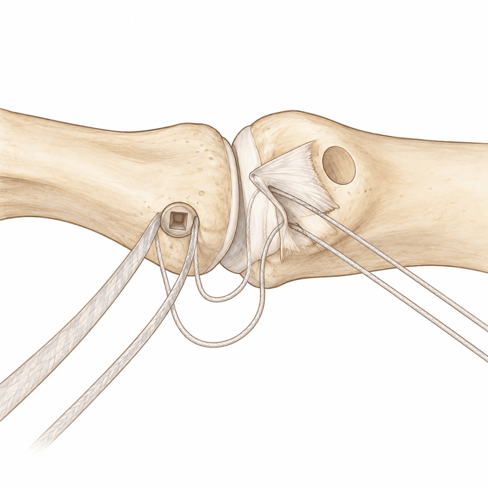
آپ کے سرجن انگوٹھے کی اس طرف ایک چھوٹا چیرا لگاتے ہیں جہاں سے لیگامنٹ اکھڑا تھا۔ اس چیرے کے ذریعے وہ لیگامنٹ کا پھٹا ہوا سرا تلاش کرتے ہیں اور ہڈی پر اس جگہ ایک چھوٹا سا حصہ تیار کرتے ہیں جہاں یہ پہلے جڑا ہوتا تھا۔ پھر ہڈی میں لگائے گئے ایک چھوٹے اینکر (anchor)، یعنی ایک چھوٹا پرزہ جو ٹانکوں کے دھاگے کو تھامتا ہے، کی مدد سے لیگامنٹ کو ٹانکوں کے ذریعے واپس اسی جگہ پر جما دیا جاتا ہے۔ یہ لیگامنٹ کو ہڈی سے دوبارہ جڑنے تک اپنی جگہ پر رکھتا ہے۔
اگر لیگامنٹ کو پھٹے ہوئے کافی عرصہ ہو گیا ہو تو ہو سکتا ہے اس کے سرے اتنے گھس چکے ہوں کہ انہیں ٹانکوں سے جوڑا نہ جا سکے۔ اس صورت میں آپ کے سرجن آپ کے اپنے انگوٹھے کے ایک قریبی ٹینڈن (tendon)، یعنی وہ مضبوط ڈوری جو پٹھے کو ہڈی سے جوڑتی ہے، کی مدد سے لیگامنٹ کو دوبارہ بنا سکتے ہیں؛ اس ٹینڈن کو موڑ کر ایک نئی پٹی بنائی جاتی ہے جو جوڑ کے دونوں طرف ہڈی سے جڑتی ہے۔ اگر لیگامنٹ ہڈی کے ایک چھوٹے ٹکڑے کے ساتھ اکھڑا ہو تو اس ٹکڑے کو ایک چھوٹی پلیٹ اور پیچوں سے واپس اپنی جگہ پر جما دیا جاتا ہے۔
کچھ مرمتوں کو لیگامنٹ کے ساتھ ساتھ سلی ہوئی فیتے جیسی ایک پٹی (tape) سے مضبوط کیا جاتا ہے۔ یہ پٹی ایک عارضی حفاظتی رسی کی طرح کام کرتی ہے، اور لیگامنٹ کے بھرنے کے دوران بوجھ بانٹتی ہے۔ چونکہ یہ کچھ کھنچاؤ خود سہتی ہے، اس لیے ہو سکتا ہے صرف مرمت کے مقابلے میں آپ کا انگوٹھا جلد حرکت کرنا شروع کر سکے۔
آپ کے سرجن جوڑ کو ساکت رکھنے اور مرمت کو ایک طرف سے دوسری طرف کے کھنچاؤ سے بچانے کے لیے انگوٹھے کے جوڑ کے آر پار دھات کا ایک چھوٹا پن بھی لگا سکتے ہیں۔ بھرنے کا عمل شروع ہو جانے کے بعد یہ پن بعد میں نکال دیا جاتا ہے۔
چیرے کو ٹانکوں سے بند کر کے اس پر زخم کی پٹی لگا دی جاتی ہے۔ آپ ریکوری روم میں جائیں گے، اور ہوش میں آتے وقت نرسوں کی ٹیم آپ کی دیکھ بھال کرتی رہے گی۔
آپریشن کے بعد
آپ ریکوری روم میں جاگیں گے، پھر وارڈ میں منتقل ہوں گے۔ آپ کا ہاتھ اسپلنٹ یا پلستر میں ہوگا، اور زخم پر پٹیاں ہوں گی۔ نرسیں آپ کو آرام میں رکھیں گی اور ضرورت ہو تو درد کی دوا دیں گی۔ جیسے ہی آپ خود کو اس قابل محسوس کریں، آپ اٹھ کر چل پھر سکتے ہیں۔ گھر پر پہلے 24 گھنٹے کسی کو آپ کے ساتھ رہنا چاہیے۔ آپ کی ٹیم آپ کو بتائے گی کہ آپ اسی دن گھر جائیں گے یا ایک رات ہسپتال میں رہیں گے۔ ہم زخم کی پٹی تقریباً 10 دن تک لگی رہنے دیتے ہیں؛ براہ کرم اس سے پہلے اسے نہ اتاریں، جب تک ہم آپ کو ایسا کرنے کو نہ کہیں۔ جب ہم آپ سے ملتے ہیں تو اسے بدل دیتے ہیں یا اتار دیتے ہیں۔
صحت یابی
پہلے چند دنوں اور ہفتوں تک آپ کے انگوٹھے میں درد اور سوجن رہے گی۔ یہ آہستہ آہستہ کم ہو جاتی ہے۔ آرام کرتے وقت ہاتھ کو اونچا رکھنا مدد دیتا ہے، اور اسی طرح آپ کی ٹیم کی دی ہوئی درد کی دوا بھی۔ آپ ایسے اسپلنٹ یا پلستر میں گھر جائیں گے جو بھرنے کے دوران مرمت کی حفاظت کرتا ہے۔
آپ کے روزمرہ معمولات آپ کی توقع سے زیادہ بدلتے ہیں۔ شروع میں آپ کو کچھ کاموں میں مدد کی ضرورت ہوگی، خاص طور پر ایسے کاموں میں جن میں مضبوط گرفت چاہیے، جیسے مرتبان کھولنا یا بھاری چیزیں اٹھا کر لے جانا۔ زیادہ تر لوگ دیکھتے ہیں کہ وہ ہلکے روزمرہ کام کافی جلد کر لیتے ہیں، اور دفتری کام اس صفحے کے جدول میں دکھائے گئے اوقات کے اندر۔ ہینڈ تھراپی (ہاتھ کی بحالی کا علاج) آپ کی صحت یابی کا ایک اہم حصہ ہے۔ آپ کی ہینڈ تھراپسٹ، Extend Rehabilitation میں Ruby Doolan، آپ کی ورزشوں کی رہنمائی کریں گی اور انگوٹھا بھرنے کے دوران ضرورت کا کوئی بھی اسپلنٹ بنائیں گی۔ ورزشیں نرمی سے شروع ہوتی ہیں اور مرمت مضبوط ہونے کے ساتھ ساتھ بڑھتی جاتی ہیں۔
جیسے جیسے سوجن کم ہوتی ہے اور حرکت واپس آتی ہے، روزمرہ کام آسان ہوتے جاتے ہیں۔ جب آپ کے سرجن مطمئن ہو جائیں کہ مرمت بھر گئی ہے، تو آپ آہستہ آہستہ بھاری کام، کھیل اور جم کی طرف واپس آئیں گے۔ درد کم ہونے اور طاقت واپس آنے کے ساتھ آخری نتیجہ کچھ عرصے تک بہتر ہوتا رہتا ہے۔
صحت یابی ہر شخص میں مختلف ہوتی ہے۔ آپ کی صحت یابی کا وقت عام اوقات سے مختلف ہو سکتا ہے، اور ہر مرحلے پر آپ کے سرجن اور تھراپسٹ آپ کی رہنمائی کریں گے۔
کیا مسائل پیدا ہو سکتے ہیں
زیادہ تر مریض ٹھیک رہتے ہیں، لیکن کبھی کبھار مسائل پیدا ہو سکتے ہیں۔ آپ کے سرجن اور ٹیم کسی بھی مسئلے کو جلد پکڑنے کے لیے آپ کی قریبی نگرانی کرتے ہیں۔
زخم میں انفیکشن ہو سکتا ہے۔ آپ کو چیرے سے باہر کی طرف پھیلتی ہوئی سرخی، اس سے رطوبت یا پیپ کا رسنا، یا گہرا، دھڑکتا ہوا درد محسوس ہو سکتا ہے جو عام درد کش دوا سے کم نہ ہو۔ اگر آپ کو ان میں سے کوئی بھی علامت نظر آئے تو اسی دن کلینک کو فون کریں۔
بعض اوقات سرجری کے دوران انگوٹھے کے قریب کسی چھوٹے عصب (nerve) میں جلن (irritation) پیدا ہو جاتی ہے۔ اس سے انگوٹھے کی پشت پر سُن پن، جھنجھناہٹ یا عجیب احساس والا ایک حصہ ہو سکتا ہے۔ اعصابی بلاک کے بعد پہلے 24 گھنٹوں میں سُن پن اور کمزوری متوقع ہے، اور بلاک کا اثر ختم ہونے کے ساتھ یہ ختم ہو جاتی ہے۔ اگر سُن پن اس سے زیادہ دیر رہے تو کلینک کو فون کریں۔
جوڑ تھوڑا ڈھیلا یا اکڑا ہوا رہ سکتا ہے۔ کچھ لوگوں کو چٹکی سے پکڑتے یا گرفت کرتے وقت انگوٹھے کی جڑ میں ہلکی دکھن محسوس ہوتی ہے، یا انگوٹھا پہلے جتنا نہیں مڑتا۔ چند لوگ محسوس کرتے ہیں کہ ان کی گرفت پہلے جتنی مضبوط نہیں۔ یہ تبدیلیاں عموماً معمولی ہوتی ہیں اور اکثر وقت اور ہینڈ تھراپی کے ساتھ ٹھیک ہو جاتی ہیں۔ اگر یہ آپ کو پریشان کریں تو اگلے معائنے پر ان کا ذکر کریں۔
خود مرمت بھی ناکام ہو سکتی ہے۔ ایسا کم ہوتا ہے، لیکن ابتدائی ہفتوں میں گرنے یا انگوٹھے پر ضرب لگنے سے بھرتا ہوا لیگامنٹ کھنچ سکتا ہے یا دوبارہ پھٹ سکتا ہے۔ آپ کو اچانک درد، سوجن اور انگوٹھے کے جواب دے جانے کا احساس ہوگا۔ اگر ایسا ہو تو کلینک کو فون کریں۔
اگر جوڑ کو ساکت رکھنے کے لیے دھات کا پن لگایا گیا ہو تو اس کے ارد گرد کی جلد میں جلن ہو سکتی ہے یا انفیکشن ہو سکتا ہے۔ پن کی جگہ کے ارد گرد سرخی، چھونے پر درد یا رطوبت کی اطلاع اسی دن کلینک کو دیں۔
اگر آپ تفصیلی اعداد و شمار جاننا چاہتے ہیں تو اس صفحے پر پیچیدگیوں کا جدول عام شرحیں دکھاتا ہے۔
ہمیں کب فون کریں
زیادہ تر لوگ بغیر کسی مسئلے کے صحت یاب ہو جاتے ہیں، لیکن کچھ علامات پر جلد توجہ دینا ضروری ہے۔
اگر آپ کی پنڈلی میں سوجن یا درد ہو، یا سانس پھولے یا سینے میں درد ہو، تو ہسپتال کی ایمرجنسی جائیں۔ یہ خون کے لوتھڑے کی علامات ہو سکتی ہیں۔ اگر آپ کا انگوٹھا، ہاتھ یا انگلیاں سفید پڑنے لگیں، ٹھنڈی ہو جائیں، بالکل سفید، نیلی یا گہرے رنگ کی ہو جائیں، تو ہسپتال کی ایمرجنسی جائیں۔
اگر آپ کو بخار ہو، زخم کے ارد گرد سرخی پھیل رہی ہو، یا اس سے رطوبت یا پیپ رس رہی ہو، تو اسی دن کلینک کو فون کریں۔ اگر درد کی دواؤں کے باوجود درد بڑھتا ہی جائے تو اسی دن کلینک کو فون کریں۔
اعصابی بلاک کے بعد پہلے 24 گھنٹوں میں سُن پن اور کمزوری متوقع ہے، اور اس کا اثر ختم ہونے کے ساتھ یہ ختم ہو جاتی ہے۔ اگر سُن پن اس سے زیادہ دیر رہے، یا بلاک کا اثر ختم ہونے کے بعد آپ اپنا انگوٹھا، ہاتھ یا انگلیاں نہ ہلا سکیں، تو کلینک کو فون کریں۔
اگر آپ کلینک سے رابطہ نہ کر سکیں، دفتری اوقات کے بعد یا ویک اینڈ پر، تو اپنے قریب ترین ہسپتال کی ایمرجنسی جائیں۔
Evidence & references
This is the clinical evidence summary written for health professionals. It is technical, and it lists the research this page was built from. You do not need to read it to understand your treatment or to make a decision about it.
Overview
- A controlled active motion therapy protocol after suture anchor repair of a ruptured UCL of the thumb is safe from a biomechanical point of view [1].
- Thumb UCL repair with suture tape augmentation demonstrates short-term outcomes comparable to what has been reported for other methods of repair [2].
- In patients with chronic UCL reconstructions, there was no significant change in pinch strength [3].
- In patients with chronic UCL reconstructions, significantly better results were achieved in grip strength, flexion, extension, ulnar and radial deviation [3].
- Acute surgical repair of the torn ulnar collateral ligament can achieve good subjective and objective functional results [4].
- Intraosseous suture anchor repair is a safe and effective method for repair of complete tears of the ulnar collateral ligament of the thumb MP joint [5].
- Late reconstruction of the ulnar collateral ligament of the thumb MP joint with free tendon graft provided good improvement in stability and strength of the thumb [6].
- Early mobilisation after robust tendon repairs is favoured to avoid tethering of the long tendons of the thumb during the early post-operative period [7].
- In a surgical cohort of active-duty soldiers with thumb MCP UCL injuries, rifle-related injury was the most commonly documented injury mechanism [8].
- Distal placement of the UCL phalangeal insertion restricted flexion motion from a mean of 57° to 47° [9].
- Avulsion of the ulnar collateral ligament of the thumb IP joint is a previously undescribed injury [10].
- At the last postoperative follow-up (range 6 weeks-6 months), 11 of 12 patients had good to excellent results subjectively and had regained 75% of the strength of the uninjured side [11].
- Arthroscopic reduction of a Stener lesion allows healing of the ulnar collateral ligament without the need for open repair [12].
- Functional and subjective outcomes were good or satisfactory in more than 90% of patients treated with functional bracing [13].
- Pure ligamentous injuries of the ulnar collateral ligament of the thumb in a child are much less common than epiphyseal fractures [14].
- Rodeo roping thumb injuries emphasize the importance of keeping the thumb upward and clear of the rope to avoid entrapment between the rope and saddle horn [15].
- After undergoing open reduction and internal fixation for thumb ulnar collateral ligament avulsion fractures, patients had relief of thumb pain and pinch strength improved from 36% of the contralateral side to 89% (p < .01) [17].
- The use of interference screws for fixation of tendon grafts to bone for hand ligament reconstructions is a promising new surgical technique [18].
Anatomy & Pathophysiology
Osseous Anatomy & Kinematics
- The skeleton of the hand consists of 27 bones, of which 19 are long bones [32].
- The thumb ray (first ray) is the shortest and consists of only three bones: a metacarpal and two phalanges [32].
- The thumb metacarpal makes an angle of about 45 degrees with the second metacarpal in the sagittal plane [32].
- The trapezium is angled out in front of the carpal plane, allowing the thumb metacarpal to oppose the other four digital rays [32].
- The epiphyseal plates are located at the proximal ends of the phalanges and the first metacarpal [32].
- The thumb metacarpal is independent and articulates with the trapezium [43].
- The metacarpophalangeal joints are the keystones of the longitudinal arches of the hand [43].
- The stability of the metacarpophalangeal joints is essential to the support of the longitudinal arch as well as the transverse metacarpal arch [43].
- The thumb ray is more mobile, shorter, and more proximal than the other rays, allowing it to project in front of the plane of the palm [32].
Ligamentous & Soft Tissue Anatomy
- The metacarpophalangeal joint is stabilized by collateral ligaments and by the thick volar articular capsule, known as the volar plate [43].
- The lateral accessory ligaments, sagittal bands of the extensor apparatus, and the first annular segment of the flexor tendon pulley insert on the volar plate [43].
- The sagittal band fibers insert onto the volar proximal phalanx and onto the lateral borders of the volar plate [31].
- The sagittal bands form a sling that allows proximal extrinsic extensor tension to be transmitted to the proximal phalanx, permitting metacarpophalangeal joint extension without a tendinous insertion onto the proximal phalanx [31].
- Rupture or attenuation of the sagittal band fibers allows the extrinsic extensor tendon to sublux to the ulnar side of the metacarpal head, causing ulnar deviation of the finger [31].
- The flexor pollicis longus inserts into the proximal base of the thumb distal phalanx and is innervated by the anterior interosseous branch of the median nerve [47].
- The flexor pollicis longus flexes both the interphalangeal and metacarpophalangeal joints of the thumb [47].
- The tenosynovial sheath of the flexor pollicis longus is continuous with the radial bursa [47].
- The princeps pollicis artery, a terminal branch of the radial artery, runs along the ulnar side of the first metacarpal bone and along the volar surface of the adductor muscle [44].
- The princeps pollicis artery emerges onto the subcutaneous palmar tissue at the level of the cutaneous flexion crease of the metacarpophalangeal joint [44].
- At the metacarpophalangeal joint level, the princeps pollicis artery divides into two terminal rami, the collateral palmar arteries of the thumb [44].
- An arcade located deep in the flexor tendon joins together the two collateral palmar arteries at the level of the distal metaphysis of the first phalanx [44].
- The dorsal arteries of the thumb originate from palmar arteries (princeps, commissural, or anastomoses of the superficial arcade) at the level of the first metacarpal [44].
- The ulnar dorso-collateral artery generally stems from the princeps pollicis onto the medial border of the neck of the first metacarpal [44].
Pathophysiology & Injury Mechanisms
- Acute injuries to the thumb ulnar collateral ligament (UCL) of the metacarpophalangeal joint are the result of a sudden, significant radial force causing abduction of the thumb [20].
- Fractures can occur at the base of the proximal phalanx in association with ruptures of the UCL of the metacarpophalangeal joint [19].
- Proximal phalanx base fractures associated with UCL rupture are caused either by avulsion of the UCL from its insertion or by shearing of the base by the palmar portion of the radial condyle [19].
- In sheared fractures, the fragment does not contain the UCL insertion, and its position is unrelated to the location of the insertion of the UCL [19].
- In avulsion fractures, the position of the bone fragment indicates the location of the end of the ligament [19].
- A displaced ligament (Stener lesion) can occur in the presence of a nondisplaced fracture [19].
- The mechanism of fracture (avulsion vs. shear) cannot be determined by radiographic appearance and is revealed only at surgery by inspecting the bone and its relationship to the end of the ligament [19].
- Supination and flexion of the thumb provided less stability when both components of the UCL were sectioned [20].
- Pronation and supination play a role in the stability of the thumb metacarpophalangeal joint [20].
- Performing a physical examination to assess instability did not create a Stener lesion if the exam was performed in a controlled, gentle manner with the thumb held without rotation [20].
- A Stener lesion was possible to create during examination only when the thumb was tested in a flexed and supinated position and there was sectioning of the proximal UCL, accessory UCL, and the origin of the ulnar sagittal band at the adductor aponeurosis [20].
- Pure ligamentous injuries of the UCL of the thumb in a child are much less common than epiphyseal fractures [14].
- Traumatic avulsion of the ulnar collateral ligament of the interphalangeal joint of the thumb is a previously undescribed injury [10].
- In a surgical cohort of active-duty soldiers with thumb metacarpophalangeal UCL injuries, rifle-related injury was the most commonly documented injury mechanism [8].
Classification
- Acute injuries of the thumb ulnar collateral ligament (UCL) of the metacarpophalangeal (MCP) joint are the result of a sudden, significant radial force causing abduction of the thumb [20].
- Fractures can occur at the base of the proximal phalanx in association with ruptures of the UCL of the MCP joint of the thumb [19].
- Proximal phalanx base fractures associated with UCL rupture are either caused by an avulsion of the UCL from its insertion on the proximal phalanx or by the shearing of the base of the proximal phalanx by the palmar portion of the radial condyle with continued displacement after UCL rupture [19].
- A sheared fragment of the proximal phalanx base does not contain the UCL insertion [19].
- If a fragment of bone has been avulsed by the UCL, its position indicates the location of the end of the ligament [19].
- A radiograph can reveal whether the insertion of the ligament is displaced, indicating a Stener lesion [19].
- A displaced ligament can occur in the presence of a nondisplaced fracture [19].
- The mechanism of fracture cannot be determined by radiographic appearance alone [19].
- The mechanism of fracture is revealed only at surgery by inspecting the bone and its relationship to the end of the ligament [19].
- Pure ligamentous injuries of the ulnar collateral ligament of the thumb in a child do occur, but they are much less common than epiphyseal fractures [14].
- Traumatic avulsion of the ulnar collateral ligament of the interphalangeal (IP) joint of the thumb is a previously undescribed injury [10].
Clinical Presentation
Injury Mechanisms and Epidemiology
- Acute thumb UCL injuries are the result of a sudden, significant radial force causing abduction of the thumb [20].
Physical Examination and Diagnostic Findings
- Performing a physical examination to assess the amount of instability of an ulnar collateral ligament injury did not create a Stener lesion if the exam was performed in a controlled, gentle manner with the thumb held without rotation [20].
- It was possible to create a Stener lesion during physical examination when the thumb was tested in a flexed and supinated position, but only if there was sectioning of the pUCL, the aUCL, and the origin of the ulnar sagittal band at the adductor aponeurosis [20].
- Thumbs with a stress angle difference of 15° should always be explored for a probable Stener lesion [34].
- Diagnostic ultrasound is a useful, noninvasive tool for assessing the extent of rupture of the ulnar collateral ligament [25].
Radiographic and Anatomical Considerations
- These fractures are either caused by an avulsion of the UCL from its insertion on the proximal phalanx or by the shearing of the base of the proximal phalanx by the palmar portion of the radial condyle with continued displacement after UCL rupture [19].
- A radiograph can reveal whether the insertion of the ligament is displaced (Stener lesion) if a fragment of bone has been avulsed by the UCL [19].
- If a fragment has been sheared, its position is unrelated to the location of the insertion of the UCL [19].
- The mechanism of fracture cannot be determined by radiographic appearance and is revealed only at surgery by inspecting the bone and its relationship to the end of the ligament [19].
Investigations
- Fluoroscopy, MRI and ultrasound are increasingly being used and evaluated as diagnostic tools for thumb UCL injuries [20].
- If a fragment of bone has been sheared rather than avulsed, its position is unrelated to the location of the insertion of the UCL [19].
- It was possible to create a Stener lesion when the thumb was tested in a flexed and supinated position, but only if there was sectioning of the pUCL, the aUCL, and the origin of the ulnar sagittal band at the adductor aponeurosis [20].
Treatment
Operative Repair Techniques
- Suture anchor and suture tape augmentation techniques preserve native ligament proprioception while providing additional stability during early healing phases [28].
- At the last postoperative follow-up (range 6 weeks-6 months), 11 of 12 patients treated with an alternative collateral ligament repair method had good to excellent results subjectively and had regained 75% of the strength of the uninjured side [11].
- Both steel wire and simple suture techniques for UCL repair were equally effective, with the steel wire offering no clinical advantage over simple suture [33].
- A novel technique of thumb collateral ligament reconstruction in acute injuries resulted in negative instability tests and satisfying results in terms of motion (ROM loss less than 20%) and grip strength (loss less than 17%) at 3-month follow-up [36].
- In a comparison of acute and chronic UCL injuries, there were no statistically significant differences between operated and contralateral healthy thumb MCP joints in flexion, extension, ulnar deviation, radial deviation, grip strength, or tip pinch strength at final follow-up [27].
- In the avulsion fracture group of a comparative study, reduction was successful and union was observed with no complications such as reduction loss, MCP osteoarthritis, bone necrosis, or non-union [27].
- Ulnar deviation and radial deviation were significantly better in the avulsion group than in the rupture group [27].
- Grip and tip pinch strengths were significantly better in the avulsion group than in the rupture group [27].
Reconstruction Techniques
- Late reconstruction of the ulnar collateral ligament of the thumb MP joint with a free tendon graft provided good improvement in stability and strength of the thumb [6].
Post-operative Management and Rehabilitation
- Early mobilization after robust tendon repairs is favored to avoid tethering of the long tendons of the thumb during the early post-operative period [7].
- Immobilization of the MCP joint with a K-wire after surgical repair of the UCL allows return to work within 7 days for 62% of patients who are not manual workers [21].
- In patients with chronic UCL reconstructions, there was no significant change in pinch strength, but significantly better results were achieved in grip strength, flexion, extension, ulnar and radial deviation [3].
Non-Operative Management
Surgical Considerations and Complications
- Immediate repair of ruptured structures at both joints in a case of traumatic avulsion of the UCL of the IP joint and MCP joint produced a satisfactory result [16].
- Fractures at the base of the proximal phalanx associated with UCL ruptures can be caused by avulsion of the UCL or by shearing of the base by the palmar portion of the radial condyle [19].
- A displaced ligament (Stener lesion) can occur in the presence of a nondisplaced fracture if the fragment was sheared rather than avulsed [19].
- The mechanism of fracture (avulsion vs. shearing) cannot be determined by radiographic appearance and is revealed only at surgery by inspecting the bone and its relationship to the end of the ligament [19].
Complications
- In a cohort of 12 patients treated with an alternative repair method, 11 had good to excellent subjective results and regained 75% of the strength of the uninjured side at follow-up ranging from 6 weeks to 6 months [11].
- After open reduction and internal fixation for thumb ulnar collateral ligament avulsion fractures, pinch strength improved from 36% of the contralateral side to 89% (p < .01) [17].
- In a comparison of acute and chronic UCL injuries, no complications such as reduction loss, MCP osteoarthritis, bone necrosis, and non-union were observed in the avulsion fracture group [27].
- In a comparison of acute and chronic UCL injuries, four patients in the avulsion group and four patients in the rupture group experienced mild intermittent pain caused by light activity [27].
Recovery
- Early active mobilization following UCL repair with a Mitek bone anchor leads to an earlier return to full hand function at 6 weeks compared to 8 weeks [29].
- Early active mobilization following UCL repair with a Mitek bone anchor leads to an earlier return to work at 7 weeks compared to 11 weeks [29].
- A policy of early mobilisation after robust tendon repairs is favoured to avoid tethering of the long tendons of the thumb during the early post-operative period [7].
Key Evidence
- [L5] A controlled active motion therapy protocol after suture anchor repair of a ruptured UCL of the thumb is safe from a biomechanical point of view. [1] (10.1016/j.jhsa.2004.04.017)
- [L4] Thumb UCL repair with suture tape augmentation demonstrates short-term outcomes comparable to what has been reported for other methods of repair. [2] (10.1142/s2424835520500046)
- [L3] There was no significant change in pinch strength in patients with chronic UCL reconstructions, but significantly better results were achieved in grip strength, flexion, extension, ulnar and radial deviation. [3] (10.1016/j.hansur.2020.03.002)
- [L4] Acute surgical repair of the torn ulnar collateral ligament can achieve good subjective and objective functional results. [4] (10.1016/0020-1383(94)90179-1)
- [L3] The authors concluded that this is a safe and effective method for repair of complete tears of the ulnar collateral ligament of the thumb MP joint. [5] (10.1016/s0363-5023(97)80113-x)
- [L1] This new technique provided good improvement in stability and strength of the thumb. [6] (10.1016/s0363-5023(03)80385-4)
- [L4] In this field of tendon surgery, we favour a policy of early mobilisation after robust tendon repairs to avoid tethering of the long tendons of the thumb during the early post-operative period. [7] (10.1197/j.jht.2005.01.008)
- [L4] In this surgical cohort of active-duty soldiers with thumb MCP UCL injuries, rifle-related injury was the most commonly documented injury mechanisms. [8] (10.1016/j.injury.2026.113711)
- [L5] Distal placement of the UCL phalangeal insertion restricted flexion motion (from a mean of 57° to 47°). [9] (10.1053/jhsu.1999.0283)
- [L5] We present a previously undescribed injury of avulsion of the ulnar collateral ligament of the thumb IP joint. [10] (10.1016/s0266-7681(05)80061-0)
- [L4] At the last postoperative follow-up (range 6 weeks-6 months) 11 of 12 patients had good to excellent results subjectively and had regained 75% of the strength of the uninjured side. [11] (10.1016/s0266-7681(97)80436-6)
- [L4] Results indicate that arthroscopic reduction of a Stener lesion allows healing of the ulnar collateral ligament without the need for open repair. [12] (10.1016/s0363-5023(05)80156-x)
- [L2] Functional and subjective outcomes were good or satisfactory in more than 90% of patients. [13] (10.1016/s0363-5023(89)80026-7)
- [L5] Pure ligamentous injuries of the ulnar collateral ligament of the thumb in a child do occur, but they are much less common than epiphyseal fractures. [14] (10.1016/s0363-5023(86)80009-0)
- [L4] These injuries emphasize the importance of keeping the thumb upward and clear of the rope to avoid entrapment between the rope and saddle horn. [15] (10.1016/s0363-5023(84)80136-7)
- [L5] Immediate repair of the ruptured structures at both joints produced a satisfactory result. [16] (10.1016/s0363-5023(87)80224-1)
- [L4] After undergoing open reduction and internal fixation, the patients had relief of thumb pain and pinch strength improved from 36% of the contralateral side to 89% (p < .01). [17] (10.1016/s0363-5023(97)80049-4)
- [L5] The use of interference screws for fixation of tendon grafts to bone for hand ligament reconstructions is a promising new surgical technique. [18] (10.1016/j.jhsa.2004.12.013)
- [L5] [19] (10.1016/s0363-5023(98)80158-5)
- [L5] [20] (10.1142/s0218810417500411)
- [L4] Immobilization of the MCP joint with a K-wire after surgical repair of the UCL is an option that allows return to work within 7 days for 62% of patients who are not manual workers. [21] (10.1016/j.hansur.2015.12.007)
- [L5] Diagnostic ultrasound is a useful, noninvasive tool for assessing the extent of rupture of the ulnar collateral ligament. [25] (10.1016/0363-5023(94)90025-6)
- [L4] [27] (10.1016/j.main.2014.10.003)
- [L5] The technique preserves native ligament proprioception while providing additional stability during early healing phases. [28] (10.1016/j.eats.2025.103957)
- [L1] Our results show that on average early active mobilization leads to an earlier return to full hand function (6 vs. 8 wk) and an earlier return to work (7 vs. 11 wk). [29] (10.1097/bth.0b013e318284dbd7)
- [L4] Both techniques were equally effective, and the use of the more expensive steel wire, while technically satisfying and easy to perform, offers no clinical advantage over simple suture. [33] (10.1016/0266-7681(92)90079-h)
- [L4] Our study shows that thumbs with a stress angle difference of 15° should always be explored for a probable Stener lesion. [34] (10.1177/1753193412455789)
- [L5] At 3 month follow up, tests for instability were negative and satisfying results in terms of motion (ROM loss less than 20%), grip strength (loss less than 17%) were observed. [36] (10.1016/j.jhse.2007.04.010)
References
[1] A biomechanical modeling of injury, repair, and rehabilitation of ulnar collateral ligament injuries of the thumb. The Journal of Hand Surgery. 2004. DOI: 10.1016/j.jhsa.2004.04.017
[2] Thumb Ulnar Collateral Ligament Repair with Suture Tape Augmentation. The Journal of Hand Surgery (Asian-Pacific Volume). 2020. DOI: 10.1142/s2424835520500046
[3] The effects of rehabilitation on the outcomes of surgically treated acute and chronic thumb metacarpophalangeal ulnar collateral ligament ruptures. Hand Surgery and Rehabilitation. 2020. DOI: 10.1016/j.hansur.2020.03.002
[4] Gamekeeper's thumb: a quantitative evaluation of acute surgical repair. Injury. 1994. DOI: 10.1016/0020-1383(94)90179-1
[5] Repair of acute ulnar collateral ligament injuries of the thumb metacarpophalangeal joint with an intraosseous suture anchor. The Journal of Hand Surgery. 1997. DOI: 10.1016/s0363-5023(97)80113-x
[6] Late reconstruction of the ulnar collateral ligament of the thumb MP joint with free tendon graft — A new technique. The Journal of Hand Surgery. 2003. DOI: 10.1016/s0363-5023(03)80385-4
[7] New Concepts in Managing the Long Tendons of the Thumb After Primary Repair. Journal of Hand Therapy. 2005. DOI: 10.1197/j.jht.2005.01.008
[8] Return to Duty After Suture Anchor Repair of Thumb Metacarpophalangeal Ulnar Collateral Ligament Injuries in Active Duty Soldiers. Injury. 2026. DOI: 10.1016/j.injury.2026.113711
[9] The effect of thumb metacarpophalangeal ulnar collateral ligament attachment site on joint range of motion: An in vitro study. The Journal of Hand Surgery. 1999. DOI: 10.1053/jhsu.1999.0283
[10] Traumatic Avulsion of the Ulnar Collateral Ligament of the IP Joint of the Thumb. Journal of Hand Surgery. 1995. DOI: 10.1016/s0266-7681(05)80061-0
[11] Alternative Method of Repairing Collateral Ligament Injuries at the Metacarpophalangeal Joints of the Thumb and Fingers. Journal of Hand Surgery. 1997. DOI: 10.1016/s0266-7681(97)80436-6
[12] Arthroscopic treatment of acute complete thumb metacarpophalangeal ulnar collateral ligament tears. The Journal of Hand Surgery. 1995. DOI: 10.1016/s0363-5023(05)80156-x
[13] Gamekeepers thumb: A prospective study of functional bracing. The Journal of Hand Surgery. 1989. DOI: 10.1016/s0363-5023(89)80026-7
[14] Ligamentous avulsion of the ulnar collateral ligament of the thumb of a child. The Journal of Hand Surgery. 1986. DOI: 10.1016/s0363-5023(86)80009-0
[15] Rodeo roping thumb injuries. The Journal of Hand Surgery. 1984. DOI: 10.1016/s0363-5023(84)80136-7
[16] An unusual variety of skier's thumb. The Journal of Hand Surgery. 1987. DOI: 10.1016/s0363-5023(87)80224-1
[17] Failure of cast immobilization for thumb ulnar collateral ligament avulsion fractures. The Journal of Hand Surgery. 1997. DOI: 10.1016/s0363-5023(97)80049-4
[18] Fixation of Tendon Grafts for Collateral Ligament Reconstructions: A Cadaveric Biomechanical Study. The Journal of Hand Surgery. 2005. DOI: 10.1016/j.jhsa.2004.12.013
[19] The stener lesion revisited: A case report. The Journal of Hand Surgery. 1998. DOI: 10.1016/s0363-5023(98)80158-5
[20] Can Physical Examination Create a Stener Lesion?. The Journal of Hand Surgery (Asian-Pacific Volume). 2017. DOI: 10.1142/s0218810417500411
[21] Duration of sick leave after surgical repair of the ulnar collateral ligament of the thumb metacarpophalangeal joint with K-wire immobilization: Prospective case series of 21 patients. Hand Surgery and Rehabilitation. 2016. DOI: 10.1016/j.hansur.2015.12.007
[25] Ultrasonographic detection of thumb ulnar collateral ligament injuries: A cadaveric study. The Journal of Hand Surgery. 1994. DOI: 10.1016/0363-5023(94)90025-6
[27] Comparison of results after surgical repair of acute and chronic ulnar collateral ligament injury of the thumb. Chirurgie de la Main. 2014. DOI: 10.1016/j.main.2014.10.003
[28] Thumb Ulnar Collateral Ligament Repair Using Suture Anchors and Suture Tape Augmentation. Arthroscopy Techniques. 2025. DOI: 10.1016/j.eats.2025.103957
[29] Early Active Mobilization Following UCL Repair With Mitek Bone Anchor. Techniques in Hand & Upper Extremity Surgery. 2013. DOI: 10.1097/bth.0b013e318284dbd7
[31] A Lange Medical Book Current Diagnosis Treatment In Orthopedics Fifth Edition. 9Hand Surgery > Image DISORDERS OF THE MUSCULATURE OF THE HAND.
[32] Exam Of The Hand Wrist 2Ed. 1.1 SKELETON OF THE HAND > The osseous skeleton.
[33] Ulnar Collateral Ligament Repair of the Metacarpo-Phalangeal Joint of the Thumb: A Study Comparing Two Methods of Repair. Journal of Hand Surgery. 1992. DOI: 10.1016/0266-7681(92)90079-h
[34] Re-evaluation of stress radiographic findings for preoperative diagnosis of Stener lesion. Journal of Hand Surgery (European Volume). 2012. DOI: 10.1177/1753193412455789
[36] A Novel Technique of Thumb Collateral Ligament Reconstruction in Acute Injuries. Journal of Hand Surgery (European Volume). 2007. DOI: 10.1016/j.jhse.2007.04.010
[43] Exam Of The Hand Wrist 2Ed. The arches of the hand > The metacarpal arch.
[44] Exam Of The Hand Wrist 2Ed. Techniques of investigation of the arterial supply by J P Melki > Vascularization of the thumb > Palmar aspect.
[47] A Lange Medical Book Current Diagnosis Treatment In Orthopedics Fifth Edition. 9Hand Surgery > FLEXOR TENDON INJURY.