Patients › Hand
Quiste mucoso
Mucous cysts – common bumps near finger joints, often linked to arthritis, and treatment options.
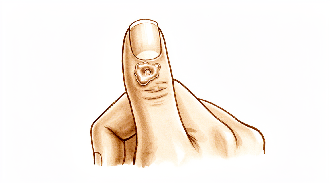
Qué está sintiendo
Un quiste mucoso es un bulto pequeño y firme que aparece en la parte dorsal del dedo, normalmente justo por detrás de la uña. Está lleno de un líquido espeso y transparente. El bulto en sí no suele doler, pero puede presionar las estructuras cercanas y causar molestias.
Es posible que note cambios en la propia uña. El quiste se encuentra cerca del lecho ungueal, por lo que, a medida que crece, puede presionar la uña. Esto suele producir un surco o una cresta a lo largo de toda la uña. La piel que cubre el quiste también puede volverse delgada y brillante. En algunos casos, la piel se rompe y el quiste pierde líquido a través de un pequeño conducto.
El bulto suele situarse justo donde se dobla el dedo, en la última articulación antes de la uña. Eso hace que algunas tareas cotidianas resulten incómodas. Meter la mano en un bolsillo estrecho, sujetar la tapa de un frasco o recoger monedas pequeñas puede resultar molesto. Escribir a mano o en el teclado puede presionar el bulto y hacer que duela.
El quiste está relacionado con la artritis por desgaste de esa misma articulación. Una pequeña protuberancia ósea, llamada osteofito, irrita la articulación y produce el líquido que llena el quiste. Por eso, la articulación puede doler después de haber usado mucho la mano, y en esos momentos el bulto puede notarse más sensible.
Si la piel que cubre el quiste se abre y sale líquido, mantenga la zona limpia y busque atención sin demora. Un conducto desde la piel hasta una articulación del dedo puede dejar entrar una infección, y una articulación infectada necesita tratamiento sin demora. Si el dedo se pone caliente, enrojecido, hinchado y doloroso, sobre todo si tiene fiebre, acuda a un servicio de urgencias ese mismo día. No necesita una derivación de su médico de cabecera.
No pinche ni apriete el quiste usted mismo. Puede dejar entrar una infección en la articulación.
Si el bulto está creciendo, la deformidad de la uña le molesta o los síntomas no mejoran con el paso de las semanas, consulte a su médico de cabecera o solicite una evaluación especializada.
¿Qué está ocurriendo realmente?
La última articulación del dedo funciona como una pequeña bisagra. Como cualquier articulación, está revestida por una funda blanda que produce un poco de líquido para que las superficies se deslicen con suavidad. En el quiste mucoso, esa articulación ha desarrollado artritis por desgaste. El revestimiento articular se irrita y empieza a producir más líquido del que la articulación necesita.
Al mismo tiempo, la artritis suele formar una pequeña protuberancia ósea en el borde de la articulación. Esa protuberancia presiona la funda articular desde dentro y acaba desgastándola. Entonces el líquido se escapa por la zona desgastada y se acumula bajo la piel, en la parte dorsal del dedo. El bulto que usted puede ver y palpar es ese líquido atrapado, situado a un lado de la articulación, justo por detrás de la uña.
El quiste en sí es una bolsa de pared delgada, no un crecimiento de tejido. Su pared está formada por fibras de tejido de sostén y no tiene un revestimiento propio. Como la pared es delgada y la piel que la cubre suele serlo aún más, un pequeño golpe puede romperla. Cuando eso ocurre, sale el líquido transparente de aspecto gelatinoso que contiene.
Esto explica los síntomas que acaba de leer. El bulto se encuentra cerca del lecho ungueal, por lo que, a medida que crece, la presión del quiste puede marcar un surco en la uña. La articulación duele porque también hay artritis, y el quiste es una señal de ella. La protuberancia del borde de la articulación es lo que mantiene el flujo de líquido, de modo que, mientras siga ahí, el quiste tiende a volver a llenarse incluso después de que salga el líquido o de que se drene.
Por eso también el tratamiento se centra en la articulación y no solo en el bulto. Extirpar la protuberancia ósea detiene la fuga en su origen, y el quiste se resuelve cuando deja de rellenarse.
Qué podemos hacer al respecto
El Dr. Kieran Hirpara, cirujano de extremidades superiores en el Mater Private Hospital Rockhampton, comienza con las opciones menos invasivas que se adapten a su condición. Por lo general, los pacientes son derivados a nuestra clínica por su médico de cabecera; si un fisioterapeuta le ha sugerido que nos consulte, igualmente necesitará una derivación de su médico de cabecera para poder acceder al reembolso de Medicare. En la consulta, tomamos su historia clínica, examinamos su dedo y, si es necesario, solicitamos pruebas de imagen. La mayoría de los quistes mucosos están relacionados con la artritis por desgaste de la articulación, por lo que normalmente probamos primero el tratamiento no quirúrgico y consideramos la cirugía cuando este no ha dado una mejoría suficiente.
El primer paso suele ser simplemente observar y esperar. Algunos quistes se mantienen pequeños y causan pocas molestias. Proteger el dedo ayuda: evite los golpes repetidos sobre el bulto y tenga cuidado con las tareas que lo presionan. Si la articulación duele después de un uso intenso, reducir esa actividad puede aliviarla. La terapia de mano puede ayudar con la rigidez y a mantener el dedo en movimiento con comodidad. Dé a estas medidas sencillas una prueba razonable durante varias semanas antes de decidir cualquier otra cosa.
El alivio del dolor es sencillo. Los analgésicos simples de la farmacia, tomados según las indicaciones, pueden calmar una articulación dolorida. Los antiinflamatorios pueden aliviar la artritis que hay detrás del quiste. Estos medicamentos tratan las molestias, no el bulto en sí.
Una opción que no ofrecemos es drenar el quiste con una aguja. Es un procedimiento cómodo que se hace en la consulta, pero después el quiste reaparece aproximadamente en la mitad de los casos. También existe el riesgo de introducir una infección en la articulación, y por eso desaconsejamos asimismo pinchar o apretar el quiste en casa.
Se plantea la cirugía cuando el quiste se sigue rellenando, la deformidad de la uña le molesta o la piel que cubre el quiste se ha vuelto delgada o se está rompiendo. La operación trata la articulación y no solo el bulto. Se extirpa la protuberancia ósea del borde de la articulación, lo que detiene la fuga de líquido en su origen, y el quiste se resuelve cuando deja de rellenarse. En algunos casos también se extirpan el quiste y un pequeño colgajo de piel local, lo que es adecuado para un quiste que ha adelgazado la piel que lo cubre. Extirpar la protuberancia ósea junto con el pedículo del quiste ofrece la menor probabilidad de que el quiste reaparezca. Hablaremos sobre qué método se adapta mejor a su dedo y lo decidiremos juntos.
Qué esperar
Si se deja sin tratamiento, un quiste mucoso tiende a seguir reapareciendo en lugar de desaparecer. Mientras la protuberancia ósea del borde de la articulación siga ahí, continúa rellenando el quiste de líquido. Algunos quistes se mantienen pequeños y causan pocas molestias durante mucho tiempo. Otros crecen lentamente, marcan más el surco en la uña o adelgazan la piel que cubre el bulto hasta que pierde líquido.
Sin tratamiento, los principales riesgos son los que ya ha leído: una uña que sigue con una cresta mientras el quiste la presiona, y una piel que se rompe y abre un conducto hacia la articulación. Ese conducto debe tomarse en serio. Si el dedo se pone caliente, enrojecido, hinchado y doloroso, sobre todo si tiene fiebre, acuda a un servicio de urgencias ese mismo día. No necesita una derivación de su médico de cabecera.
Con un tratamiento dirigido a la articulación, el pronóstico es claro. Extirpar la protuberancia ósea detiene la fuga de líquido en su origen, y el quiste se resuelve cuando deja de rellenarse. Cuando se extirpa la protuberancia ósea, es raro que el quiste reaparezca. Cuando además se extirpa el propio quiste, la recurrencia sigue siendo baja: reaparecen unos 2 de cada 100 quistes tratados quirúrgicamente, y alrededor de 1,4 de cada 100 cuando se utiliza un colgajo de piel local para cerrar la zona. Las personas tratadas de esta forma suelen estar satisfechas con el aspecto de la cicatriz y volverían a pasar por ello.
La recuperación no es una gran empresa, pero tampoco es insignificante. El dedo suele estar rígido y sensible durante unas semanas, y las tareas cotidianas como agarrar o pellizcar tardan un tiempo en volver a sentirse normales. La terapia de mano tras la cirugía se realiza con Ruby Doolan, de Extend Rehabilitation. Ruby es terapeuta de mano: guía sus ejercicios y le fabrica cualquier férula que necesite. La mayoría de las personas vuelven a usar la mano con normalidad en un plazo de unas semanas a unos pocos meses.
El surco de la uña también suele mejorar una vez que deja de haber presión sobre el lecho ungueal. Tenga paciencia con esa parte: la porción deformada de la uña tiene que crecer hasta salir, lo que lleva varios meses.
Si los síntomas no mejoran con el paso de las semanas, o empeoran, consulte a su médico de cabecera o solicite una evaluación especializada.
¿Cuándo consultar a un especialista?
La mayoría de los quistes mucosos no son urgentes, y muchos mejoran con cuidados sencillos. Algunos signos sí requieren actuar.
Acuda a un servicio de urgencias ese mismo día si el dedo se pone caliente, enrojecido, hinchado y doloroso, sobre todo si tiene fiebre. Eso indica una infección en la articulación, que necesita tratamiento sin demora. Lo mismo se aplica si la piel que cubre el quiste se ha abierto y está saliendo líquido, porque esa abertura puede dejar entrar una infección.
Consulte a su médico de cabecera o solicite una evaluación especializada si el bulto sigue creciendo, el surco de la uña le molesta o la piel que cubre el quiste se ha vuelto delgada. Estas son las señales de que vale la pena hablar del tratamiento. Si los síntomas no mejoran con el paso de las semanas, o empeoran, su médico de cabecera puede gestionar una derivación.
En mayor profundidad
Advanced reading: the deeper science (optional)
Esta sección profundiza más de lo necesario para que usted tome decisiones sobre su propio tratamiento. Vale la pena leer más sobre los quistes mucosos, ya que existe un hallazgo quirúrgico bien fundamentado: el quiste no es el problema, y la intervención que funciona mejor no implica necesariamente su extirpación.
El espolón óseo es la causa, no el quiste
El quiste mucoso se origina en una articulación de la punta del dedo afectada por artritis. El espolón óseo, o osteofito, irrita y perfora la cápsula articular; el líquido sinovial escapa por dicha perforación y se acumula bajo la piel. El quiste es el resultado visible de este proceso, no su causa inicial.
Esta comprensión tiene una consecuencia quirúrgica directa: en una serie de casos, la extirpación del osteofito sin extirpar el quiste condujo a la resolución completa en la mayoría de los pacientes, siendo considerada una buena opción terapéutica que implica un procedimiento menos invasivo [1].
Al eliminar el espolón óseo se cierra la vía de escape del líquido sinovial; el quiste, al dejar de recibir este líquido, desaparece. Esta misma lógica rige en el caso de los quistes ganglionares de la muñeca: es el tallo del quiste, y no el saco en sí, el que determina su recurrencia. Asimismo, explica por qué el simple drenaje o punción de un quiste mucoso fracasa sistemáticamente.
En los casos en que se extirpa el quiste, los resultados también son buenos
El otro método consiste en extirpar el quiste junto con un colgajo cutáneo local para cerrar el defecto. Este método también resulta fiable: en 69 pacientes, la extirpación quirúrgica con un colgajo de avance local arrojó una tasa de recurrencia del 1,4%, además de una alta satisfacción por parte de los pacientes respecto a la cicatriz y su disposición a someterse nuevamente al procedimiento [2].
Ambos métodos son efectivos y atacan la articulación afectada. La diferencia práctica radica en la cantidad de piel implicada: un quiste de larga evolución adelgaza la piel que lo cubre, a veces hasta el punto de producir secreción; en tales casos, esa piel adelgazada debe ser extirpada y reemplazada, independientemente de lo que se haga con el hueso.
¿Por qué la uña se deforma y si puede recuperarse?
Es común que aparezca un surco o relieve a lo largo de toda la uña; esto preocupa más a las personas que el propio bulto. La causa es mecánica: el quiste se ubica justo encima de la matriz germinativa, es decir, la parte del lecho ungueal que genera la uña, y ejerce presión sobre ella, por lo que la uña crece con un defecto.
Lo positivo es que se trata de presión y no de destrucción. Una vez que se descomprime el quiste, la uña suele volver a crecer de forma normal; sin embargo, pueden pasar varios meses hasta que la parte deformada se vaya desprendiendo por el extremo. Por lo tanto, la deformidad de la uña es un motivo para tratar el quiste, no una consecuencia permanente de él.
Motivo por el cual hay que tener cuidado al punzar una de ellas
El quiste mucoso se comunica directamente con la articulación. Punzarlo, ya sea de forma intencionada o por la ruptura de la piel sobre un quiste de gran tamaño, crea un canal desde el exterior hacia la articulación fingeriana; la artritis séptica en una articulación pequeña constituye un problema mucho más grave que el propio quiste.
Este es el argumento práctico en contra del drenaje casero de un quiste que parece ser simplemente una ampolla de líquido, y la razón por la cual un quiste que se drena espontáneamente debe tratarse con cierta urgencia en lugar de simplemente observarse.
Referencias
[1] Lee H, Kim P, Jeon I, Kyung H, Ra I, Kim T. Excisión de osteofitos sin extirpación del quiste en el tratamiento de los quistes mucosos digitales. J Hand Surg Eur Vol. 2013;39(3):258-61. https://doi.org/10.1177/1753193413478549
[2] Johnson SM, Treon K, Thomas S, Cox QGN. Un tratamiento quirúrgico eficaz para los quistes mucosos digitales. J Hand Surg Eur Vol. 2013;39(8):856-60. https://doi.org/10.1177/1753193413508540
Evidence & references
This is the clinical evidence summary written for health professionals. It is technical, and it lists the research this page was built from. You do not need to read it to understand your treatment or to make a decision about it.
Overview
- The scientific data regarding mucous cysts consist almost entirely of retrospective studies, and much of what is done or recommended is based on expert opinion [3].
- Surgical treatment of mucous cysts is not a totally benign procedure, and complications do occur contrary to literature reporting glowing results [5].
- In-office excision is a safe and effective option for treating digital mucous cysts [6].
- Complications are rare and occur only in cysts associated with fistula, justifying their early surgical treatment [7].
- Surgery provided definitive treatment with no major long-term problems [10].
- Surgical excision was the primary treatment, with no reported recurrences [11].
- Incomplete excisions will invariably result in recurrence [12].
- Surgical excision with a local advancement skin flap is a reliable treatment for digital mucous cysts, demonstrating a low recurrence rate of 1.4% and high patient satisfaction regarding the scar and willingness to undergo the procedure again [13].
- The 2% recurrence rate (2 of 113) obtained in this series illustrates the advantage of this procedure [16].
- Osteophyte excision without cyst excision may be a good treatment choice for mucous cyst of the finger, providing a less invasive method with complete resolution in most cases [20].
- The final recurrence rate of less than 1.5% may be acceptable and comparable with other techniques [21].
- Excision with local flap cover offers benefit in terms of a reduced recurrence rate and is particularly valuable where the cyst has caused marked thinning of the skin [30].
- Excision with local flap cover would seem to offer benefit in terms of a reduced recurrence rate, and is particularly valuable where the cyst has caused marked thinning of the skin [34].
- Excision of the cyst and complete removal of the marginal osteophyte eradicates mucous cysts with extremely rare recurrence [1].
- A total dorsal capsulectomy alone was a simple treatment for mucous cysts and did not lead to any recurrence [8].
Anatomy & Pathophysiology
Clinical Presentation and Location
- Mucous cysts are small, firm, cystic masses that appear just distal to the distal interphalangeal joint [23].
- The lesion is always located to one side of the midline [23].
- Mucous cysts are rarely greater than fifteen millimeters in diameter [23].
- The skin over the mucous cyst is thinned out and occasionally may be ulcerated [23].
- Pressure on the mucous cyst does not usually result in a decrease in its size [23].
- Longitudinal grooving of the nail may be noted, occasionally occurring prior to the appearance of the cyst [23].
- Mucous cysts are usually small and located to one side as they emerge from the joint beside the extensor mechanism [28, 29].
- Occasionally, mucous cysts may spread across the whole of the dorsum of the digit between the distal interphalangeal joint and nail fold [28, 29].
- The overlying skin of a mucous cyst is frequently thin, and minimal trauma may result in rupture [28, 29].
Histology and Ultrastructure
- The histological appearance of a mucous cyst is essentially identical to that of ganglions, synovial cysts, and similar lesions appearing on other areas of the extremities [23].
- The mucous cyst is surrounded by an undemarcated fibrous capsule [23].
- The basic structure of the mucous cyst is myxomatous with interspersed fibroblasts [23].
- Areas of myxomatous degeneration tend to coalesce to form a multiloculated cyst [23].
- An epithelial lining has not been reported for mucous cysts [23].
- Digital mucous cysts and wrist ganglia show no anatomic difference at the light and scanning electron microscopic levels [19].
- The wall of a digital mucous cyst consists of multidirectional strata of collagen fibers with rare fibroblast-like cells on the inner surface [19].
- There are no signs of myxomatous degeneration in the wall of a digital mucous cyst [19].
- The surface of the digital mucous cyst wall is generally flat, with some areas showing localized elevations [19].
- Multifunctional mesenchymal cells are located beneath the lining of the ganglion and digital mucous cyst [19].
- These multifunctional mesenchymal cells contain abundant rough endoplasmic reticulum, smooth-walled vacuoles, and Golgi complexes [19].
- The ganglion fluid may be produced by these multifunctional cells and reaches the ganglion cavity by flowing through the porous collagen matrix of the ganglion wall [19].
- A digital mucous cyst has a large globular dome-shaped interconnecting cavity having a wall of collagen fibers covered by a fenestrated pseudomembrane [19].
- A porous network of collagen fibers is present in the wall of the digital mucous cyst on cross section [19].
Etiology and Pathogenesis
- Degenerative arthritis of the distal interphalangeal joint is a frequent associated finding with mucous cysts [23].
- An incidence of 78 percent for degenerative arthritis of the distal interphalangeal joint has been reported in association with mucous cysts [23].
- The etiology indicated by evidence is that the lesion arises from the joint capsule [23].
- Mucous cysts are often associated with degenerative changes at the distal interphalangeal joints [28, 29].
- Theories on the pathogenesis of mucous cysts include myxoid degeneration, repeated local trauma, vascular insufficiency, and synovial herniation [28, 29].
- Studies suggest that mucous cysts arise due to excessive hyaluronic acid production from a degenerate distal interphalangeal joint [28, 29].
Classification
- The lesion is a small, firm, cystic mass which appears just distal to the distal interphalangeal joint [23].
- The lesion is always located to one side of the mid-line [23].
- The lesion is rarely greater than fifteen millimeters in diameter [23].
- The skin over the lesion is thinned out and occasionally may be ulcerated [23].
- Pressure on the lesion does not usually result in a decrease in its size [23].
- Degenerative arthritis of the distal interphalangeal joint is a frequent associated finding [23].
- An incidence of 78 per cent has been reported for degenerative arthritis of the distal interphalangeal joint associated with mucous cysts [23].
- The histological appearance is essentially identical to that of ganglions, synovial cysts, and similar lesions appearing on other areas of the extremities [23].
- The lesion is surrounded by an undemarcated fibrous capsule [23].
- The basic structure is myxomatous with interspersed fibroblasts [23].
- An epithelial lining has not been reported [23].
- In spite of their different clinical settings, digital mucous cysts and wrist ganglia show no anatomic difference at the light and scanning electron microscopic levels [19].
- The wall of the digital mucous cyst consisted of multidirectional strata of collagen fibers with rare fibroblast-like cells on the inner surface [19].
- There were no signs of myxomatous degeneration in the wall of the digital mucous cyst [19].
- The surface of the digital mucous cyst was generally flat, with some areas showing localized elevations [19].
- Multifunctional mesenchymal cells beneath the lining of the ganglion are postulated to produce collagen fibers, elastic fibers, and the interfibrillary mucopolysaccharide matrix [19].
- The porous nature of the ganglion wall was confirmed in the ultrastructural study of digital mucous cysts [19].
- A previous ultrastructural study of digital mucous cysts demonstrated a large globular dome-shaped interconnecting cavity having a wall of collagen fibers covered by a fenestrated pseudomembrane [19].
- The current study confirmed the anatomic organization of a large globular dome-shaped interconnecting cavity having a wall of collagen fibers covered by a fenestrated pseudomembrane [19].
- A porous network of collagen fibers in the wall was revealed on cross section [19].
Clinical Presentation
- The mucous cyst presents as a small, firm, cystic mass located just distal to the distal interphalangeal joint [23].
- The lesion is easily distinguished from a Heberden's node by its placement distal to the distal interphalangeal joint [23].
- The histological appearance of the mucous cyst is essentially identical to that of ganglions, synovial cysts, and similar lesions appearing on other areas of the extremities [23].
- The basic structure of the lesion is myxomatous with interspersed fibroblasts [23].
- An epithelial lining has not been reported for the mucous cyst [23].
- The wall of the digital mucous cyst consists of a porous network of collagen fibers [19].
- The fluid within the digital mucous cyst is produced by underlying mesenchymal cells [19].
Investigations
- Ultrasound is a powerful modality for evaluation of pathologic conditions in the hand and wrist, providing a cost-effective and expedient alternative and/or adjunct to MRI [33].
- Ultrasound is best used when there is a specific clinical question regarding a well-localized abnormality [33].
- Ultra-sonography provides a new ability to study the complex anatomical structures of the hand, with the technique being quick and simple to perform [38].
- A careful physical examination is essential to direct care and future testing if indicated [35].
- Diagnostic tests such as imaging and serum laboratory studies are useful in this determination but can be expensive, time consuming, and often nonspecific [35].
Treatment
Operative Techniques
- Total dorsal capsulectomy alone is a simple treatment for mucous cysts that did not lead to any recurrence [8].
- Osteophyte-sparing treatment of mucous cysts has a final recurrence rate of less than 1.5% [21].
- Surgical excision was the primary treatment in a series with no reported recurrences [11].
- Surgery provided definitive treatment for mucous cysts with no major long-term problems [10].
- The 2% recurrence rate (2 of 113) obtained in a series of 191 excised cysts illustrates the advantage of the procedure [4, 16].
- The authors of a study on osteophyte excision without cyst excision state that their principle is not to do more damage to the friable skin because the skin is not the culprit of the cyst [15].
Outcomes and Complications
- Complications of surgical treatments include infection, recurrence, nail deformity, swelling, stiffness and pain [28].
- This report provides additional basis for prophylactic extirpation of mucous cyst of the finger [2].
- This case emphasizes the utility of a pathohistological analysis in cases where doubts arise about the initial diagnosis of a benign tumorous lesion [14].
Non-Operative and Adjunctive Considerations
- Aspiration with injection of steroid is a convenient technique that can be performed in an outpatient setting but is followed by a recurrence rate of 50% [28].
- Surgical treatment has a higher cure rate than aspiration with injection of steroid [28].
- Radical treatment with complete excision of the stalk and underlying osteophytes seems to reduce recurrence rates [28].
Complications
- Complications are rare and occur only in cysts associated with fistula [7].
- Aspiration with injection of steroid is followed by a recurrence rate of 50% [28].
- The 2% recurrence rate (2 of 113) was obtained in a series of surgically treated mucous cysts [16].
- Surgical excision with a local advancement skin flap demonstrated a low recurrence rate of 1.4% [13].
- A total dorsal capsulectomy alone did not lead to any recurrence [8].
- Eccrine porocarcinomas have a substantial risk of metastasis, high risk of local recurrence, and are potentially fatal [24].
Recovery
- Surgical excision with a local advancement skin flap demonstrated a low recurrence rate of 1.4% and high patient satisfaction regarding the scar and willingness to undergo the procedure again [13].
Key Evidence
- [L4] Excision of the cyst and complete removal of the marginal osteophyte eradicates mucous cysts with extremely rare recurrence. [1] (10.2106/00004623-197355030-00013)
- [L4] This report provides additional basis for prophylactic extirpation of mucous cyst of the finger. [2] (10.1016/s0363-5023(84)80152-5)
- [L4] The scientific data regarding mucous cysts consist almost entirely of retrospective studies, and much of what is done or recommended is based on expert opinion. [3] (10.1016/j.jhsa.2010.01.029)
- [L5] Of the 191 mucous cysts excised, 113 had at least 6 months of follow-up time. [4] (10.1016/j.jhsa.2010.07.028)
- [L4] Surgical treatment of mucous cysts is not a totally benign procedure, and complications do occur contrary to literature reporting glowing results. [5] (10.1016/s0266-7681(97)80067-8)
- [Paper] In-office excision is a safe and effective option for treating digital mucous cysts. [6] (10.1177/15589447251350168)
- [L4] Complications are rare and occur only in cysts associated with fistula, justifying their early surgical treatment. [7] (10.1016/j.main.2015.06.001)
- [L4] A total dorsal capsulectomy alone was a simple treatment for mucous cysts and did not lead to any recurrence. [8] (10.1016/j.jhsa.2014.03.004)
- [L4] Surgery provided definitive treatment with no major long-term problems. [10] (10.1053/jhsu.2003.50088)
- [L5] Incomplete excisions will invariably result in recurrence. [12] (10.1016/0363-5023(93)90302-j)
- [L4] Surgical excision with a local advancement skin flap is a reliable treatment for digital mucous cysts, demonstrating a low recurrence rate of 1.4% and high patient satisfaction regarding the scar and willingness to undergo the procedure again. [13] (10.1177/1753193413508540)
- [L5] This case emphasizes the utility of a pathohistological analysis in cases where doubts arise about the initial diagnosis of a benign tumorous lesion. [14] (10.1007/s00402-008-0794-4)
- [L5] The authors state that their principle is not to do more damage to the friable skin because the skin is not the culprit of the cyst. [15] (10.1177/1753193414546990)
- [L4] The 2% recurrence rate (2 of 113) obtained in this series illustrates the advantage of this procedure. [16] (10.1016/0363-5023(94)90071-x)
- [L5] [19] (10.1016/s0363-5023(88)80143-6)
- [L4] Osteophyte excision without cyst excision may be a good treatment choice for mucous cyst of the finger, providing a less invasive method with complete resolution in most cases. [20] (10.1177/1753193413478549)
- [L4] The final recurrence rate of less than 1.5% may be acceptable and comparable with other techniques. [21] (10.5435/jaaosglobal-d-21-00164)
- [L4] [23] (10.2106/00004623-197254070-00008)
- [L4] Prompt recognition and appropriate treatment are critical because eccrine porocarcinomas have a substantial risk of metastasis, high risk of local recurrence, and are potentially fatal. [24] (10.1016/j.jhsa.2016.07.112)
- [L4] [28] (10.1177/1753193408103498)
- [L4] [29] (10.1177/1753193409103498)
- [L4] Excision with local flap cover offers benefit in terms of a reduced recurrence rate and is particularly valuable where the cyst has caused marked thinning of the skin. [30] (10.1016/0266-7681(90)90064-b)
- [L5] Ultrasound is a powerful modality for evaluation of pathologic conditions in the hand and wrist, providing a cost-effective and expedient alternative and/or adjunct to MRI, best used when there is a specific clinical question regarding a well-localized abnormality. [33] (10.1016/j.jhsa.2009.02.010)
- [L4] Excision with local flap cover would seem to offer benefit in terms of a reduced recurrence rate, and is particularly valuable where the cyst has caused marked thinning of the skin. [34] (10.1016/0266-7681_90_90064-b)
- [L4] Ultra-sonography provides a new ability to study the complex anatomical structures of the hand, with the technique being quick and simple to perform. [38] (10.1016/0266-7681(90)90133-o)
References
[1] Marginal Osteophyte Excision in Treatment of Mucous Cysts. The Journal of Bone & Joint Surgery. 1973. DOI: 10.2106/00004623-197355030-00013
[2] Infected mucous cyst of the finger. The Journal of Hand Surgery. 1984. DOI: 10.1016/s0363-5023(84)80152-5
[3] Mucous Cysts. The Journal of Hand Surgery. 2010. DOI: 10.1016/j.jhsa.2010.01.029
[4] Mucous Cysts. The Journal of Hand Surgery. 2010. DOI: 10.1016/j.jhsa.2010.07.028
[5] Complications Following Mucous Cyst Excision. Journal of Hand Surgery. 1997. DOI: 10.1016/s0266-7681(97)80067-8
[6] Safety and Cost-Effectiveness of In-Office Digital Mucous Cyst Excisions. HAND. 2025. DOI: 10.1177/15589447251350168
[7] Surgical treatment of mucous cysts by subcutaneous excision and osteophyte resection: Results in 68 cases at a mean 6.63 years’ follow-up. Chirurgie de la Main. 2015. DOI: 10.1016/j.main.2015.06.001
[8] Total Dorsal Capsulectomy for the Treatment of Mucous Cysts. The Journal of Hand Surgery. 2014. DOI: 10.1016/j.jhsa.2014.03.004
[10] Treatment of mucous cysts of the fingers: Review of 134 cases with minimum 2-year follow-up evaluation. The Journal of Hand Surgery. 2003. DOI: 10.1053/jhsu.2003.50088
[11] 10.1177-15589447261433068. n.d..
[12] Juvenile hyaline fibromatosis of the hand in an adult. The Journal of Hand Surgery. 1993. DOI: 10.1016/0363-5023(93)90302-j
[13] A reliable surgical treatment for digital mucous cysts. Journal of Hand Surgery (European Volume). 2013. DOI: 10.1177/1753193413508540
[14] Malignant Natural-Killer cell neoplasm presenting as a mucous cyst on the distal interphalangeal joint of the finger. Archives of Orthopaedic and Trauma Surgery. 2008. DOI: 10.1007/s00402-008-0794-4
[15] Re: Lee, H.-J., Kim, P.-T., Jeon, I.-H., Kyung, H.-S., Ra, I.-H. and Kim, T.-K. Osteophyte excision without cyst excision for a mucous cyst of the finger. J Hand Surg Eur. 2014, 39: 258–61. Journal of Hand Surgery (European Volume). 2014. DOI: 10.1177/1753193414546990
[16] Outcome of surgically treated mucous cysts of the hand. The Journal of Hand Surgery. 1994. DOI: 10.1016/0363-5023(94)90071-x
[19] A surface ultrastructure study of ganglia and digital mucous cysts. The Journal of Hand Surgery. 1988. DOI: 10.1016/s0363-5023(88)80143-6
[20] Osteophyte excision without cyst excision for a mucous cyst of the finger. Journal of Hand Surgery (European Volume). 2013. DOI: 10.1177/1753193413478549
[21] Osteophyte-Sparing Treatment of Mucous Cysts: Case Analysis and Surgical Technique. JAAOS: Global Research and Reviews. 2021. DOI: 10.5435/jaaosglobal-d-21-00164
[23] Etiology and Treatment of the So-Called Mucous Cyst of the Finger. The Journal of Bone & Joint Surgery. 1972. DOI: 10.2106/00004623-197254070-00008
[24] Eccrine Porocarcinoma Presenting as a Hand Cyst. The Journal of Hand Surgery. 2016. DOI: 10.1016/j.jhsa.2016.07.112
[28] Use of Wolfe Graft for the Treatment of Mucous Cysts. Journal of Hand Surgery (European Volume). 2009. DOI: 10.1177/1753193408103498
[29] Use of Wolfe Graft for the Treatment of Mucous Cysts. Journal of Hand Surgery (European Volume). 2009. DOI: 10.1177/1753193409103498
[30] Mucous cyst of the distal interphalangeal joint: Treatment by simple excision or excision and rotation flap. The Journal of Hand Surgery: Journal of the British Society for Surgery of the Hand. 1990. DOI: 10.1016/0266-7681(90)90064-b
[33] Ultrasound of the Hand and Wrist. The Journal of Hand Surgery. 2009. DOI: 10.1016/j.jhsa.2009.02.010
[34] Mucous Cyst of the Distal Interphalangeal Joint: Treatment by Simple Excision or Excision and Rotation Flap. Journal of Hand Surgery. 1990. DOI: 10.1016/0266-7681_90_90064-b
[35] Orthopaedic Knowledge Update 13 Ebook Without Multimedia. Anatomy, Evaluation, Clinical Examination, and Imaging > Evaluation and Clinical Examination: Current Concepts.
[38] Diagnostic medical ultrasound in the management of hand injuries. The Journal of Hand Surgery: Journal of the British Society for Surgery of the Hand. 1990. DOI: 10.1016/0266-7681(90)90133-o