Patients › Elbow
Síndrome del túnel radial
Radial tunnel syndrome — causes forearm pain, weakness straightening fingers, and is distinct from tennis elbow.
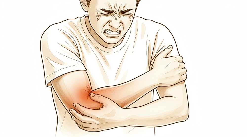
Qué está sintiendo
El síntoma principal es el dolor en la parte externa del antebrazo, cerca del codo. Suele ser más intenso en un punto sensible situado unos pocos centímetros por debajo del bulto óseo en el exterior del codo. Presionar en ese punto suele doler. El dolor puede extenderse por la parte posterior del antebrazo hacia la muñeca.
Ciertos movimientos lo empeoran. Girar la palma hacia arriba contra resistencia, o extender el dedo medio contra resistencia, puede provocar el dolor. También puede hacerlo la actividad que carga el antebrazo de forma continuada. Muchas personas notan que el dolor se aviva después del trabajo o del deporte, más que en reposo.
Esta afección afecta a un nervio que inerva músculos y no la piel. Eso significa que es poco probable que tenga entumecimiento u hormigueo, y el propio nervio suele funcionar con normalidad. El problema es el dolor, no la debilidad ni la pérdida de sensibilidad.
En el día a día, el dolor tiende a aparecer en las tareas que exigen agarrar y girar. Levantar un hervidor, girar el pomo de una puerta, usar un destornillador o escurrir un paño pueden doler. Como el punto sensible está cerca de la parte externa del codo, esta afección se confunde a menudo con el codo de tenista, y ambos pueden darse a la vez.
No existe una única prueba que confirme esta afección. Su cirujano llega al diagnóstico a partir de su historia y de una exploración, buscando ese punto sensible concreto y el patrón de su dolor.
Esté atento a estas señales. Acuda a su médico de cabecera o solicite una valoración por parte de un especialista si sus síntomas no mejoran, empeoran a lo largo de las semanas, le despiertan por la noche o le impiden trabajar o usar el brazo. Acuda a urgencias el mismo día si el brazo se pone caliente, rojo, hinchado y doloroso, sobre todo si tiene fiebre, o si la mano se vuelve pálida, fría, blanca o azulada. Si no puede contactar con la clínica fuera del horario de atención o durante el fin de semana, acuda al servicio de urgencias más cercano.
¿Qué está ocurriendo realmente?
El dolor procede de un nervio, no de la propia articulación del codo. Un nervio es un cable que transmite señales entre el cerebro y los músculos. Una rama del nervio radial recorre la parte externa del antebrazo, pasando junto al codo, para inervar los músculos que extienden la muñeca y los dedos.
A lo largo de ese tramo, varias estructuras distintas quedan muy cerca del nervio. El borde de un músculo, una banda fibrosa o un pequeño grupo de vasos sanguíneos que lo cruzan pueden, cada uno, presionarlo. No se trata de un único paso estrecho. Se parece más a un sendero con varios posibles puntos de pinzamiento a lo largo del camino, cualquiera de los cuales puede apretar el cable que discurre a su lado.
Cuando una de esas estructuras presiona el nervio, este se irrita y duele. Por eso el dolor se localiza en un punto sensible por debajo del bulto óseo en el exterior del codo, y por eso girar la palma hacia arriba o extender el dedo medio contra resistencia lo empeora. Esos movimientos tiran de los músculos que inerva este nervio, y el nervio irritado se queja.
El nervio sigue funcionando, de modo que los músculos siguen trabajando y usted no nota entumecimiento ni hormigueo. El problema es la irritación y el dolor, no la pérdida de señal. Como el punto doloroso está tan cerca de la parte externa del codo, el dolor se confunde fácilmente con el codo de tenista, que afecta a un tendón de la misma zona. Ambos pueden incluso darse a la vez.
¿Qué podemos hacer al respecto?
El Dr. Kieran Hirpara, cirujano de extremidades superiores en el Mater Private Hospital Rockhampton, comienza con las opciones menos invasivas que se adapten a su condición. Por lo general, los pacientes son derivados a nuestra clínica por su médico de cabecera; si un fisioterapeuta le ha sugerido que nos consulte, igualmente necesitará una derivación de su médico de cabecera para poder acceder al reembolso de Medicare. En la clínica tomamos su historia clínica, examinamos su brazo y solicitamos pruebas de imagen si van a ser útiles. Para esta afección, normalmente probamos primero el tratamiento no quirúrgico, y consideramos la cirugía cuando este no ha dado una mejoría suficiente.
El primer paso es cambiar la forma en que usa el brazo. Reducir las tareas de agarre y giro que avivan el dolor da al nervio irritado la oportunidad de calmarse. La terapia de mano tiene como objetivo calmar el dolor y reconstruir su tolerancia a la carga en el antebrazo, para que el trabajo y las tareas cotidianas vuelvan a resultar cómodos. Le pedimos que dé a esto una oportunidad razonable antes de pensar en cualquier otra cosa.
Los analgésicos y los antiinflamatorios pueden ayudarle a superar una crisis de dolor. Calman el dolor mientras las demás medidas surten efecto. No resuelven la presión sobre el propio nervio.
Si el tratamiento no quirúrgico no ha dado una mejoría suficiente, la cirugía es una opción. La operación se llama liberación del túnel radial. Consiste en liberar el nervio en cada uno de los lugares donde una estructura cercana lo está presionando. Antes de recomendarla, comprobamos con cuidado dónde se está comprimiendo el nervio, para que la operación se dirija al punto correcto y altere el menor tejido circundante posible. Como el diagnóstico se basa en su historia y en la exploración, y no en una prueba clara, consideramos la decisión de operar como una decisión compartida, que tomamos juntos una vez que usted entiende lo que la cirugía puede y no puede resolver.
Qué esperar
En la mayoría de las personas, esta afección no daña el nervio. El nervio sigue funcionando, de modo que los músculos siguen trabajando y usted no pierde sensibilidad. El problema es el dolor, y el dolor de un nervio irritado puede tardar mucho tiempo en calmarse. Rara vez desaparece de un día para otro, y a menudo se aviva con las tareas de agarre y giro antes de remitir.
Para ser sinceros, esta afección es difícil de predecir. Algunas personas mejoran con reposo, cambios en las actividades y terapia de mano. Otras siguen teniendo dolor a pesar de un buen tratamiento no quirúrgico. No existe una prueba fiable que confirme el diagnóstico, lo que hace más difícil saber de antemano en qué grupo estará usted. Lo que sí podemos decir es que el dolor tiende a ir y venir según cuánto cargue el antebrazo, en lugar de aumentar de forma constante.
Si el tratamiento no quirúrgico funciona, el dolor disminuye gradualmente a lo largo de semanas o meses, a medida que el nervio se irrita menos. Lo esperable es que primero le resulten menos dolorosas las tareas cotidianas, como levantar un hervidor o girar el pomo de una puerta, antes de que el trabajo más pesado vuelva a sentirse normal. Si se opera después de que el tratamiento no quirúrgico no haya ayudado lo suficiente, la recuperación también es gradual y no inmediata, y algunas personas siguen teniendo algo de dolor después.
Si no se trata la afección, puede calmarse por sí sola, pero también puede persistir o seguir avivándose cada vez que vuelva a las tareas que la agravan. No hay forma de saber de antemano qué ocurrirá. Lo que suele importar es dar al tratamiento una oportunidad razonable y no forzar a pesar de un dolor que no se calma.
Si sus síntomas no mejoran, empeoran a lo largo de las semanas, le despiertan por la noche o le impiden trabajar o usar el brazo, acuda a su médico de cabecera o solicite una valoración por parte de un especialista. Acuda a urgencias el mismo día si el brazo se pone caliente, rojo, hinchado y doloroso, sobre todo si tiene fiebre, o si la mano se vuelve pálida, fría, blanca o azulada. Si no puede contactar con la clínica fuera del horario de atención o durante el fin de semana, acuda al servicio de urgencias más cercano.
¿Cuándo consultar a un especialista?
Las señales de alarma de esta afección tienen que ver con un dolor que no se calma, no con un peligro. Acuda a su médico de cabecera o solicite una valoración por parte de un especialista si el dolor del antebrazo no se ha calmado tras una prueba razonable de reposo y cambios en las actividades, o si sigue impidiéndole trabajar o usar el brazo. Consulte más pronto que tarde si el punto sensible por debajo de la parte externa del codo es un rasgo destacado, o si girar la palma hacia arriba o extender el dedo medio contra resistencia le provoca el dolor de forma constante. Estos patrones apuntan a esta afección más que al codo de tenista, y merece la pena aclararlos bien.
Esta afección no suele dañar el nervio, por lo que una debilidad repentina de la muñeca o de los dedos no forma parte típica de ella. Si nota una debilidad nueva al extender la muñeca, los dedos o el pulgar, solicite sin demora una valoración por parte de un especialista en lugar de esperar, porque ese patrón sugiere un problema nervioso distinto que se valora con sus propios plazos.
Acuda a urgencias el mismo día si el brazo se pone caliente, rojo, hinchado y doloroso, sobre todo si tiene fiebre, o si la mano se vuelve pálida, fría, blanca o azulada. Si no puede contactar con la clínica fuera del horario de atención o durante el fin de semana, acuda al servicio de urgencias más cercano.
En mayor profundidad
Advanced reading: the deeper science (optional)
Esta sección profundiza más de lo necesario para que usted tome sus propias decisiones terapéuticas. El síndrome del túnel radial merece ser leído con atención, aunque sea por una razón desagradable: se trata de la afección en esta zona con la base de evidencia más débil; no existe ninguna prueba que la confirme, y conocer abiertamente el grado de incertidumbre debe formar parte de cualquier decisión quirúrgica.
No existe una prueba confirmatoria
La mayoría de las compresiones nerviosas pueden demostrarse. El síndrome del túnel carpiano provoca una disminución en la conducción nerviosa a nivel de la muñeca; el síndrome del túnel cubital, a nivel del codo. En cambio, el síndrome del túnel radial, por lo general, no presenta este fenómeno: la rama nerviosa afectada inerva músculos y no piel, por lo que no hay entumecimiento que pueda localizarse; además, los estudios de conducción nerviosa suelen ser normales.
Por consiguiente, el diagnóstico se basa en el patrón de dolor, en la sensibilidad al tacto a unos pocos centímetros distal al epicóndilo lateral (no justo sobre él) y en la respuesta al bloqueo anestésico local. Cada uno de estos signos resulta sugestivo, pero ninguno es concluyente. Revisiones recientes señalan la controversia persistente en torno al diagnóstico y los resultados [1].
La consecuencia práctica es una alta tasa de confusión con el codo de tenista, que se localiza justo al lado y puede coexistir con el síndrome del túnel radial. El “codo de tenista” crónico que no responde a un tratamiento adecuado constituye una de las situaciones más frecuentes en las que finalmente se sospecha del síndrome del túnel radial.
La evidencia sobre su tratamiento es realmente escasa
Una revisión sistemática de las distintas intervenciones reveló una tendencia a pensar que la descompresión quirúrgica podría ser efectiva; además, el hallazgo más destacado es que la eficacia de los tratamientos conservadores es desconocida, ya que para la mayoría de ellos no existen estudios en absoluto [2].
Esta no es la típica formulación de “la evidencia es limitada”. En el caso de la mayoría de los tratamientos no quirúrgicos disponibles para esta afección, simplemente no se han realizado estudios. Las revisiones actuales siguen recomendando el tratamiento no quirúrgico como primera opción; la descompresión quirúrgica se considera una alternativa viable para casos refractarios [1]. Este orden de prioridades refleja una prudencia razonable, más que una superioridad demostrada de alguna opción sobre las demás.
Qué implica esto a la hora de tomar una decisión
Se derivan dos consecuencias, que van en direcciones opuestas.
En contra de la cirugía temprana: no se puede confirmar el diagnóstico, por lo que la operación podría estar realizándose para descomprimir un nervio que no es el causante del problema. Cuando la descompresión fracasa, a menudo resulta imposible determinar si la intervención fue insuficiente o si el diagnóstico estaba equivocado.
Para considerar la cirugía en el paciente adecuado: tampoco existen bases científicas que respalden las alternativas; por lo tanto, la espera no es la opción respaldada por la evidencia como muchos suponen. Simplemente es la opción de menor riesgo.
La postura razonable es que, aquí más que en ningún otro punto de este sitio, un diagnóstico fiable es fundamental: se requiere una historia clínica coherente, un examen que localice el problema en el túnel radial y no en el epicóndilo, estudios de imagen que descarten la presencia de una lesión ocupante de espacio, y, a ser posible, una respuesta convincente al bloqueo diagnóstico antes de llevar a cabo una intervención irreversible.
Un problema relacionado pero distinto
La parálisis del nervio interóseo posterior, caracterizada por debilidad en la extensión de los dedos y el pulgar en lugar de dolor, constituye una entidad clínica distinta con un manejo más definido. Cuando las imágenes no revelan ninguna lesión compresiva, se debe intentar primero el tratamiento conservador; la cirugía queda reservada para casos en que se confirme la existencia de dichas lesiones o cuando el tratamiento conservador fracase [3]. En ausencia de lesiones que ocupen espacio, resulta recomendable optar por un manejo no quirúrgico; se aconseja realizar una exploración quirúrgica si no se observan signos de recuperación muscular tras 6 semanas de seguimiento, o si la debilidad progresa [4].
Estos son umbrales concretos, y su existencia ilustra claramente la diferencia: cuando el nervio deja de funcionar visiblemente, la literatura médica indica qué hacer y cuándo hacerlo. Cuando solo hay dolor, no puede hacerlo.
Referencias
[1] Wolf JM, Patel R, Ghosh K. Síndrome del túnel radial: revisión y mejores evidencias. J Am Acad Orthop Surg. 2023;31(15):813-9. https://doi.org/10.5435/JAAOS-D-23-00314
[2] Huisstede B, Miedema HS, van Opstal T, de Ronde MT, Verhaar JA, Koes BW. Intervenciones para el tratamiento del síndrome del túnel radial: una revisión sistemática de estudios observacionales. J Hand Surg Am. 2008;33(1):72.e1-72.e10. https://doi.org/10.1016/j.jhsa.2007.10.001
[3] McGraw I. Parálisis espontánea aislada del nervio interóseo posterior: revisión de su etiología y tratamiento. J Hand Surg Eur Vol. 2018;44(3):310-6. https://doi.org/10.1177/1753193418813788
[4] Sigamoney KV, Rashid A, Ng CY. Tratamiento de la parálisis no traumática del nervio interóseo posterior. J Hand Surg Am. 2017;42(10):826-30. https://doi.org/10.1016/j.jhsa.2017.07.026
Evidence & references
This is the clinical evidence summary written for health professionals. It is technical, and it lists the research this page was built from. You do not need to read it to understand your treatment or to make a decision about it.
Overview
- Radial tunnel syndrome is relatively uncommon but is an important cause of lateral forearm pain [1].
- Clinical examination is a crucial part of the diagnosis of radial tunnel syndrome [1].
- The duration of symptoms averaged 2.3 years before a definitive diagnosis of radial tunnel syndrome was made [2].
- The diagnosis of radial tunnel syndrome remains clinical [5].
- Adherence to a defined protocol is useful in diagnosing radial tunnel syndrome [5].
- The issue surrounding radial tunnel syndrome traditionally has been properly identifying it clinically [7].
- Compression of the radial nerve at the elbow is of moderate frequency compared to the ulnar tunnel syndrome [9].
- A case of bilateral radial tunnel syndrome with signs discordant with traditionally used clinical diagnostic tests was successfully relieved with surgical decompression [6].
- The Rule-of-Nine test effectively specifies the site of tenderness for the diagnosis of radial tunnel syndrome [11].
Anatomy & Pathophysiology
Clinical Presentation & Diagnostic Challenges
- Radial tunnel syndrome is a relatively uncommon but important cause of lateral forearm pain [1].
- Clinical examination is a crucial component of the diagnosis for radial tunnel syndrome [1].
- The average duration of symptoms before a definitive diagnosis of radial tunnel syndrome was 2.3 years [2].
- Prominent focal tenderness in the area of the radial tunnel is a principal diagnostic criterion for radial tunnel syndrome [20].
- In radial tunnel syndrome, symptoms do not occur in the distribution of the purportedly affected nerve, which distinguishes focal tenderness from a positive Phalen's test in carpal tunnel syndrome [20].
- Skeptics note that radial tunnel syndrome presents with normal neurologic function and no confirmatory electrodiagnostic evidence of nerve dysfunction, contrasting with other well-described entrapment neuropathies [20].
Etiology & Pathophysiology
- There is dispute over the etiology of radial tunnel syndrome, with some skeptics questioning its status as a viable entrapment neuropathy [20].
- The posterior interosseous nerve carries unmyelinated (group IV) afferent fibers from the wrist capsule and small myelinated (group IIA) afferent fibers from muscles along its distribution [20].
- Unmyelinated group IV fibers are associated with nociception and pain [20].
- The current best evidence for the conservative management of radial tunnel syndrome consists primarily of expert opinion and inferences from studies on other nerve compressions [3].
Relevant Elbow Anatomy
- The elbow is a trocho-ginglymoid joint consisting of medial and lateral articulations that afford bony stability [63].
- The ulnohumeral joint is formed by the articulation of the trochlea with the ulna within the greater sigmoid notch [63].
- The radiocapitellar joint is formed by the articulation of the capitellum and radial head [63].
- The radius is held in close approximation to the ulna at the proximal radioulnar joint by the annular ligament [63].
- The lateral epicondyle is the origin of the lateral extensor musculature [63].
- The radial nerve enters the interval between the brachialis and brachioradialis muscles in the proximal angle of the lateral approach to the elbow [66].
- The deep branch of the radial nerve enters the supinator muscle [66].
- The posterior interosseous nerve is located just superficial to the anterior joint capsule at the level of the radiocapitellar joint [75].
- At the level of the radial neck, the posterior interosseous nerve may come in direct contact with the joint capsule [75].
- Supination increases the linear distance between the posterior interosseous nerve and the radial head [107].
Classification
- Radial tunnel syndrome (RTS) and posterior interosseous nerve (PIN) compression are distinct entities with different clinical presentations but share identical potential sites of nerve interference [29].
- The author of [29] proposes unifying RTS and PIN compression as mild and severe forms of one disease to simplify nomenclature [29].
- The authors of [52] recommend viewing radial tunnel syndrome and posterior interosseous nerve syndrome as a single condition presenting along a spectrum of nerve entrapment [52].
- Radial tunnel syndrome is a pain syndrome caused by compression of the posterior interosseous nerve at the proximal forearm [22].
- The diagnosis of radial tunnel syndrome remains clinical, and adherence to a defined protocol is useful in diagnosing this condition [5].
- A differential latency of ≥0.30 ms was considered indicative of radial tunnel syndrome [16].
- Radial tunnel syndrome is an illness construct based on a speculative pathophysiology with no verifiable pathophysiology or accepted reference standard for diagnosis [14].
- Skeptics note that the signs and symptoms of RTS contrast from other well-described entrapment neuropathies in that there is prominent focal tenderness, normal neurologic function, and no confirmatory electrodiagnostic evidence of nerve dysfunction [20].
- Prominent focal tenderness in the area of the radial tunnel remains one of the principal diagnostic criteria for RTS [20].
- Focal tenderness at the radial tunnel in RTS differs from a positive Phalen's test in carpal tunnel syndrome in that the symptoms do not occur in the distribution of the purportedly affected nerve [20].
- There has been no randomized controlled trial that compares surgical with nonsurgical treatment or with a placebo for RTS [20].
- Skeptics of RTS point to the great variability of surgical results reported in the literature as one of the characteristics of placebo surgery [20].
Clinical Presentation
- Radial tunnel syndrome is a relatively uncommon cause of lateral forearm pain [1].
- Radial tunnel syndrome and posterior interosseous nerve compression are distinct entities with different clinical presentations [29].
- The author proposes unifying radial tunnel syndrome and posterior interosseous nerve compression as mild and severe forms of one disease to simplify nomenclature [29].
- The authors recommend viewing radial tunnel syndrome and posterior interosseous nerve syndrome as a single condition presenting along a spectrum of nerve entrapment [52].
- Prominent focal tenderness in the area of the radial tunnel is one of the principal diagnostic criteria for radial tunnel syndrome [20].
- In radial tunnel syndrome, symptoms do not occur in the distribution of the purportedly affected nerve, which differs from a positive Phalen's test in carpal tunnel syndrome [20].
- A diagnosis of radial tunnel syndrome should be considered in patients with forearm and wrist pain that has not responded to more conventional treatment [13].
- The diagnosis of radial tunnel syndrome remains clinical, and adherence to a defined protocol is useful in diagnosing the condition [5].
- The issue surrounding radial tunnel syndrome has traditionally been properly identifying it clinically [7].
- A case of bilateral radial tunnel syndrome presented with signs discordant with traditionally used clinical diagnostic tests [6].
- Muscle denervation edema or atrophy along the distribution of the posterior interosseous nerve is the most common MR finding in radial tunnel syndrome [32].
- A symptomatic double-level entrapment of the posterior interosseous branch of the radial nerve has been described [51].
- Compression of the radial nerve at the elbow is of moderate frequency compared to ulnar tunnel syndrome [9].
- The annual incidence rate of posterior interosseous nerve entrapment is estimated to be 0.03% [88].
- The most frequent location of radial nerve entrapment is around the elbow, specifically involving the posterior interosseous nerve branch [88].
- The radial tunnel spans 5 cm, extending from the humero-radial joint and running distally between the deep and superficial heads of the supinator [88].
- The radial tunnel is bounded laterally by extensor carpi radialis longus and extensor carpi radialis brevis muscles and medially by biceps tendon and the brachialis [88].
- The roof of the radial tunnel is formed by brachioradialis [88].
- The floor of the radial tunnel is the elbow-joint capsule [88].
- Potential sites of entrapment in the radial tunnel include capsular tissue of the radiocapitellar joint, hypertrophic crossing branches of leash of Henry, the leading proximal tendinous and medial edge of ECRB, the arcade of Frohse, and the distal border of the supinator between its two heads [88].
- The arcade of Frohse is noted to be the most common site of entrapment in the radial tunnel [88].
- Clinical criteria for radial tunnel syndrome include activity-related pain, maximal tenderness 3–5 cm distal to the lateral epicondyle, pain exacerbation with forearm supination, radiation to the dorsoradial aspect of the forearm, and a positive Lister test [81].
- A clinical diagnosis of radial tunnel syndrome requires 4 of 5 diagnostic signs and symptoms, with one being maximal tenderness 3–5 cm distal to the lateral epicondyle [81].
- Physical examination for radial tunnel syndrome includes wrist flexion and forearm pronation, the Rule of Nines test, and assessment of weakness and pain with resisted long finger extension [86].
- The history for radial tunnel syndrome includes extensor musculature "forearm aching" [86].
Investigations
Clinical Diagnosis
- A diagnosis of radial tunnel syndrome should always be born in mind when dealing with patients with forearm and wrist pain that has not responded to more conventional treatment [13].
- It is important to understand the anatomic course and distribution of the radial nerve in order to make an accurate diagnosis [24].
Electrodiagnostic Studies
Imaging
- Neuroimaging should be considered as a complementary diagnostic method in posterior interosseous neuropathy [132].
- Dynamic ultrasonographic assessment of changes in the AP diameter and CSA is an effective diagnostic tool for identifying radial tunnel syndrome [121].
- Ultrasonography, computed tomography, and magnetic resonance imaging revealed the location of the ganglion in every patient with radial nerve palsy caused by a ganglion [124].
- The authors acknowledge that their study does not establish diagnostic accuracy or a reference standard for radial tunnel syndrome [49].
Treatment
Non-Operative Management
- Current best evidence for the conservative management of radial tunnel syndrome consists primarily of expert opinion and inferences taken from studies on other nerve compressions and related syndromes [3].
- The effectiveness of conservative treatments for radial tunnel syndrome is unknown because, for most treatments, no studies are available [48].
- Prospective evaluation of a single corticosteroid injection demonstrated improvement in standardized outcomes measures of pain and function at one year in 75% of patients presenting with symptoms of radial tunnel syndrome [8].
- Nonsurgical management is first-line for radial tunnel syndrome [4].
- The two most common nerve entrapment disorders about the elbow, including radial tunnel, should be initially managed conservatively before considering surgical intervention [80].
Operative Management
- Surgical decompression remains a viable option for refractory cases of radial tunnel syndrome despite ongoing controversy regarding diagnosis and outcomes [4].
- There is a tendency that surgical decompression of the radial tunnel might be effective in patients with radial tunnel syndrome [48].
- The authors advocate for surgery in high radial nerve entrapment neuropathy cases resistant to conservative treatment, emphasizing the importance of dissecting the entire length of the fibrous tunnel [17].
- This study indicates that pain in patients with radial tunnel syndrome may be treated successfully by surgical decompression of the superficial branch of the radial nerve [104].
- The results of radial tunnel release have been sufficiently good thus far as to warrant some attention, with an overall significant improvement in 99 of 108 cases (91.7%) when combined with other series [19].
- Nineteen patients (20 extremities) felt satisfied and helped and believed they gained improved function because of surgical decompression of the radial tunnel [27].
- The authors believe that a high rate of morbidity is associated with both the disease and its treatment, suggesting that great caution has to be taken before performing radial tunnel release [26].
- The authors suggest that great caution be taken before performing radial tunnel release and strict adherence to the indications noted during the preoperative examination [50].
Evidence Limitations and Controversy
- There has been no randomized controlled trial that compares surgical with nonsurgical treatment or with a placebo for radial tunnel syndrome [20].
- Skeptics of radial tunnel syndrome point to the great variability of surgical results reported in the literature as one of the characteristics of placebo surgery [20].
- Although radial tunnel syndrome is classically described as a nerve compression and entrapment syndrome, there is dispute over its etiology [20].
Complications
- A high rate of morbidity is associated with both radial tunnel syndrome and its treatment [26].
- In a case of congenital compression of the radial nerve, the patient was followed for an additional 3 months without clinical improvement in radial nerve function [12].
- Nerve injury is an uncommon though troublesome complication of fractures or dislocations of the elbow [39].
Recovery
- A patient with congenital compression of the radial nerve was followed for an additional 3 months without clinical improvement in radial nerve function [12].
- In a patient with posterior interosseous-nerve syndrome secondary to rheumatoid synovitis where the duration of entrapment was more than two years, a tendon transfer was used as treatment [139].
- Isolated posterior interosseous nerve neurectomy has shown excellent clinical outcomes, with few patients experiencing recurrent pain at long-term follow-up [123].
Key Evidence
- [L5] Radial tunnel syndrome is relatively uncommon but is an important cause of lateral forearm pain, with clinical examination being a crucial part of the diagnosis. [1] (10.1197/j.jht.2006.02.005)
- [L4] The duration of symptoms averaged 2.3 years before a definitive diagnosis of radial tunnel syndrome was made. [2] (10.1016/s0363-5023(83)80201-9)
- [L5] Current best evidence for the conservative management of radial tunnel syndrome (RTS) consists primarily of expert opinion and inferences taken from studies on other nerve compressions and related syndromes. [3] (10.1197/j.jht.2006.02.020)
- [L4] The article reviews the anatomy, diagnosis, and treatment of radial tunnel syndrome, noting that while nonsurgical management is first-line, surgical decompression remains a viable option for refractory cases despite ongoing controversy regarding diagnosis and outcomes. [4] (10.5435/jaaos-d-23-00314)
- [L4] The diagnosis of radial tunnel syndrome remains clinical, and adherence to a defined protocol is useful in diagnosing this condition. [5] (10.1016/s0266-7681(98)80015-6)
- [L5] A case of bilateral radial tunnel syndrome with signs discordant with traditionally used clinical diagnostic tests was successfully relieved with surgical decompression. [6] (10.1177/15589447211029045)
- [L5] The issue surrounding radial tunnel syndrome traditionally has been properly identifying it clinically. [7] (10.1097/01.bth.0000231580.32406.71)
- [L4] Prospective evaluation of corticosteroid injection demonstrated improvement in standardized outcomes measures of pain and function at one year in 75% of patients presenting with symptoms of radial tunnel syndrome. [8] (10.1016/j.jhsa.2017.06.095)
- [Paper] Compression of the radial nerve at the elbow is of moderate frequency compared to the ulnar tunnel syndrome. [9] (10.1016/j.main.2004.10.006)
- [L4] The Rule-of-Nine test effectively specifies the site of tenderness for the diagnosis of radial tunnel syndrome. [11] (10.1177/230949900401200115)
- [L5] The patient was followed for an additional 3 months, without clinical improvement in radial nerve function. [12] (10.1016/s0363-5023(89)80099-1)
- [L4] A diagnosis of radial tunnel syndrome should always be born in mind when dealing with patients with forearm and wrist pain that has not responded to more conventional treatment. [13] (10.1016/s0266-7681(05)80152-4)
- [L5] Radial tunnel syndrome is an illness construct based on a speculative pathophysiology with no verifiable pathophysiology or accepted reference standard for diagnosis. [14] (10.1016/j.jhsa.2010.03.020)
- [L4] A differential latency of ≥0.30 ms was considered indicative of radial tunnel syndrome. [16] (10.1016/s0363-5023(98)80163-9)
- [Case_report] The authors advocate for surgery in high radial nerve entrapment neuropathy cases resistant to conservative treatment, emphasizing the importance of dissecting the entire length of the fibrous tunnel. [17] (10.1016/j.jse.2025.02.060)
- [L4] The results of radial tunnel release have been sufficiently good thus far as to warrant some attention, with an overall significant improvement in 99 of 108 cases (91.7%) when combined with other series. [19] (10.1016/s0363-5023(79)80105-7)
- [L5] [20] (10.1016/j.jhsa.2009.10.016)
- [L5] Radial tunnel syndrome is a pain syndrome caused by compression of the posterior interosseous nerve at the proximal forearm. [22] (10.1016/j.ocl.2012.07.022)
- [L5] It is important to understand the anatomic course and distribution of the radial nerve in order to make an accurate diagnosis. [24] (10.1007/s11420-011-9238-8)
- [L5] The authors believe that a high rate of morbidity is associated with both the disease and its treatment, suggesting that great caution has to be taken before performing radial tunnel release. [26] (10.1097/00130911-200212000-00010)
- [L4] Nineteen patients (20 extremities), however, felt satisfied and helped and believed they gained improved function because of surgical decompression of the radial tunnel. [27] (10.1016/s0363-5023(97)80086-x)
- [L5] Radial tunnel syndrome (RTS) and posterior interosseous nerve (PIN) compression are distinct entities with different clinical presentations but share identical potential sites of nerve interference; the author proposes unifying them as mild (RTS) and severe (PIN compression) forms of one disease to simplify nomenclature. [29] (10.1177/1753193420953990)
- [L4] Muscle denervation edema or atrophy along the distribution of the posterior interosseous nerve is the most common MR finding in radial tunnel syndrome. [32] (10.1148/radiol.2401050028)
- [L4] Nerve injury is an uncommon though troublesome complication of fractures or dislocations of the elbow. [39] (10.1016/s0020-1383(79)80015-7)
- [L4] [48] (10.1016/j.jhsa.2007.10.001)
- [Paper] The authors acknowledge that their study does not establish diagnostic accuracy or a reference standard for radial tunnel syndrome. [49] (10.1177/17531934261463150)
- [L4] The authors suggest that great caution be taken before performing radial tunnel release and strict adherence to the indications noted during the preoperative examination. [50] (10.1053/jhsu.1999.0566)
- [L5] A symptomatic double-level entrapment of the posterior interosseous branch of the radial nerve is described. [51] (10.1016/s0363-5023(83)80202-0)
- [L5] The authors advocate for consistent use of the terminology distinguishing the deep branch of the radial nerve (DBRN) and the posterior interosseous nerve (PIN), and recommend viewing radial tunnel syndrome and posterior interosseous nerve syndrome as a single condition presenting along a spectrum of nerve entrapment. [52] (10.1177/17531934241254706)
- [L2] [81] (10.1016/j.jhsa.2024.09.023)
- [L5] [88] (10.1016/j.jisako.2024.03.001)
- [L4] This study indicates that pain in patients with radial tunnel syndrome may be treated successfully by surgical decompression of the superficial branch of the radial nerve. [104] (10.1177/1753193408099832)
- [L5] Supination increases the linear distance between the PIN and radial head and should be considered to increase the safe working volume whenever intra-articular procedures are performed on the anterolateral aspect of the elbow. [107] (10.1016/j.jse.2018.08.019)
- [L4] Dynamic ultrasonographic assessment of changes in the AP diameter and CSA is an effective diagnostic tool for identifying radial tunnel syndrome. [121] (10.1177/17531934261443138)
- [L4] Isolated PINN have shown excellent clinical outcomes, with few patients experiencing recurrent pain at long-term follow-up. [123] (10.1177/1558944717692093)
- [L4] Ultrasonography, computed tomography, and magnetic resonance imaging revealed the location of the ganglion in every patient. [124] (10.1016/s0363-5023(10)80102-9)
- [L4] Neuroimaging should be considered as a complementary diagnostic method in PINS. [132] (10.1212/wnl.0000000000003287)
- [L4] In the third patient, in whom the duration of entrapment was more than two years, a tendon transfer was used as treatment. [139] (10.2106/00004623-197355040-00009)
References
[1] Radial Tunnel Syndrome: A Surgeon's Perspective. Journal of Hand Therapy. 2006. DOI: 10.1197/j.jht.2006.02.005
[2] Radial tunnel syndrome: A spectrum of clinical presentations. The Journal of Hand Surgery. 1983. DOI: 10.1016/s0363-5023(83)80201-9
[3] Management of Radial Tunnel Syndrome: A Therapist's Clinical Perspective. Journal of Hand Therapy. 2006. DOI: 10.1197/j.jht.2006.02.020
[4] Radial Tunnel Syndrome: Review and Best Evidence. Journal of the American Academy of Orthopaedic Surgeons. 2023. DOI: 10.5435/jaaos-d-23-00314
[5] Radial Tunnel Syndrome: Diagnosis and Management. Journal of Hand Surgery. 1998. DOI: 10.1016/s0266-7681(98)80015-6
[6] Radial Tunnel Syndrome: Case Report and Comprehensive Critical Review of a Compression Neuropathy Surrounded by Controversy. HAND. 2021. DOI: 10.1177/15589447211029045
[7] A Unified Approach to Radial Tunnel Syndrome and Lateral Tendinosis. Techniques in Hand & Upper Extremity Surgery. 2006. DOI: 10.1097/01.bth.0000231580.32406.71
[8] Prospective Evaluation of Single Corticosteroid Injection in Radial Tunnel Syndrome. The Journal of Hand Surgery. 2017. DOI: 10.1016/j.jhsa.2017.06.095
[9] Compressions du nerf radial au coude. Chirurgie de la Main. 2004. DOI: 10.1016/j.main.2004.10.006
[11] A New Clinical Test for Radial Tunnel Syndrome—The Rule-of-Nine Test: A Cadaveric Study. Journal of Orthopaedic Surgery. 2001. DOI: 10.1177/230949900401200115
[12] Congenital compression of the radial nerve. The Journal of Hand Surgery. 1989. DOI: 10.1016/s0363-5023(89)80099-1
[13] Radial Tunnel Syndrome. Journal of Hand Surgery. 1995. DOI: 10.1016/s0266-7681(05)80152-4
[14] Radial Tunnel Syndrome. The Journal of Hand Surgery. 2010. DOI: 10.1016/j.jhsa.2010.03.020
[16] Differential latency testing: A more sensitive test for radial tunnel syndrome. The Journal of Hand Surgery. 1998. DOI: 10.1016/s0363-5023(98)80163-9
[17] High radial nerve entrapment neuropathy: an anatomical cadaver study and case report. Journal of Shoulder and Elbow Surgery. 2025. DOI: 10.1016/j.jse.2025.02.060
[19] The radial tunnel syndrome. The Journal of Hand Surgery. 1979. DOI: 10.1016/s0363-5023(79)80105-7
[20] Unusual Compression Neuropathies of the Forearm, Part I: Radial Nerve. The Journal of Hand Surgery. 2009. DOI: 10.1016/j.jhsa.2009.10.016
[22] Radial Tunnel Syndrome. Orthopedic Clinics of North America. 2012. DOI: 10.1016/j.ocl.2012.07.022
[24] Posterior Interosseous Neuropathy: Electrodiagnostic Evaluation. HSS Journal®: The Musculoskeletal Journal of Hospital for Special Surgery. 2012. DOI: 10.1007/s11420-011-9238-8
[26] Radial Tunnel Syndrome. Techniques in Hand and Upper Extremity Surgery. 2002. DOI: 10.1097/00130911-200212000-00010
[27] Radial tunnel syndrome: Long-term results of surgical decompression. The Journal of Hand Surgery. 1997. DOI: 10.1016/s0363-5023(97)80086-x
[29] Radial tunnel syndrome: definition, distinction and treatments. Journal of Hand Surgery (European Volume). 2020. DOI: 10.1177/1753193420953990
[32] MR Imaging Features of Radial Tunnel Syndrome: Initial Experience. Radiology. 2006. DOI: 10.1148/radiol.2401050028
[39] Acute nerve injury as a complication of closed fractures or dislocations of the elbow. Injury. 1979. DOI: 10.1016/s0020-1383(79)80015-7
[48] Interventions for Treating the Radial Tunnel Syndrome: A Systematic Review of Observational Studies. The Journal of Hand Surgery. 2008. DOI: 10.1016/j.jhsa.2007.10.001
[49] Re: Role of high-resolution dynamic ultrasonography in the evaluation of posterior interosseous nerve compression at radial tunnel: a prospective case-control study. Journal of Hand Surgery (European Volume). 1934. DOI: 10.1177/17531934261463150
[50] Results of surgical treatment for radial tunnel syndrome. The Journal of Hand Surgery. 1999. DOI: 10.1053/jhsu.1999.0566
[51] Double-entrapment radial tunnel syndrome. The Journal of Hand Surgery. 1983. DOI: 10.1016/s0363-5023(83)80202-0
[52] Nomenclature of the radial nerve: distinguishing between the deep branch of the radial nerve and the posterior interosseous nerve. Journal of Hand Surgery (European Volume). 2024. DOI: 10.1177/17531934241254706
[63] Orthopaedic Knowledge Update 13 Ebook Without Multimedia. Anatomy, Biomechanics, Physical Examination, and Imaging of the Elbow > Anatomy > Bony Anatomy.
[66] Campbell S Operative Orthopaedics 4 Volume Set. LATERAL APPROACHES.
[75] Green S Operative Hand Surgery. PERTINENT ANATOMY AND PORTALS.
[80] Orthopaedic Knowledge Update 13 Ebook Without Multimedia. Elbow Degenerative Conditions and Nerve Disorders > Summary.
[81] Investigating the Effect of Triamcinolone Local Injection on Clinical Outcomes of Patients With Radial Tunnel Syndrome: A Placebo-Controlled Clinical Trial. The Journal of Hand Surgery. 2025. DOI: 10.1016/j.jhsa.2024.09.023
[86] Orthopaedic Knowledge Update 13 Ebook Without Multimedia. Anatomy, Biomechanics, Physical Examination, and Imaging of the Elbow > Biomechanics > Clinical Examination.
[88] Current concepts of surgical approach for radial nerve entrapment around the elbow. Journal of ISAKOS. 2024. DOI: 10.1016/j.jisako.2024.03.001
[104] Radial Tunnel Syndrome: Emphasis on the Superficial Branch of the Radial Nerve. Journal of Hand Surgery (European Volume). 2009. DOI: 10.1177/1753193408099832
[107] The posterior interosseous nerve crosses the radial head midline and increases its distance from bony structures with supination of the forearm. Journal of Shoulder and Elbow Surgery. 2019. DOI: 10.1016/j.jse.2018.08.019
[121] Role of high-resolution dynamic ultrasonography in the evaluation of posterior interosseous nerve compression at radial tunnel: a prospective case-control study. Journal of Hand Surgery (European Volume). 2026. DOI: 10.1177/17531934261443138
[123] Outcomes Following Isolated Posterior Interosseous Nerve Neurectomy: A Systematic Review. HAND. 2017. DOI: 10.1177/1558944717692093
[124] Diagnosis of radial nerve palsy caused by ganglion with use of different imaging techniques. The Journal of Hand Surgery. 1991. DOI: 10.1016/s0363-5023(10)80102-9
[132] Posterior interosseous neuropathy. Neurology. 2016. DOI: 10.1212/wnl.0000000000003287
[139] Posterior Interosseous-Nerve Syndrome Secondary to Rheumatoid Synovitis. The Journal of Bone & Joint Surgery. 1973. DOI: 10.2106/00004623-197355040-00009