Patients › Hand
Reparación del ligamento colateral ulnar del pulgar
¿Por qué se ha recomendado esta operación?
El Dr. Kieran Hirpara, cirujano de extremidad superior en el Mater Private Hospital Rockhampton, adapta el tratamiento a su lesión específica. El ligamento afectado está en el lado interno del pulgar, en la articulación donde el pulgar se une con la mano. Esta operación repara ese ligamento para que la articulación vuelva a ser estable.
Por lo general, empezamos con un tratamiento no quirúrgico. En un desgarro parcial, un yeso o una férula pueden mantener el pulgar inmovilizado mientras cicatriza, y esto a menudo deja un pulgar estable y sin dolor. Pero cuando el ligamento está roto por completo, a menudo no cicatriza por sí solo. Alrededor del 75% de los desgarros completos no llegan a cicatrizar solo con férula. El extremo roto del ligamento también puede desplazarse de su sitio, lo que le impide volver a unirse al hueso. Si un desgarro así no se trata, puede causar dolor a largo plazo y dificultades para usar el pulgar.
La cirugía puede recomendarse de entrada en un desgarro completo, o después de que la férula no haya logrado una mejoría suficiente. El objetivo es un pulgar estable, sin dolor y lo bastante fuerte para agarrar y hacer pinza. El dolor y la función referidos por los propios pacientes mejoran a los tres y a los 12 meses de la reparación en comparación con antes de la cirugía.
Antes de la operación
Su cirujano planificará la operación a partir de pruebas de imagen del pulgar. Normalmente se empieza con una radiografía, que puede mostrar cualquier pequeña fractura en el punto donde se arrancó el ligamento. También puede usarse una resonancia magnética o una ecografía para ver el propio ligamento y confirmar si el extremo roto se ha desplazado de su sitio.
Antes de la cirugía, recibirá instrucciones claras. No debe comer durante las siete horas previas a su operación. Esto permite adelantar su cirugía si la lista de quirófano avanza antes de lo previsto. Puede que tenga que suspender algunos medicamentos, y su cirujano le indicará cuáles y cuándo. Lleve una lista de todo lo que toma. Organice que alguien lo lleve a casa después, y use ropa holgada y cómoda.
Si padece otras enfermedades, es posible que necesite análisis de sangre o una consulta con el anestesista.
El día de la intervención
Llegará a la unidad de admisiones quirúrgicas del hospital, donde se le registrará y se le preparará para la cirugía. Allí conocerá al anestesista. Esta intervención se realiza bajo anestesia general. En ocasiones, se añade un bloqueo nervioso regional para aliviar el dolor postoperatorio; el anestesista hablará con usted al respecto ese mismo día.
A continuación, se le llevará al quirófano, donde se realizará la operación. Después, despertará en la sala de recuperación, donde las enfermeras le vigilarán mientras desaparece el efecto de la anestesia. Cuando su estado sea estable, pasará a la planta del hospital o se irá a casa, dependiendo del procedimiento y de su recuperación.
Qué implica la operación
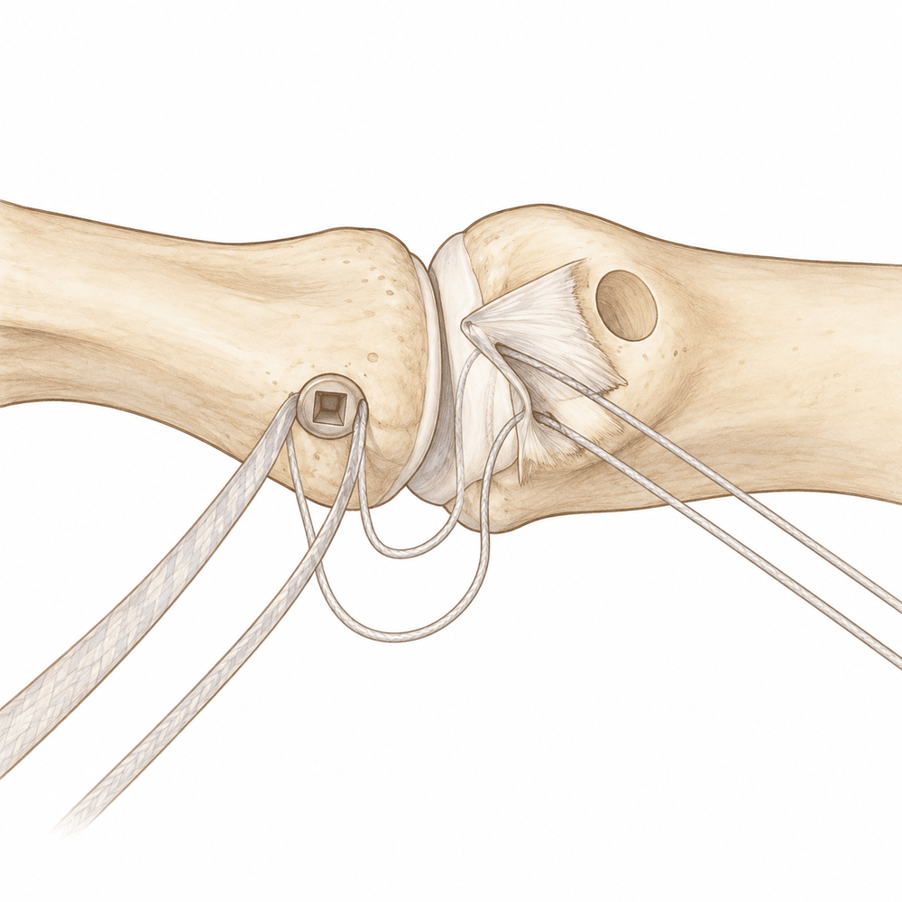
El cirujano realiza una pequeña incisión en el lateral del pulgar, en el punto donde se arrancó el ligamento. A través de esta incisión, localiza el extremo desgarrado del ligamento y prepara un pequeño punto en el hueso donde antes estaba unido. Después, el ligamento se vuelve a coser sobre ese punto mediante un pequeño anclaje colocado en el hueso. Esto mantiene el ligamento en su lugar mientras cicatriza de nuevo sobre el hueso.
Si el ligamento lleva mucho tiempo desgarrado, los extremos pueden estar demasiado desgastados para coserlos. En ese caso, el cirujano puede reconstruir el ligamento con un tendón cercano de su propio pulgar, plegado para formar una nueva banda que se une al hueso a cada lado de la articulación. Si el ligamento se arrancó junto con un pequeño fragmento de hueso, ese fragmento se vuelve a fijar en su sitio con una pequeña placa y tornillos.
Algunas reparaciones se refuerzan con una cinta, similar a un lazo, que se cose junto al ligamento. Esta cinta actúa como una cuerda de seguridad temporal y comparte la carga mientras el ligamento cicatriza. Como soporta parte de la tensión, puede que el pulgar empiece a moverse antes que con una reparación sola.
El cirujano también puede colocar un pequeño pin metálico a través de la articulación del pulgar para mantenerla inmovilizada y proteger la reparación de las tensiones laterales. El pin se retira más adelante, cuando la cicatrización ya está en marcha.
La incisión se cierra con puntos y se cubre con un apósito. Pasará a la sala de recuperación, y el equipo de enfermería le vigilará mientras se despierta.
Después de la operación
Despertará en la sala de recuperación y después pasará a la planta. Su mano estará en una férula o un yeso, con apósitos sobre la herida. Las enfermeras le mantendrán cómodo y le darán analgésicos si los necesita. Puede levantarse y moverse en cuanto se sienta capaz. Alguien debe quedarse con usted en casa durante las primeras 24 horas. Su equipo médico le indicará si podrá irse a casa el mismo día o si deberá permanecer una noche en el hospital. Dejamos el vendaje puesto durante unos 10 días; por favor, no lo retire antes de ese plazo a menos que se lo indiquemos. Lo cambiaremos o lo retiraremos cuando le veamos.
Recuperación
Durante los primeros días y semanas, su pulgar estará adolorido e hinchado. Esto mejora gradualmente. Ayuda mantener la mano elevada mientras descansa, al igual que los analgésicos que le da su equipo. Se irá a casa con una férula o un yeso que protege la reparación mientras cicatriza.
Su rutina diaria cambia más de lo que podría esperar. Al principio necesitará ayuda con algunas tareas, sobre todo con todo lo que requiera agarrar con fuerza, como abrir frascos o llevar cosas pesadas. La mayoría de las personas descubren que pueden hacer tareas diarias ligeras bastante pronto, y trabajo de oficina dentro de los plazos que se muestran en la tabla de esta página. La terapia de la mano es una parte importante de su recuperación. Su terapeuta de la mano, Ruby Doolan, de Extend Rehabilitation, le guiará en los ejercicios y le confeccionará cualquier férula que necesite mientras el pulgar cicatriza. Los ejercicios empiezan de forma suave y van aumentando a medida que la reparación se fortalece.
A medida que disminuye la hinchazón y vuelve el movimiento, las tareas cotidianas se vuelven más fáciles. Cuando su cirujano considere que la reparación ha cicatrizado, irá volviendo poco a poco a trabajos más pesados, al deporte y al gimnasio. El resultado final sigue mejorando durante un tiempo, a medida que el dolor se calma y vuelve la fuerza.
La recuperación varía de una persona a otra. Sus plazos pueden ser distintos de los rangos habituales, y su cirujano y su terapeuta le orientarán en cada etapa.
Qué puede salir mal
La mayoría de los pacientes evolucionan bien, pero en ocasiones pueden surgir problemas. Su cirujano y el equipo lo vigilarán de cerca para detectar cualquier incidencia a tiempo.
La herida puede infectarse. Podría notar un enrojecimiento que se extiende desde la incisión, líquido o pus que sale de ella, o un dolor profundo y palpitante que no cede con analgésicos comunes. Llame a la clínica ese mismo día si nota alguna de estas señales.
A veces, un pequeño nervio cercano al pulgar se irrita durante la cirugía. Esto puede causar entumecimiento, hormigueo o una zona de sensibilidad extraña en el dorso del pulgar. El entumecimiento y la debilidad durante las primeras 24 horas después de un bloqueo nervioso son esperables y desaparecen a medida que pasa el efecto del bloqueo. Si el entumecimiento dura más allá de ese plazo, llame a la clínica.
La articulación puede quedar algo floja o rígida. Algunas personas notan un dolor leve en la base del pulgar al hacer pinza o agarrar, o que el pulgar no se dobla tanto como antes. Unas pocas notan que su agarre no es tan fuerte. Estos cambios suelen ser leves y a menudo mejoran con el tiempo y la terapia de la mano. Coméntelos en su próxima revisión si le molestan.
La propia reparación puede fallar. Es poco frecuente, pero una caída o un golpe en el pulgar durante las primeras semanas puede forzar o volver a romper el ligamento que está cicatrizando. Notaría un dolor repentino, hinchazón y la sensación de que el pulgar cede. Llame a la clínica si esto ocurre.
Si se ha usado un pin metálico para mantener la articulación inmovilizada, la piel alrededor puede irritarse o infectarse. El enrojecimiento, el dolor a la presión o el líquido alrededor del punto de salida del pin deben comunicarse a la clínica ese mismo día.
En la tabla de complicaciones de esta página se detallan las tasas típicas, por si desea conocer los datos específicos.
¿Cuándo deben llamarnos?
La mayoría de las personas se recuperan sin problemas, pero algunas señales requieren atención rápida.
Acuda a urgencias si tiene hinchazón o dolor en la pantorrilla, o dificultad para respirar o dolor en el pecho. Pueden ser signos de un coágulo de sangre. Acuda a urgencias si el pulgar, la mano o los dedos se ponen pálidos, fríos, blancos, azules u oscuros.
Llame a la clínica ese mismo día si tiene fiebre, un enrojecimiento que se extiende alrededor de la herida, o líquido o pus que sale de ella. Llame a la clínica ese mismo día si el dolor sigue empeorando a pesar de los analgésicos.
El entumecimiento y la debilidad durante las primeras 24 horas después de un bloqueo nervioso son esperables y desaparecen a medida que pasa su efecto. Si el entumecimiento dura más allá de ese plazo, o no puede mover el pulgar, la mano o los dedos una vez que ha pasado el efecto del bloqueo, llame a la clínica.
Si no puede contactar con la clínica, fuera del horario de atención o en fin de semana, acuda al servicio de urgencias más cercano.
Evidence & references
This is the clinical evidence summary written for health professionals. It is technical, and it lists the research this page was built from. You do not need to read it to understand your treatment or to make a decision about it.
Overview
- A controlled active motion therapy protocol after suture anchor repair of a ruptured UCL of the thumb is safe from a biomechanical point of view [1].
- Thumb UCL repair with suture tape augmentation demonstrates short-term outcomes comparable to what has been reported for other methods of repair [2].
- In patients with chronic UCL reconstructions, there was no significant change in pinch strength [3].
- In patients with chronic UCL reconstructions, significantly better results were achieved in grip strength, flexion, extension, ulnar and radial deviation [3].
- Acute surgical repair of the torn ulnar collateral ligament can achieve good subjective and objective functional results [4].
- Intraosseous suture anchor repair is a safe and effective method for repair of complete tears of the ulnar collateral ligament of the thumb MP joint [5].
- Late reconstruction of the ulnar collateral ligament of the thumb MP joint with free tendon graft provided good improvement in stability and strength of the thumb [6].
- Early mobilisation after robust tendon repairs is favoured to avoid tethering of the long tendons of the thumb during the early post-operative period [7].
- In a surgical cohort of active-duty soldiers with thumb MCP UCL injuries, rifle-related injury was the most commonly documented injury mechanism [8].
- Distal placement of the UCL phalangeal insertion restricted flexion motion from a mean of 57° to 47° [9].
- Avulsion of the ulnar collateral ligament of the thumb IP joint is a previously undescribed injury [10].
- At the last postoperative follow-up (range 6 weeks-6 months), 11 of 12 patients had good to excellent results subjectively and had regained 75% of the strength of the uninjured side [11].
- Arthroscopic reduction of a Stener lesion allows healing of the ulnar collateral ligament without the need for open repair [12].
- Functional and subjective outcomes were good or satisfactory in more than 90% of patients treated with functional bracing [13].
- Pure ligamentous injuries of the ulnar collateral ligament of the thumb in a child are much less common than epiphyseal fractures [14].
- Rodeo roping thumb injuries emphasize the importance of keeping the thumb upward and clear of the rope to avoid entrapment between the rope and saddle horn [15].
- After undergoing open reduction and internal fixation for thumb ulnar collateral ligament avulsion fractures, patients had relief of thumb pain and pinch strength improved from 36% of the contralateral side to 89% (p < .01) [17].
- The use of interference screws for fixation of tendon grafts to bone for hand ligament reconstructions is a promising new surgical technique [18].
Anatomy & Pathophysiology
Osseous Anatomy & Kinematics
- The skeleton of the hand consists of 27 bones, of which 19 are long bones [32].
- The thumb ray (first ray) is the shortest and consists of only three bones: a metacarpal and two phalanges [32].
- The thumb metacarpal makes an angle of about 45 degrees with the second metacarpal in the sagittal plane [32].
- The trapezium is angled out in front of the carpal plane, allowing the thumb metacarpal to oppose the other four digital rays [32].
- The epiphyseal plates are located at the proximal ends of the phalanges and the first metacarpal [32].
- The thumb metacarpal is independent and articulates with the trapezium [43].
- The metacarpophalangeal joints are the keystones of the longitudinal arches of the hand [43].
- The stability of the metacarpophalangeal joints is essential to the support of the longitudinal arch as well as the transverse metacarpal arch [43].
- The thumb ray is more mobile, shorter, and more proximal than the other rays, allowing it to project in front of the plane of the palm [32].
Ligamentous & Soft Tissue Anatomy
- The metacarpophalangeal joint is stabilized by collateral ligaments and by the thick volar articular capsule, known as the volar plate [43].
- The lateral accessory ligaments, sagittal bands of the extensor apparatus, and the first annular segment of the flexor tendon pulley insert on the volar plate [43].
- The sagittal band fibers insert onto the volar proximal phalanx and onto the lateral borders of the volar plate [31].
- The sagittal bands form a sling that allows proximal extrinsic extensor tension to be transmitted to the proximal phalanx, permitting metacarpophalangeal joint extension without a tendinous insertion onto the proximal phalanx [31].
- Rupture or attenuation of the sagittal band fibers allows the extrinsic extensor tendon to sublux to the ulnar side of the metacarpal head, causing ulnar deviation of the finger [31].
- The flexor pollicis longus inserts into the proximal base of the thumb distal phalanx and is innervated by the anterior interosseous branch of the median nerve [47].
- The flexor pollicis longus flexes both the interphalangeal and metacarpophalangeal joints of the thumb [47].
- The tenosynovial sheath of the flexor pollicis longus is continuous with the radial bursa [47].
- The princeps pollicis artery, a terminal branch of the radial artery, runs along the ulnar side of the first metacarpal bone and along the volar surface of the adductor muscle [44].
- The princeps pollicis artery emerges onto the subcutaneous palmar tissue at the level of the cutaneous flexion crease of the metacarpophalangeal joint [44].
- At the metacarpophalangeal joint level, the princeps pollicis artery divides into two terminal rami, the collateral palmar arteries of the thumb [44].
- An arcade located deep in the flexor tendon joins together the two collateral palmar arteries at the level of the distal metaphysis of the first phalanx [44].
- The dorsal arteries of the thumb originate from palmar arteries (princeps, commissural, or anastomoses of the superficial arcade) at the level of the first metacarpal [44].
- The ulnar dorso-collateral artery generally stems from the princeps pollicis onto the medial border of the neck of the first metacarpal [44].
Pathophysiology & Injury Mechanisms
- Acute injuries to the thumb ulnar collateral ligament (UCL) of the metacarpophalangeal joint are the result of a sudden, significant radial force causing abduction of the thumb [20].
- Fractures can occur at the base of the proximal phalanx in association with ruptures of the UCL of the metacarpophalangeal joint [19].
- Proximal phalanx base fractures associated with UCL rupture are caused either by avulsion of the UCL from its insertion or by shearing of the base by the palmar portion of the radial condyle [19].
- In sheared fractures, the fragment does not contain the UCL insertion, and its position is unrelated to the location of the insertion of the UCL [19].
- In avulsion fractures, the position of the bone fragment indicates the location of the end of the ligament [19].
- A displaced ligament (Stener lesion) can occur in the presence of a nondisplaced fracture [19].
- The mechanism of fracture (avulsion vs. shear) cannot be determined by radiographic appearance and is revealed only at surgery by inspecting the bone and its relationship to the end of the ligament [19].
- Supination and flexion of the thumb provided less stability when both components of the UCL were sectioned [20].
- Pronation and supination play a role in the stability of the thumb metacarpophalangeal joint [20].
- Performing a physical examination to assess instability did not create a Stener lesion if the exam was performed in a controlled, gentle manner with the thumb held without rotation [20].
- A Stener lesion was possible to create during examination only when the thumb was tested in a flexed and supinated position and there was sectioning of the proximal UCL, accessory UCL, and the origin of the ulnar sagittal band at the adductor aponeurosis [20].
- Pure ligamentous injuries of the UCL of the thumb in a child are much less common than epiphyseal fractures [14].
- Traumatic avulsion of the ulnar collateral ligament of the interphalangeal joint of the thumb is a previously undescribed injury [10].
- In a surgical cohort of active-duty soldiers with thumb metacarpophalangeal UCL injuries, rifle-related injury was the most commonly documented injury mechanism [8].
Classification
- Acute injuries of the thumb ulnar collateral ligament (UCL) of the metacarpophalangeal (MCP) joint are the result of a sudden, significant radial force causing abduction of the thumb [20].
- Fractures can occur at the base of the proximal phalanx in association with ruptures of the UCL of the MCP joint of the thumb [19].
- Proximal phalanx base fractures associated with UCL rupture are either caused by an avulsion of the UCL from its insertion on the proximal phalanx or by the shearing of the base of the proximal phalanx by the palmar portion of the radial condyle with continued displacement after UCL rupture [19].
- A sheared fragment of the proximal phalanx base does not contain the UCL insertion [19].
- If a fragment of bone has been avulsed by the UCL, its position indicates the location of the end of the ligament [19].
- A radiograph can reveal whether the insertion of the ligament is displaced, indicating a Stener lesion [19].
- A displaced ligament can occur in the presence of a nondisplaced fracture [19].
- The mechanism of fracture cannot be determined by radiographic appearance alone [19].
- The mechanism of fracture is revealed only at surgery by inspecting the bone and its relationship to the end of the ligament [19].
- Pure ligamentous injuries of the ulnar collateral ligament of the thumb in a child do occur, but they are much less common than epiphyseal fractures [14].
- Traumatic avulsion of the ulnar collateral ligament of the interphalangeal (IP) joint of the thumb is a previously undescribed injury [10].
Clinical Presentation
Injury Mechanisms and Epidemiology
- Acute thumb UCL injuries are the result of a sudden, significant radial force causing abduction of the thumb [20].
Physical Examination and Diagnostic Findings
- Performing a physical examination to assess the amount of instability of an ulnar collateral ligament injury did not create a Stener lesion if the exam was performed in a controlled, gentle manner with the thumb held without rotation [20].
- It was possible to create a Stener lesion during physical examination when the thumb was tested in a flexed and supinated position, but only if there was sectioning of the pUCL, the aUCL, and the origin of the ulnar sagittal band at the adductor aponeurosis [20].
- Thumbs with a stress angle difference of 15° should always be explored for a probable Stener lesion [34].
- Diagnostic ultrasound is a useful, noninvasive tool for assessing the extent of rupture of the ulnar collateral ligament [25].
Radiographic and Anatomical Considerations
- These fractures are either caused by an avulsion of the UCL from its insertion on the proximal phalanx or by the shearing of the base of the proximal phalanx by the palmar portion of the radial condyle with continued displacement after UCL rupture [19].
- A radiograph can reveal whether the insertion of the ligament is displaced (Stener lesion) if a fragment of bone has been avulsed by the UCL [19].
- If a fragment has been sheared, its position is unrelated to the location of the insertion of the UCL [19].
- The mechanism of fracture cannot be determined by radiographic appearance and is revealed only at surgery by inspecting the bone and its relationship to the end of the ligament [19].
Investigations
- Fluoroscopy, MRI and ultrasound are increasingly being used and evaluated as diagnostic tools for thumb UCL injuries [20].
- If a fragment of bone has been sheared rather than avulsed, its position is unrelated to the location of the insertion of the UCL [19].
- It was possible to create a Stener lesion when the thumb was tested in a flexed and supinated position, but only if there was sectioning of the pUCL, the aUCL, and the origin of the ulnar sagittal band at the adductor aponeurosis [20].
Treatment
Operative Repair Techniques
- Suture anchor and suture tape augmentation techniques preserve native ligament proprioception while providing additional stability during early healing phases [28].
- At the last postoperative follow-up (range 6 weeks-6 months), 11 of 12 patients treated with an alternative collateral ligament repair method had good to excellent results subjectively and had regained 75% of the strength of the uninjured side [11].
- Both steel wire and simple suture techniques for UCL repair were equally effective, with the steel wire offering no clinical advantage over simple suture [33].
- A novel technique of thumb collateral ligament reconstruction in acute injuries resulted in negative instability tests and satisfying results in terms of motion (ROM loss less than 20%) and grip strength (loss less than 17%) at 3-month follow-up [36].
- In a comparison of acute and chronic UCL injuries, there were no statistically significant differences between operated and contralateral healthy thumb MCP joints in flexion, extension, ulnar deviation, radial deviation, grip strength, or tip pinch strength at final follow-up [27].
- In the avulsion fracture group of a comparative study, reduction was successful and union was observed with no complications such as reduction loss, MCP osteoarthritis, bone necrosis, or non-union [27].
- Ulnar deviation and radial deviation were significantly better in the avulsion group than in the rupture group [27].
- Grip and tip pinch strengths were significantly better in the avulsion group than in the rupture group [27].
Reconstruction Techniques
- Late reconstruction of the ulnar collateral ligament of the thumb MP joint with a free tendon graft provided good improvement in stability and strength of the thumb [6].
Post-operative Management and Rehabilitation
- Early mobilization after robust tendon repairs is favored to avoid tethering of the long tendons of the thumb during the early post-operative period [7].
- Immobilization of the MCP joint with a K-wire after surgical repair of the UCL allows return to work within 7 days for 62% of patients who are not manual workers [21].
- In patients with chronic UCL reconstructions, there was no significant change in pinch strength, but significantly better results were achieved in grip strength, flexion, extension, ulnar and radial deviation [3].
Non-Operative Management
Surgical Considerations and Complications
- Immediate repair of ruptured structures at both joints in a case of traumatic avulsion of the UCL of the IP joint and MCP joint produced a satisfactory result [16].
- Fractures at the base of the proximal phalanx associated with UCL ruptures can be caused by avulsion of the UCL or by shearing of the base by the palmar portion of the radial condyle [19].
- A displaced ligament (Stener lesion) can occur in the presence of a nondisplaced fracture if the fragment was sheared rather than avulsed [19].
- The mechanism of fracture (avulsion vs. shearing) cannot be determined by radiographic appearance and is revealed only at surgery by inspecting the bone and its relationship to the end of the ligament [19].
Complications
- In a cohort of 12 patients treated with an alternative repair method, 11 had good to excellent subjective results and regained 75% of the strength of the uninjured side at follow-up ranging from 6 weeks to 6 months [11].
- After open reduction and internal fixation for thumb ulnar collateral ligament avulsion fractures, pinch strength improved from 36% of the contralateral side to 89% (p < .01) [17].
- In a comparison of acute and chronic UCL injuries, no complications such as reduction loss, MCP osteoarthritis, bone necrosis, and non-union were observed in the avulsion fracture group [27].
- In a comparison of acute and chronic UCL injuries, four patients in the avulsion group and four patients in the rupture group experienced mild intermittent pain caused by light activity [27].
Recovery
- Early active mobilization following UCL repair with a Mitek bone anchor leads to an earlier return to full hand function at 6 weeks compared to 8 weeks [29].
- Early active mobilization following UCL repair with a Mitek bone anchor leads to an earlier return to work at 7 weeks compared to 11 weeks [29].
- A policy of early mobilisation after robust tendon repairs is favoured to avoid tethering of the long tendons of the thumb during the early post-operative period [7].
Key Evidence
- [L5] A controlled active motion therapy protocol after suture anchor repair of a ruptured UCL of the thumb is safe from a biomechanical point of view. [1] (10.1016/j.jhsa.2004.04.017)
- [L4] Thumb UCL repair with suture tape augmentation demonstrates short-term outcomes comparable to what has been reported for other methods of repair. [2] (10.1142/s2424835520500046)
- [L3] There was no significant change in pinch strength in patients with chronic UCL reconstructions, but significantly better results were achieved in grip strength, flexion, extension, ulnar and radial deviation. [3] (10.1016/j.hansur.2020.03.002)
- [L4] Acute surgical repair of the torn ulnar collateral ligament can achieve good subjective and objective functional results. [4] (10.1016/0020-1383(94)90179-1)
- [L3] The authors concluded that this is a safe and effective method for repair of complete tears of the ulnar collateral ligament of the thumb MP joint. [5] (10.1016/s0363-5023(97)80113-x)
- [L1] This new technique provided good improvement in stability and strength of the thumb. [6] (10.1016/s0363-5023(03)80385-4)
- [L4] In this field of tendon surgery, we favour a policy of early mobilisation after robust tendon repairs to avoid tethering of the long tendons of the thumb during the early post-operative period. [7] (10.1197/j.jht.2005.01.008)
- [L4] In this surgical cohort of active-duty soldiers with thumb MCP UCL injuries, rifle-related injury was the most commonly documented injury mechanisms. [8] (10.1016/j.injury.2026.113711)
- [L5] Distal placement of the UCL phalangeal insertion restricted flexion motion (from a mean of 57° to 47°). [9] (10.1053/jhsu.1999.0283)
- [L5] We present a previously undescribed injury of avulsion of the ulnar collateral ligament of the thumb IP joint. [10] (10.1016/s0266-7681(05)80061-0)
- [L4] At the last postoperative follow-up (range 6 weeks-6 months) 11 of 12 patients had good to excellent results subjectively and had regained 75% of the strength of the uninjured side. [11] (10.1016/s0266-7681(97)80436-6)
- [L4] Results indicate that arthroscopic reduction of a Stener lesion allows healing of the ulnar collateral ligament without the need for open repair. [12] (10.1016/s0363-5023(05)80156-x)
- [L2] Functional and subjective outcomes were good or satisfactory in more than 90% of patients. [13] (10.1016/s0363-5023(89)80026-7)
- [L5] Pure ligamentous injuries of the ulnar collateral ligament of the thumb in a child do occur, but they are much less common than epiphyseal fractures. [14] (10.1016/s0363-5023(86)80009-0)
- [L4] These injuries emphasize the importance of keeping the thumb upward and clear of the rope to avoid entrapment between the rope and saddle horn. [15] (10.1016/s0363-5023(84)80136-7)
- [L5] Immediate repair of the ruptured structures at both joints produced a satisfactory result. [16] (10.1016/s0363-5023(87)80224-1)
- [L4] After undergoing open reduction and internal fixation, the patients had relief of thumb pain and pinch strength improved from 36% of the contralateral side to 89% (p < .01). [17] (10.1016/s0363-5023(97)80049-4)
- [L5] The use of interference screws for fixation of tendon grafts to bone for hand ligament reconstructions is a promising new surgical technique. [18] (10.1016/j.jhsa.2004.12.013)
- [L5] [19] (10.1016/s0363-5023(98)80158-5)
- [L5] [20] (10.1142/s0218810417500411)
- [L4] Immobilization of the MCP joint with a K-wire after surgical repair of the UCL is an option that allows return to work within 7 days for 62% of patients who are not manual workers. [21] (10.1016/j.hansur.2015.12.007)
- [L5] Diagnostic ultrasound is a useful, noninvasive tool for assessing the extent of rupture of the ulnar collateral ligament. [25] (10.1016/0363-5023(94)90025-6)
- [L4] [27] (10.1016/j.main.2014.10.003)
- [L5] The technique preserves native ligament proprioception while providing additional stability during early healing phases. [28] (10.1016/j.eats.2025.103957)
- [L1] Our results show that on average early active mobilization leads to an earlier return to full hand function (6 vs. 8 wk) and an earlier return to work (7 vs. 11 wk). [29] (10.1097/bth.0b013e318284dbd7)
- [L4] Both techniques were equally effective, and the use of the more expensive steel wire, while technically satisfying and easy to perform, offers no clinical advantage over simple suture. [33] (10.1016/0266-7681(92)90079-h)
- [L4] Our study shows that thumbs with a stress angle difference of 15° should always be explored for a probable Stener lesion. [34] (10.1177/1753193412455789)
- [L5] At 3 month follow up, tests for instability were negative and satisfying results in terms of motion (ROM loss less than 20%), grip strength (loss less than 17%) were observed. [36] (10.1016/j.jhse.2007.04.010)
References
[1] A biomechanical modeling of injury, repair, and rehabilitation of ulnar collateral ligament injuries of the thumb. The Journal of Hand Surgery. 2004. DOI: 10.1016/j.jhsa.2004.04.017
[2] Thumb Ulnar Collateral Ligament Repair with Suture Tape Augmentation. The Journal of Hand Surgery (Asian-Pacific Volume). 2020. DOI: 10.1142/s2424835520500046
[3] The effects of rehabilitation on the outcomes of surgically treated acute and chronic thumb metacarpophalangeal ulnar collateral ligament ruptures. Hand Surgery and Rehabilitation. 2020. DOI: 10.1016/j.hansur.2020.03.002
[4] Gamekeeper's thumb: a quantitative evaluation of acute surgical repair. Injury. 1994. DOI: 10.1016/0020-1383(94)90179-1
[5] Repair of acute ulnar collateral ligament injuries of the thumb metacarpophalangeal joint with an intraosseous suture anchor. The Journal of Hand Surgery. 1997. DOI: 10.1016/s0363-5023(97)80113-x
[6] Late reconstruction of the ulnar collateral ligament of the thumb MP joint with free tendon graft — A new technique. The Journal of Hand Surgery. 2003. DOI: 10.1016/s0363-5023(03)80385-4
[7] New Concepts in Managing the Long Tendons of the Thumb After Primary Repair. Journal of Hand Therapy. 2005. DOI: 10.1197/j.jht.2005.01.008
[8] Return to Duty After Suture Anchor Repair of Thumb Metacarpophalangeal Ulnar Collateral Ligament Injuries in Active Duty Soldiers. Injury. 2026. DOI: 10.1016/j.injury.2026.113711
[9] The effect of thumb metacarpophalangeal ulnar collateral ligament attachment site on joint range of motion: An in vitro study. The Journal of Hand Surgery. 1999. DOI: 10.1053/jhsu.1999.0283
[10] Traumatic Avulsion of the Ulnar Collateral Ligament of the IP Joint of the Thumb. Journal of Hand Surgery. 1995. DOI: 10.1016/s0266-7681(05)80061-0
[11] Alternative Method of Repairing Collateral Ligament Injuries at the Metacarpophalangeal Joints of the Thumb and Fingers. Journal of Hand Surgery. 1997. DOI: 10.1016/s0266-7681(97)80436-6
[12] Arthroscopic treatment of acute complete thumb metacarpophalangeal ulnar collateral ligament tears. The Journal of Hand Surgery. 1995. DOI: 10.1016/s0363-5023(05)80156-x
[13] Gamekeepers thumb: A prospective study of functional bracing. The Journal of Hand Surgery. 1989. DOI: 10.1016/s0363-5023(89)80026-7
[14] Ligamentous avulsion of the ulnar collateral ligament of the thumb of a child. The Journal of Hand Surgery. 1986. DOI: 10.1016/s0363-5023(86)80009-0
[15] Rodeo roping thumb injuries. The Journal of Hand Surgery. 1984. DOI: 10.1016/s0363-5023(84)80136-7
[16] An unusual variety of skier's thumb. The Journal of Hand Surgery. 1987. DOI: 10.1016/s0363-5023(87)80224-1
[17] Failure of cast immobilization for thumb ulnar collateral ligament avulsion fractures. The Journal of Hand Surgery. 1997. DOI: 10.1016/s0363-5023(97)80049-4
[18] Fixation of Tendon Grafts for Collateral Ligament Reconstructions: A Cadaveric Biomechanical Study. The Journal of Hand Surgery. 2005. DOI: 10.1016/j.jhsa.2004.12.013
[19] The stener lesion revisited: A case report. The Journal of Hand Surgery. 1998. DOI: 10.1016/s0363-5023(98)80158-5
[20] Can Physical Examination Create a Stener Lesion?. The Journal of Hand Surgery (Asian-Pacific Volume). 2017. DOI: 10.1142/s0218810417500411
[21] Duration of sick leave after surgical repair of the ulnar collateral ligament of the thumb metacarpophalangeal joint with K-wire immobilization: Prospective case series of 21 patients. Hand Surgery and Rehabilitation. 2016. DOI: 10.1016/j.hansur.2015.12.007
[25] Ultrasonographic detection of thumb ulnar collateral ligament injuries: A cadaveric study. The Journal of Hand Surgery. 1994. DOI: 10.1016/0363-5023(94)90025-6
[27] Comparison of results after surgical repair of acute and chronic ulnar collateral ligament injury of the thumb. Chirurgie de la Main. 2014. DOI: 10.1016/j.main.2014.10.003
[28] Thumb Ulnar Collateral Ligament Repair Using Suture Anchors and Suture Tape Augmentation. Arthroscopy Techniques. 2025. DOI: 10.1016/j.eats.2025.103957
[29] Early Active Mobilization Following UCL Repair With Mitek Bone Anchor. Techniques in Hand & Upper Extremity Surgery. 2013. DOI: 10.1097/bth.0b013e318284dbd7
[31] A Lange Medical Book Current Diagnosis Treatment In Orthopedics Fifth Edition. 9Hand Surgery > Image DISORDERS OF THE MUSCULATURE OF THE HAND.
[32] Exam Of The Hand Wrist 2Ed. 1.1 SKELETON OF THE HAND > The osseous skeleton.
[33] Ulnar Collateral Ligament Repair of the Metacarpo-Phalangeal Joint of the Thumb: A Study Comparing Two Methods of Repair. Journal of Hand Surgery. 1992. DOI: 10.1016/0266-7681(92)90079-h
[34] Re-evaluation of stress radiographic findings for preoperative diagnosis of Stener lesion. Journal of Hand Surgery (European Volume). 2012. DOI: 10.1177/1753193412455789
[36] A Novel Technique of Thumb Collateral Ligament Reconstruction in Acute Injuries. Journal of Hand Surgery (European Volume). 2007. DOI: 10.1016/j.jhse.2007.04.010
[43] Exam Of The Hand Wrist 2Ed. The arches of the hand > The metacarpal arch.
[44] Exam Of The Hand Wrist 2Ed. Techniques of investigation of the arterial supply by J P Melki > Vascularization of the thumb > Palmar aspect.
[47] A Lange Medical Book Current Diagnosis Treatment In Orthopedics Fifth Edition. 9Hand Surgery > FLEXOR TENDON INJURY.