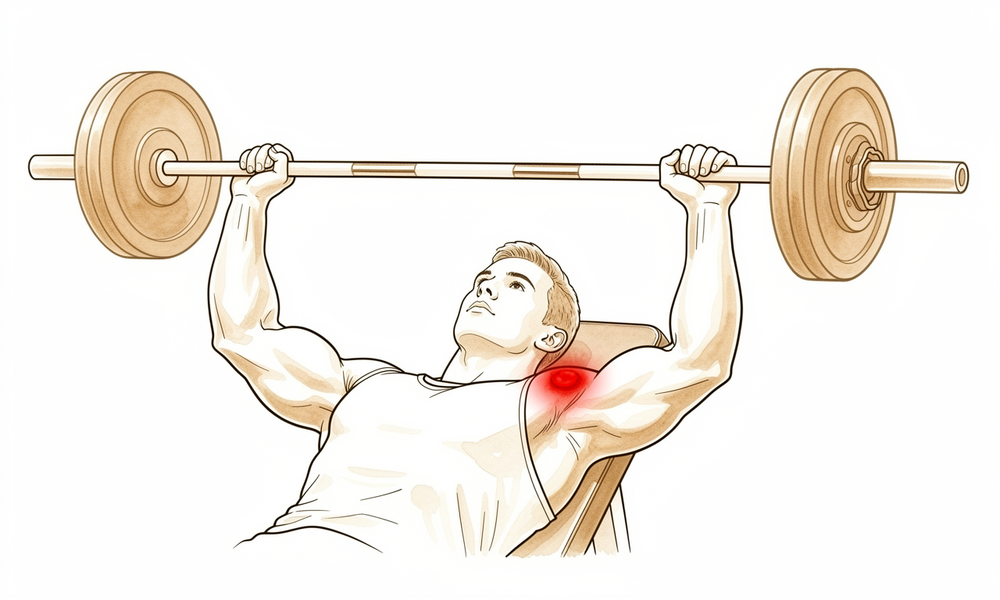
Qué está sintiendo
El dolor se localiza en el extremo externo de la clavícula, justo donde esta se une a la parte superior del hombro. Ese punto se denomina articulación acromioclavicular. Suele doler más cuando usa el brazo, sobre todo al empujar, levantar peso o hacer movimientos de press, y normalmente se calma cuando descansa.
El entrenamiento con pesas es un desencadenante frecuente. El press de banca y otros levantamientos de mucho peso cargan directamente esta articulación, y el dolor suele ir aumentando a lo largo de semanas de entrenamiento en lugar de aparecer con una lesión concreta. Algunas personas también lo notan después de una lesión en el hombro, cuando el dolor persiste mucho después de que el resto del hombro se haya recuperado.
La molestia puede empeorar por la noche o después de una sesión en el gimnasio. Cruzar el brazo por delante del cuerpo, empujar con los brazos para levantarse de una silla, llevar la compra en ese lado o empujar un cortacésped pueden empeorarla. Como la molestia se localiza en la parte más alta del hombro, al principio es fácil confundirla con una distensión muscular.
Esta afección puede parecerse mucho a otros problemas del hombro, así que se utilizan pruebas de imagen para confirmar lo que ocurre. Una radiografía especial de la articulación, tomada en ambos lados para compararlos, y una resonancia magnética pueden mostrar cambios en el extremo de la clavícula.
Esté atento a estas señales y actúe ante ellas:
Acuda al servicio de urgencias ese mismo día si el brazo se pone caliente, rojo, hinchado y doloroso, sobre todo si tiene fiebre. Acuda ese mismo día si pierde de repente la sensibilidad o el movimiento del brazo después de una lesión.
Consulte a su médico de cabecera o pida una valoración por un especialista si el dolor no mejora, empeora a lo largo de las semanas, le despierta por la noche o le impide trabajar o usar el brazo. Si no puede comunicarse con la clínica, fuera del horario de atención o en fin de semana, acuda al servicio de urgencias más cercano.
¿Qué está ocurriendo realmente?
La articulación acromioclavicular es una pequeña articulación situada en la parte superior del hombro, donde el extremo externo de la clavícula se une a un saliente óseo llamado acromion. La clavícula actúa como el puntal que mantiene el brazo separado del pecho, y esta pequeña articulación es uno de los puntos donde se une al omóplato. Una banda resistente de ligamentos situada por debajo mantiene los dos huesos alineados mientras levanta peso o empuja.
En esta afección, el extremo externo de la clavícula empieza a deteriorarse. El término médico para este deterioro del hueso es osteólisis. Normalmente, el cuerpo sustituye el hueso desgastado por hueso nuevo, pero aquí el deterioro va más rápido que la reparación, de modo que el hueso del extremo de la clavícula se ablanda y se erosiona. La parte de la articulación situada por encima se mantiene sana; el problema se limita al lado de la clavícula.
El desencadenante más frecuente es la carga intensa repetida, sobre todo el press de banca y el entrenamiento con pesas. Cada repetición de press empuja los dos huesos de la articulación uno contra otro, un poco como pisar una y otra vez el mismo trozo pequeño de suelo hasta que se desgasta. El mismo proceso puede aparecer después de una lesión en el hombro, cuando el dolor persiste semanas o meses después del golpe inicial. Con menos frecuencia, puede desencadenarlo una infección de bajo grado o una placa metálica utilizada para sujetar la clavícula hacia abajo tras una luxación.
A medida que el hueso se erosiona, la articulación se inflama y se hincha, y por eso la molestia aumenta a lo largo de semanas de entrenamiento y empeora por la noche. El extremo erosionado también pierde su encaje suave con el acromion, de modo que al empujar, levantar peso o cruzar el brazo por delante del cuerpo el hueso en carne viva roza contra el otro. Ese es el dolor que siente en la parte más alta del hombro.
Qué podemos hacer al respecto
El Dr. Kieran Hirpara, cirujano de extremidades superiores en el Mater Private Hospital Rockhampton, comienza con las opciones menos invasivas que se adapten a su condición. Por lo general, los pacientes son derivados a nuestra clínica por su médico de cabecera; si un fisioterapeuta le ha sugerido consultarnos, igualmente necesitará una derivación de su médico de cabecera para poder acceder al reembolso de Medicare. Una vez confirmado el diagnóstico con su historia clínica, una exploración y pruebas de imagen, vamos pasando por los tratamientos en orden, empezando por el más sencillo.
El primer paso es cambiar la forma en que carga la articulación. Como es el press intenso repetido lo que desgasta el hueso, reducir o sustituir los ejercicios que lo agravan da al hueso la oportunidad de recuperarse. La fisioterapia busca calmar la inflamación y mantener el hombro fuerte y con movilidad mientras tanto. En deportistas en plena temporada, este enfoque le permite seguir jugando mientras el tratamiento hace efecto. Cuánto tiempo lleva depende de si puede cambiar de verdad su forma de entrenar, y hablaremos con usted de lo que eso supone para su deporte o su trabajo.
La medicación es el siguiente nivel. Los antiinflamatorios en comprimidos, a veces llamados AINE, reducen la hinchazón y la molestia en la articulación. También podemos inyectar una combinación de anestésico local y cortisona, un antiinflamatorio potente, directamente en la articulación. Esta inyección cumple dos funciones: confirma que la articulación es el origen de su dolor y puede calmar ese dolor durante un tiempo después.
Si estos pasos no le han aliviado lo suficiente, podemos hablar de la cirugía. La operación extirpa una pequeña lámina de hueso del extremo externo de la clavícula, para que las superficies erosionadas y en carne viva dejen de rozar entre sí. Solo se extirpan unos pocos milímetros de hueso, y si un espolón óseo presiona sobre un tendón cercano, lo limpiamos en la misma operación. Le explicaremos en qué consiste la operación, cómo es la recuperación y qué puede y qué no puede cambiar, para que decida junto con nosotros si es adecuada para usted.
Qué esperar
En la mayoría de las personas, esta afección mejora una vez que se interrumpe la carga que la provocó. El hueso del extremo de la clavícula puede regenerarse cuando se reducen los press y los levantamientos repetidos. Esa es, con honestidad, la condición: el tratamiento funciona cuando puede cambiar de verdad su forma de entrenar o de trabajar. Si sigue cargando la articulación de la misma manera, el dolor tiende a persistir o a reaparecer una y otra vez.
La recuperación sin operación no es una solución rápida. La molestia aumenta a lo largo de semanas, y normalmente hacen falta semanas o meses de reducir la carga antes de que se calme. Algunas personas notan el dolor después de una lesión en el hombro, y en ese caso puede persistir mucho después de que el resto del hombro haya curado. Si el dolor sigue presente tras un periodo razonable de reposo y de cambios en el entrenamiento, merece la pena que lo revisen en lugar de aguantarlo.
Si el tratamiento no quirúrgico no le alivia lo suficiente, la cirugía es una opción. La operación extirpa una pequeña lámina de hueso del extremo externo de la clavícula para que las superficies en carne viva dejen de rozar. Las personas a las que se les hace refieren menos dolor y mejor movilidad del hombro, y en general están satisfechas con el resultado. La versión artroscópica tiende a dejar menos dolor residual que la versión abierta, y con ella las personas vuelven antes a sus actividades.
La cirugía no es una promesa de un hombro sin dolor. Un pequeño número de personas necesita otra operación más adelante, entre el 4% y el 10%. Esto puede ocurrir si se extirpó demasiado poco o demasiado hueso, si había otro problema en el hombro que pasó desapercibido o si se forma hueso nuevo en la articulación mientras cicatriza. Su cirujano hablará con usted de estos riesgos antes de que decida nada.
Sea cual sea el camino que elija, el objetivo es el mismo: calmar la inflamación, recuperar un movimiento suave y que vuelva a usar el brazo sin ese dolor agudo en la parte superior del hombro. Si sus síntomas no mejoran, empeoran a lo largo de las semanas, le despiertan por la noche o le impiden trabajar o usar el brazo, consulte a su médico de cabecera o pida una valoración por un especialista.
Cuándo consultar a un especialista
Esta afección es fácil de confundir con una distensión muscular, por lo que al principio a menudo pasa desapercibida. Consulte a su médico de cabecera si el dolor en el extremo externo de la clavícula persiste después de una lesión en el hombro, una vez descartadas una fractura o una articulación inestable. Pida una valoración por un especialista si la molestia sigue aumentando con el entrenamiento, o si no mejora a pesar del reposo y de los cambios en la carga. Las pruebas de imagen pueden confirmar el diagnóstico: una radiografía especial tomada de ambos hombros para compararlos y una resonancia magnética que muestra hinchazón en el interior del extremo de la clavícula. Acertar con el diagnóstico es importante, porque el mismo dolor puede deberse a otros problemas del hombro, y un tratamiento dirigido a la causa equivocada no servirá de nada. Si sus síntomas no mejoran, empeoran a lo largo de las semanas, le despiertan por la noche o le impiden trabajar o usar el brazo, consulte a su médico de cabecera o pida una valoración por un especialista. Si no puede comunicarse con la clínica, fuera del horario de atención o en fin de semana, acuda al servicio de urgencias más cercano.
En profundidad
Advanced reading: the deeper science (optional)
Esta sección va más allá de lo que necesita para tomar sus propias decisiones de tratamiento. Merece la pena leer más sobre la osteólisis distal de la clavícula porque su causa casi siempre puede identificarse, la mayoría de las personas se recuperan sin operarse, y lo difícil es saber si la carga que la provocó puede cambiarse realmente.
La carga suele poder identificarse, y suele ser el press de banca
Una revisión exploratoria de 2026 agrupó 8 estudios con 483 pacientes y halló que el press de banca era la actividad desencadenante más frecuente, con un 49.1%, y el entrenamiento con pesas en general, un 24.4% adicional [1]. El dolor en el extremo externo de la clavícula o en la articulación acromioclavicular fue el síntoma de presentación en el 69.9% de los pacientes [1]. La serie que definió por primera vez la afección encontró el mismo patrón: de 46 hombres diagnosticados de osteólisis sin antecedentes de lesión aguda, 45 levantaban pesas como parte de su entrenamiento [2].
La mayoría mejora sin cirugía, pero solo si la carga cambia de verdad
En la revisión agrupada, la mayoría de los pacientes se recuperó con tratamiento conservador, y un 16% acabó operándose tras el fracaso del tratamiento conservador [1]. Un grupo más pequeño, el 4.4% (3 pacientes), llegó a la cirugía porque la enfermedad progresó estructuralmente o porque no estaban dispuestos a modificar su carga por motivos deportivos o laborales [1]. Ese último grupo refleja con honestidad el problema: el tratamiento no quirúrgico funciona cuando la actividad desencadenante se interrumpe de verdad, y la dificultad rara vez está en hacer el diagnóstico.
Cuando se hace la resección, se extirpa poco hueso y la recuperación es rápida
En una serie de levantadores de pesas, una resección artroscópica limitada de una media de 4.5 mm permitió volver al deporte en una media de 3.2 días y al programa de entrenamiento con pesas previo a la operación en 9.1 días; todos los pacientes siguieron entrenando y aumentaron sus cargas después [3]. Una serie prospectiva moderna de 59 pacientes con un seguimiento de al menos dos años describió un descenso del dolor de 8.20 a 1.36 sobre 10 y de la discapacidad según el SPADI de 62.65 a 6.13 a los 24 meses, con vuelta al deporte a los 1.72 meses y al trabajo a los 3.02 meses [4]. En la serie original, la resección se realizó en 21 pacientes y alivió los síntomas en los 19 a los que se hizo seguimiento [2].
Las pruebas de imagen la confirman; el cuadro clínico por sí solo no
La propia conclusión de la revisión es que los signos y síntomas clínicos de esta afección no se podían distinguir de los de otras patologías del hombro [1]. La resonancia magnética fue la prueba más utilizada, y mostró edema de la médula ósea y fractura subcondral en el extremo de la clavícula; los autores señalan explícitamente que debe interpretarse junto con la exploración física y no en su lugar [1]. En la práctica, por eso la historia clínica —qué levanta, con qué frecuencia y cuánto peso— tiene tanta importancia como la prueba de imagen.
Referencias
[1] Wilkinson M, Groch N, Freestone C, et al. Factores de riesgo y tratamiento de la osteólisis atraumática de la clavícula distal: una revisión exploratoria. Shoulder Elbow. 2026. https://doi.org/10.1177/17585732261479715 [2] Cahill BR. Osteólisis de la porción distal de la clavícula en atletas varones. J Bone Joint Surg Am. 1982;64(7):1053-58. https://doi.org/10.2106/00004623-198264070-00015 [3] Auge WK, Fischer RA. Resección artroscópica de la clavícula distal en la osteólisis atraumática aislada en levantadores de pesas. Am J Sports Med. 1998;26(2):189-92. https://doi.org/10.1177/03635465980260020701 [4] Leon JV, Hermans D, Venkatesha V, et al. Resultados de los pacientes tras la escisión artroscópica de la clavícula distal: una serie de casos prospectiva. JSES Int. 2023;7(6):2400-05. https://doi.org/10.1016/j.jseint.2023.07.014
Evidence & references
This is the clinical evidence summary written for health professionals. It is technical, and it lists the research this page was built from. You do not need to read it to understand your treatment or to make a decision about it.
Overview
Clinical Presentation and Diagnosis
- Clinical signs and symptoms for atraumatic distal clavicular osteolysis are not distinguishable from other shoulder pathologies [1].
- Post-traumatic osteolysis of the distal clavicle should be considered if there is persistent pain after a shoulder injury and bony lesions or instabilities have been excluded [3].
- Osteolysis of the distal end of the clavicle is a rare condition that can occur during judo [5].
- Special care must be taken to properly identify the acromioclavicular joint and rule out pre-existing distal clavicle stress fracture or osteolysis before performing the arthroscopic Mumford procedure [22].
Etiology and Pathogenesis
- Propionibacterium acnes–mediated distal clavicular osteolysis is a documented etiology [2].
- Heterotopic bone formation is a more common cause of failure of acromioplasty or distal claviculectomy than is acknowledged by the literature [13].
- Subacromial osteolysis can occur following hook plate fixation for acromioclavicular dislocation [4].
- The risk of subacromial osteolysis following hook plate fixation is minimized by removing the implant within 5.5 months and preventing severe osteolysis by removal no more than 11.9 months after surgery [15].
- Maintaining the acromion-hook angle at 10 degrees or less is recommended to minimize the risk of subacromial osteolysis following hook plate fixation [15].
Surgical Management
- The combination of distal clavicle resection and antibiotics halted osteolysis in a patient with Propionibacterium acnes–mediated disease, who remained symptom-free at 10 months after surgery [2].
- Arthroscopic distal clavicle excision results in statistically and clinically significant improvements in range of motion and patient-reported outcome measures in carefully selected patients with isolated acromioclavicular joint pathology [7].
- Arthroscopic and open distal clavicle excisions both provide significant pain reduction at 1 year with no significant difference in outcome measures between groups, except for VAS pain score improvement [9].
- Patients undergoing arthroscopic distal clavicle excision for acromioclavicular joint pathology can expect a faster return to activities while obtaining similar long-term outcomes compared with the open procedure [11].
- Limited distal clavicle excision of patients with acromioclavicular joint osteoarthritis resistant to conservative treatment reduced pain and improved shoulder function at midterm follow-up [14].
- Open and arthroscopic distal clavicle excision are both effective surgeries to treat recalcitrant acromioclavicular joint pain, providing similarly good to excellent results regarding patient satisfaction and shoulder function at intermediate-term follow-up [32].
- Less residual pain was found using the arthroscopic technique compared to the open technique for distal clavicle excision [32].
- Both the direct superior approach and the indirect subacromial approach to arthroscopic distal clavicle resection result in successful clinical outcome with clinically insignificant difference at final follow-up [24].
- A well-performed distal clavicle excision will likely perform better than a poorly performed one, regardless of whether an open or arthroscopic approach is chosen [21].
- Portal placement remains paramount in both facilitating surgery and avoiding injury to adjacent extra-articular structures regardless of the technique chosen for distal clavicle resection [19].
- Intraoperative use of ultrasound and cannulated dilators allows surgeons to perform distal clavicle excisions in a more efficient, reproducible, and safer manner [33].
- The supraspinatus fossa portal technique provides a technical option for distal clavicle resection when the patient is in the lateral decubitus position, allowing convenient removal of the posterior edge of the distal clavicle to prevent impingement [38].
- Coracoclavicular ligament reconstruction is an effective surgical approach for decreasing the incidence of subacromial osteolysis following hook plate fixation [4].
Surgical Technique and Biomechanics
- A 5 mm resection of the distal clavicle guaranteed no bone-to-bone abutment in a cadaver model, whereas excision with 2.5 mm of bone was successful in many specimens [16].
- Surgery for osteolysis of the distal clavicle progressing to osteoarthritic change in the acromioclavicular joint may involve resection of the distal clavicle and capsule, transfer of the acromial attachment, and repair of the supraspinatus tendon [10].
Indications and Contraindications
- Routine distal clavicle resection in the setting of rotator cuff repair does not result in improved outcomes for patients with no difference being observed at 24 months post surgery [23].
- Distal clavicle resection in patients with rotator cuff tears did not result in better clinical outcome scores or shoulder range of motion and was not associated with a lower risk of reoperation [26].
- Routine distal clavicle excision is not absolutely necessary, even in patients with symptomatic acromioclavicular joint osteoarthritis [29].
Anatomy & Pathophysiology
Bony Anatomy
- The clavicle is the only long bone to ossify by intramembranous ossification [39].
- The clavicle serves as the primary stabilizer between the axial skeleton via the sternoclavicular joint and the appendicular skeleton via the acromioclavicular joint [39].
- The clavicle forms a unique S-shaped curve on the axial view [39].
- The distal clavicle is flat in the AP plane [39].
- The primary blood supply to the clavicle is periosteal, with no nutrient blood supply [39].
- The clavicle is subcutaneous, and its muscular envelope includes the platysma, pectoralis major, deltoid, and some of the strap muscles of the neck [39].
- In the transverse plane, the clavicle resembles an italic S [52].
- The greater radius of curvature of the clavicle occurs at its medial curve, which is anteriorly convex [52].
- The smaller lateral curve of the clavicle is posteriorly convex [52].
- The bone is somewhat rounded in its midsection and medially and relatively flat laterally [52].
- The medial end of the clavicle has a 30% incidence of a rhomboid fossa on its inferior surface where the costoclavicular ligaments insert [52].
- The medial end of the clavicle has a 2.5% incidence of actual articular surface facing inferiorly toward the first rib [52].
- The middle portion of the clavicle contains the subclavian groove where the subclavius muscle has a fleshy insertion [52].
- The lateral portion of the clavicle has the coracoclavicular process when present [52].
- The conoid ligament attaches to the clavicle at the conoid tubercle [52].
- The trapezoid ligament attaches at the trapezoid line, which lies in an anteroposterior direction just lateral to the conoid tubercle [52].
- The distance from the lateral edge of the clavicle to the medial edge of the conoid tubercle is approximately 45 mm in male and female specimens [52].
- The distance from the lateral edge of the clavicle to the center of the trapezoid tuberosity is approximately 25 mm in male and female specimens [52].
- The trapezius inserts on the posterosuperior surface of the distal end of the clavicle [52].
- The subclavius muscle has a fleshy insertion on the inferior surface of the middle third of the clavicle [52].
- The deltoid originates on the anterior portion of the inner surface of the lateral curve of the clavicle [52].
- The pectoralis major originates from the anterior portion of the medial two-thirds of the clavicle [52].
- The sternocleidomastoid largely originates on the posterior portion of the middle third of the clavicle [52].
- The sternohyoid originates on the clavicle just medial to the origin of the sternocleidomastoid [52].
- The subclavian vein and artery and brachial plexus lie posterior to the clavicle [52].
- The medial anterior curve of the clavicle is often described as an accommodation for the subclavian vein and artery and brachial plexus [52].
- The clavicle is one of the first bones to ossify, beginning from two primary ossification centers (medial and lateral) by 5 to 6 weeks of gestation [51].
- By 7 to 8 weeks of gestation, the clavicle has already assumed its overall contour and “S” shape [51].
- Most growth (80%) of the clavicle occurs from the medial physis [51].
- The lateral epiphysis of the clavicle forms and fuses at around 18 to 19 years of age [51].
- The medial epiphysis of the clavicle is the last in the body to ossify, at the age of 18 to 20 years [51].
- The medial epiphysis of the clavicle is the last to complete ossification, at the age of 23 to 25 years [51].
Ligaments and Soft Tissue Stabilizers
- The coracoclavicular ligaments consist of the conoid (medial) and trapezoid (lateral) components [39].
- The coracoclavicular ligaments are the primary stabilizers to superior (vertical) translation of the distal clavicle [39].
- The superior shoulder suspensory complex is a bone–soft-tissue ring that provides a stable connection of the glenoid and scapula to the clavicle [39].
- The superior shoulder suspensory complex is composed of four bony landmarks: distal clavicle, acromion, coracoid process, and glenoid neck [39].
- The superior shoulder suspensory complex includes the supporting ligamentous complexes of the AC joint and the CC ligaments [39].
- The acromion has three ossification centers: the meta-acromion (base), the mesoacromion (mid), and the preacromion (tip) [50].
- The coracoid process is the site of ligament attachments for the CC ligaments [50].
- The superior shoulder suspensory complex plays an important role in shoulder stability and biomechanics [50].
- The pectoralis major muscle originates from the clavicular shaft anteroinferiorly [56].
- The sternocleidomastoid originates superiorly on the clavicle [56].
- The pectoralis origin merges with the origin of the anterior deltoid laterally [56].
- The trapezius insertion blends superiorly with the deltoid origin at the lateral margin of the clavicle [56].
- The subclavius muscle inserts on the undersurface of the clavicle [56].
- The platysma usually envelopes the anterior and superior aspects of the clavicle and runs in the subcutaneous tissues [56].
- The supraclavicular nerves originate from cervical roots C3 and C4 [58].
- The supraclavicular nerves exit from a common trunk behind the posterior border of the sternocleidomastoid muscle [58].
- There are typically three major branches of the supraclavicular nerves (anterior, middle, and posterior) that cross the clavicle superficially from medial to lateral [58].
- The subclavian vein runs directly below the subclavius muscle and above the first rib [58].
- The subclavian artery and the brachial plexus lie more posteriorly than the subclavian vein, separated from the vein and clavicle by the scalenus anterior muscle medially [58].
- The brachial plexus is closest to the clavicle in its midportion [58].
- In the middle third of the clavicle, the subclavian artery is a mean of 17 mm from the clavicle [58].
- In the middle third of the clavicle, the subclavian vein is a mean of 13 mm from the clavicle [58].
- In the middle third of the clavicle, the subclavian vessels are located at an approximate angle of 60 degrees to the horizontal [58].
- Laterally, the subclavian artery is a mean of 63 mm from the clavicle [58].
- Laterally, the subclavian vein is a mean of 76 mm from the clavicle [58].
Pathophysiology
- Clinical signs and symptoms for atraumatic distal clavicular osteolysis were not distinguishable from other shoulder pathologies [1].
- Post-traumatic osteolysis of the distal clavicle occurs from several weeks to several months after injury to the acromioclavicular joint [17].
- In post-traumatic osteolysis, radiographs show no luxation or fracture initially [17].
- Shoulder pain in post-traumatic osteolysis develops gradually with swelling of the acromioclavicular joint and limitation of shoulder motion [17].
- The process of osteolysis in post-traumatic distal clavicle osteolysis occurs only in the distal clavicle, and there is no osteolytic lesion in the acromion [17].
- Post-traumatic osteolysis of the distal clavicle was first described by Werder in 1950 [17].
- More than 100 cases of post-traumatic osteolysis have been reported [17].
- The pathogenesis of post-traumatic osteolysis remains unclear [17].
- Osteoclastic resorption has been hypothesized as a pathogenesis for post-traumatic osteolysis [17].
- Nervous system dysfunction has been hypothesized as a pathogenesis for post-traumatic osteolysis [17].
- Avascular necrosis has been hypothesized as a pathogenesis for post-traumatic osteolysis [17].
- Microfracture has been hypothesized as a pathogenesis for post-traumatic osteolysis [17].
- Synovial invasion has been hypothesized as a pathogenesis for post-traumatic osteolysis [17].
- Non-traumatic osteolysis of the acromial end of the clavicle should be considered in cases of pain in the shoulder in athletes [6].
- It is important to consider the possibility of post-traumatic osteolysis of the distal clavicle if there is persistent pain after a shoulder injury, and bony lesions or instabilities have been excluded [3].
- The combination of distal clavicle resection and antibiotics halted the osteolysis in a case of Propionibacterium acnes–mediated distal clavicular osteolysis [2].
- The patient with Propionibacterium acnes–mediated distal clavicular osteolysis remained symptom free at 10 months after surgery [2].
- For all patients with atraumatic osteolysis of the distal clavicle, return to sports activities was dependent on changes in the way of training [8].
- Distal clavicle excision is a widely accepted surgical treatment for symptomatic acromioclavicular joint pathology, including osteoarthritis, post-traumatic degeneration, and osteolysis [47].
- Mechanisms of failure following distal clavicle excision include inadequate resection [47].
- Mechanisms of failure following distal clavicle excision include excessive bone removal resulting in instability [47].
- Mechanisms of failure following distal clavicle excision include unrecognized concomitant pathology [47].
- Mechanisms of failure following distal clavicle excision include iatrogenic disruption of the coracoclavicular ligaments [47].
- The reported incidence of reoperation following distal clavicle excision ranges from 4% to 10% [47].
- A 5-mm distal clavicle resection guaranteed no abutment but decreased joint stiffness [35].
- If the resection was 2.5 mm or less, abutment was likely to occur between the acromion and the clavicle, particularly inferiorly and posteriorly [53].
- Joint stiffness diminished with the amount of bone resected [53].
- Resection of more than 7.6 mm of the distal clavicle in men can violate the superior AC ligament [53].
- Resection of more than 5.2 mm of the distal clavicle in women can violate the superior AC ligament [53].
- Resection of less than 11.0 mm should never violate any portion of the trapezoid ligament in 98% of men or women [53].
- Resection of less than 24.0 mm should never violate any of the conoid ligament [53].
- Patients with a resection of more than 1.0 cm have more pain than patients with less than 1 cm of resection [53].
- The anterior-posterior load to clinical failure of the AC joint after 5 mm of resection from the distal clavicle and medial acromion is significantly greater than 1 cm of the resected distal clavicle alone [77].
- Intact acromioclavicular ligaments protect the coracoclavicular reconstruction by decreasing the in situ graft force [75].
- Inaccurate resection from lack of depth perception and inadequate visualization has been reported to lead to failure and revision surgery [28].
Classification
- Distal clavicular osteolysis presents as isolated pain at the distal clavicle and acromioclavicular joint that tends to worsen with activity [27].
- There are two forms of distal clavicular osteolysis: posttraumatic distal clavicular osteolysis caused by a direct traumatic injury, and atraumatic distal clavicular osteolysis attributable to repetitive stress [27].
- In adults, the most common risk factor for stress-induced distal clavicular osteolysis is weightlifting [27].
- Post-traumatic osteolysis of the distal clavicle is an uncommon condition that occurs more often than is generally recognized, with many cases potentially overlooked or misinterpreted [17].
- In post-traumatic osteolysis, radiographs initially show no luxation or fracture, followed by gradual development of shoulder pain, swelling of the acromioclavicular joint, and limitation of shoulder motion [17].
- The process of post-traumatic osteolysis occurs only in the distal clavicle, with no osteolytic lesion in the acromion [17].
- Hypothesized pathogenesis for post-traumatic osteolysis includes osteoclastic resorption, nervous system dysfunction, avascular necrosis, microfracture, and synovial invasion [17].
- Propionibacterium acnes–mediated distal clavicular osteolysis is a recognized etiology for the condition [2].
- Aseptic foreign-body reaction to Gore-Tex grafts used for clavicle-to-coracoid stabilization can result in clavicle osteolysis [20].
- Prolonged implant retention and higher-grade fracture types significantly increase the risk of subacromial osteolysis following hook plate fixation [78].
Clinical Presentation
Symptoms and Signs
- Patients with distal clavicular osteolysis present with isolated pain at the distal clavicle and acromioclavicular joint that tends to worsen with activity [27, 65].
- Post-traumatic osteolysis of the distal clavicle is characterized by shoulder pain that develops gradually with swelling of the acromioclavicular joint and limitation of shoulder motion [17].
- In post-traumatic osteolysis, radiographs initially show no luxation or fracture [17].
- Persistent pain after a shoulder injury, after bony lesions or instabilities have been excluded, should prompt consideration of post-traumatic osteolysis of the distal clavicle [3].
- Non-traumatic osteolysis of the acromial end of the clavicle should be considered in cases of shoulder pain in athletes [6].
- Osteolysis of the distal end of the clavicle can occur during judo [5].
- A 58-year-old male carpenter presented with osteolysis of the distal clavicle progressing to osteoarthritic change in the acromioclavicular joint [10].
Imaging Findings
- Radiographic findings for distal clavicular osteolysis may range from mild osteopenia to overt osteolysis [27, 65].
- The bilateral Zanca view is helpful for diagnosing distal clavicular osteolysis as it profiles the acromioclavicular joint and allows for comparison with the contralateral side [27, 65].
- MRI characteristics of distal clavicular osteolysis include distal clavicular bone marrow edema that is out of proportion to the edema at the acromion [27, 65].
- MRI characteristics of distal clavicular osteolysis include subchondral cystic change [27, 65].
- In advanced cases of distal clavicular osteolysis, periostitis may be observed at the distal clavicle on MRI [27, 65].
- The process of osteolysis in post-traumatic cases occurs only in the distal clavicle, with no osteolytic lesion in the acromion [17].
Risk Factors and Etiology
- Posttraumatic distal clavicular osteolysis is caused by a direct traumatic injury [27, 65].
- Atraumatic distal clavicular osteolysis is attributable to repetitive stress [27, 65].
- Post-traumatic osteolysis of the distal clavicle is an uncommon condition that occurs more often than generally recognized, with many cases potentially overlooked or misinterpreted [17].
- More than 100 cases of post-traumatic osteolysis have been reported since Werder first described the condition in 1950 [17].
- Propionibacterium acnes infection has been identified as a cause of distal clavicular osteolysis [2].
- Aseptic foreign-body reaction to Gore-Tex grafts can cause clavicle osteolysis [20].
- Bone osteolysis can occur following acromioclavicular joint reconstruction using synthetic ligament (Surgilig™) [31].
Investigations
Clinical Presentation and Diagnosis
- In post-traumatic osteolysis, radiographs typically show no luxation or fracture initially [17].
- The process of osteolysis in post-traumatic distal clavicle injury occurs only in the distal clavicle, with no osteolytic lesion in the acromion [17].
- A high index of suspicion is needed to diagnose complications such as osteolysis early before irretrievable bone loss [31].
Imaging and Anatomical Landmarks
- Special care must be taken to properly identify the AC joint and rule out pre-existing distal clavicle stress fracture or osteolysis before performing the arthroscopic Mumford procedure [22].
- The cross-sectional A-frame morphology of the superior cortex of the distal clavicle provides a reproducible landmark that is eliminated approximately 1.0 cm medial to the distal, lateral end of the clavicle [49].
- The A-frame morphology of the distal clavicle can be used intraoperatively to determine when adequate resection has been completed [49].
Treatment
Non-Operative
- Activity modification, NSAIDs, and physical therapy are first-line treatments for distal clavicular osteolysis [27].
- Intra-articular injection of lidocaine and corticosteroids into the acromioclavicular joint is both diagnostic and therapeutic for distal clavicular osteolysis [27].
- Nonoperative treatment with injections, medication, and physical therapy is a good option for in-season athletes with distal clavicular osteolysis [27].
- The combination of distal clavicle resection and antibiotics halted osteolysis in a case of Propionibacterium acnes–mediated distal clavicular osteolysis [2].
Operative
- Distal clavicle resection is the definitive operative treatment for distal clavicular osteolysis failing nonoperative management [27].
- Patients undergoing arthroscopic distal clavicle excision through the direct approach can expect a faster return to activities while obtaining similar long-term outcomes compared with the open procedure [11].
- Open and arthroscopic distal clavicle excision provide similarly good to excellent results regarding patient satisfaction and shoulder function at intermediate-term follow-up, though less residual pain was found using the arthroscopic technique [32].
- A 5 mm resection of the distal clavicle guaranteed no bone-to-bone abutment in a cadaver model [16].
- A 5-mm distal clavicle resection guaranteed no abutment but decreased joint stiffness in a cadaveric model [35].
- Anterior acromioplasty is needed in cases with subacromial impingement during distal clavicle resection [18].
- SLAP lesion repair after initial distal clavicle resection successfully treated 15 patients, indicating that associated or underlying superior labral detachment may cause similar symptoms leading to misdiagnosis [25].
- The use of intraoperative ultrasound and cannulated dilators allows surgeons to perform distal clavicle excisions in a more efficient, reproducible, and safer manner [33].
- The introduction of the supraspinatus fossa portal technique provides a special technical option for distal clavicle resection when the patient is in the lateral decubitus position, allowing convenient removal of the posterior edge of the distal clavicle to prevent impingement [38].
- Coracoclavicular ligament reconstruction is an effective surgical approach for decreasing the incidence of subacromial osteolysis following hook plate fixation for acromioclavicular dislocation [4].
- Removing the implant within 5.5 months minimizes osteolysis risk and no more than 11.9 months prevents severe osteolysis, while maintaining the acromion-hook angle at 10 degrees or less [15].
- Foreign-body reaction and osteolysis have been reported with the use of Gore-Tex graft to stabilize the clavicle to the coracoid [20].
Complications
Diagnostic and Pathologic Complications
- Post-traumatic osteolysis of the distal clavicle is an uncommon condition that occurs more often than generally recognized, with many cases overlooked or misinterpreted [17].
- In post-traumatic osteolysis, radiographs initially show no luxation or fracture, and shoulder pain develops gradually with swelling of the acromioclavicular joint and limitation of shoulder motion [17].
- Heterotopic bone formation may be a more common cause of failure of acromioplasty or distal clavicectomy than acknowledged in the literature [30].
- Aseptic foreign-body reaction and clavicle osteolysis can occur following acromioclavicular reconstruction using Gore-Tex graft [20].
- A high index of suspicion is needed to diagnose complications such as osteolysis early before irretrievable bone loss occurs [31].
- Distal clavicle fracture is a complication of arthroscopic distal clavicle resection [22].
- Pre-existing distal clavicle stress fracture or osteolysis must be ruled out before performing the arthroscopic Mumford procedure [22].
Operative Complications and Failure Mechanisms
- Multiple mechanisms of failure following distal clavicle excision include inadequate resection, excessive bone removal resulting in instability, unrecognized concomitant pathology, and iatrogenic disruption of the coracoclavicular ligaments [47].
- A 5 mm resection of the distal clavicle guaranteed no bone-to-bone abutment in a cadaver model, whereas 2.5 mm resection was successful in many specimens [16].
- SLAP lesions can cause continued symptoms and failure of distal clavicle resection due to misdiagnosis of associated or underlying superior labral detachment [25].
- Resection of the lateral end of the clavicle following osteolysis is a described surgical management [6].
- Surgery for osteolysis of the distal clavicle progressing to osteoarthritic change can involve resection of the distal clavicle and capsule, transfer of the acromial attachment, and repair of the supraspinatus tendon [10].
Outcomes and Prognosis
- A subset of patients experience persistent pain, residual instability, or functional limitation following primary distal clavicle excision that ultimately requires additional surgical intervention [47].
- Outcomes following revision surgery for persistent pain and/or instability after distal clavicle excision remain poorly characterized and are primarily composed of small cohort studies [47].
- Return to sports activities for patients with atraumatic osteolysis of the distal clavicle was dependent on changes in the way of training [8].
- In carefully selected patients with isolated acromioclavicular joint pathology, arthroscopic distal clavicle excision results in statistically and clinically significant improvements in range of motion and patient-reported outcome measures [7].
Recovery
Post-operative Outcomes and Return to Activity
- Patients undergoing arthroscopic distal clavicle excision, specifically through the direct approach, can expect a faster return to activities while obtaining similar long-term outcomes compared with the open procedure [11].
Surgical Technique and Biomechanics
Complications and Failure Modes
- The successful treatment of 15 patients by SLAP lesion repair after initial distal clavicle resection indicates that there may be misdiagnosis due to associated or underlying superior labral detachment causing similar symptoms [25].
- A high index of suspicion is needed to diagnose complications such as bone osteolysis following acromioclavicular joint reconstruction using synthetic ligament early before irretrievable bone loss [31].
- The combination of distal clavicle resection and antibiotics halted osteolysis in a case of Propionibacterium acnes–mediated distal clavicular osteolysis, with the patient remaining symptom free at 10 months after surgery [2].
Key Evidence
- [L4] Clinical signs and symptoms for atraumatic distal clavicular osteolysis (ADCO) were not distinguishable from other shoulder pathologies. [1] (10.1177/17585732261479715)
- [Case_report] The combination of distal clavicle resection and antibiotics halted the osteolysis, and the patient has remained symptom free at 10 months after surgery. [2] (10.1016/j.jse.2015.03.004)
- [L4] It is important to consider the possibility of post-traumatic osteolysis of the distal clavicle if there is persistent pain after a shoulder injury, and bony lesions or instabilities have been excluded. [3] (10.1007/bf00573456)
- [L1] The current analysis suggests coracoclavicular ligament reconstruction as an effective surgical approach for decreasing the incidence of subacromial osteolysis. [4] (10.1016/j.jse.2024.03.018)
- [L5] Osteolysis of the distal end of the clavicle is a rare condition and it can occur during judo. [5] (10.1016/s1058-2746(96)80543-x)
- [L4] Non-traumatic osteolysis of the acromial end of the clavicle should be borne in mind in cases of pain in the shoulder in athletes. [6] (10.1016/0020-1383(87)90010-6)
- [L4] In carefully selected patients with isolated ACJ pathology, arthroscopic distal clavicle excision results in statistically and clinically significant improvements in range of motion and patient-reported outcome measures. [7] (10.1016/j.jseint.2023.07.014)
- [L4] For all patients, return to sports activities was dependent on changes in the way of training. [8] (10.1016/s1058-2746(96)80377-6)
- [L1] Arthroscopic and open distal clavicle excisions both provide significant pain reduction at 1 year with no significant difference in outcome measures between groups, except for VAS pain score improvement. [9] (10.1016/j.jse.2006.10.006)
- [L5] A 58-year-old male carpenter with osteolysis of the distal clavicle progressing to osteoarthritic change in the acromioclavicular joint underwent surgery involving resection of the distal clavicle and capsule, transfer of the acromial attachment, and repair of the supraspinatus tendon. [10] (10.1016/s1058-2746(95)80313-0)
- [L3] Among patients undergoing distal clavicle excision for acromioclavicular joint pathology, those having an arthroscopic procedure, specifically through the direct approach, can expect a faster return to activities while obtaining similar long-term outcomes compared with the open procedure. [11] (10.1016/j.arthro.2009.12.007)
- [L4] This retrospective series suggests that heterotopic bone formation is a more common cause of failure of acromioplasty or distal claviculectomy than is acknowledged by the literature. [13] (10.1016/s1058-2746(05)80050-3)
- [L4] Limited distal clavicle excision of patients with AC joint osteoarthritis resistant to conservative treatment reduced pain and improved shoulder function at midterm follow-up. [14] (10.1016/j.otsr.2016.01.008)
- [L3] The authors recommend removing the implant within 5.5 months to minimize osteolysis risk and no more than 11.9 months to prevent severe osteolysis, while maintaining the acromion-hook angle at 10 degrees or less. [15] (10.1016/j.jse.2024.09.027)
- [Abstract] Although distal clavicle excision with 2.5 mm of bone was successful in many specimens, a 5 mm resection guaranteed no bone-to-bone abutment. [16] (10.1016/j.jse.2007.02.105)
- [L5] [17] (10.1067/mse.2002.122229)
- [L4] Additionally, the anterior acromioplasty is needed in the cases with subacromial impingement. [18] (10.1016/s1058-2746(96)80520-9)
- [Case_report] Regardless of the technique chosen for distal clavicle resection, portal placement remains paramount in both facilitating surgery and avoiding injury to adjacent extra-articular structures. [19] (10.1016/j.jse.2010.08.032)
- [L5] The case presented reports foreign-body reaction and osteolysis with the use of Gore-Tex graft to stabilize the clavicle to the coracoid. [20] (10.1016/j.jse.2004.01.027)
- [L5] A well-performed distal clavicle excision will likely perform better than a poorly performed one, regardless of whether an open or arthroscopic approach is chosen. [21] (10.1016/j.arthro.2018.03.004)
- [L4] Special care must be taken to properly identify the AC joint and rule out pre-existing distal clavicle stress fracture or osteolysis before performing the arthroscopic Mumford procedure. [22] (10.1016/j.arthro.2009.02.008)
- [L2] Routine distal clavicle resection in the setting of rotator cuff repair does not result in improved outcomes for patients with no difference being observed at 24 months post surgery. [23] (10.1177/1758573217741124)
- [L2] Both the direct superior approach and the indirect subacromial approach to the arthroscopic distal clavicle resection result in successful clinical outcome with clinically insignificant difference at final follow-up. [24] (10.1177/0363546506294855)
- [L4] The successful treatment of 15 patients by SLAP lesion repair after initial distal clavicle resection indicates that there may be misdiagnosis due to associated or underlying superior labral detachment causing similar symptoms. [25] (10.1016/s1058-2746(96)80319-3)
- [L1] Distal clavicle resection in patients with rotator cuff tears did not result in better clinical outcome scores or shoulder ROM and was not associated with a lower risk of reoperation. [26] (10.1097/corr.0000000000000424)
- [Paper] [28] (10.1016/j.eats.2020.10.010)
- [L2] Routine distal clavicle excision is not absolutely necessary, even in patients with symptomatic ACJ osteoarthritis. [29] (10.1007/s00167-020-06098-y)
- [L4] Heterotopic bone formation may be a more common cause of failure of acromioplasty or distal clavicectomy than acknowledged in the literature. [30] (10.1016/s1058-2746(96)80126-1)
- [L4] A high index of suspicion is needed to diagnose such complications early before irretrievable bone loss to osteolysis. [31] (10.1111/sae.12035)
- [L3] Open and arthroscopic distal clavicle excision are both effective surgeries to treat recalcitrant acromioclavicular joint pain, providing similarly good to excellent results regarding patient satisfaction and shoulder function at intermediate-term follow-up, though less residual pain was found using the arthroscopic technique. [32] (10.1177/0363546511419633)
- [L5] The technique will allow surgeons to perform distal clavicle excisions in a more efficient, reproducible and safer manner. [33] (10.1016/j.eats.2024.103331)
- [L5] A 5-mm distal clavicle resection guaranteed no abutment but decreased joint stiffness. [35] (10.1016/j.arthro.2007.07.004)
- [Paper] The introduction of this technique provides a special technical option for distal clavicle resection when the patient is in the lateral decubitus position, allowing convenient removal of the posterior edge of the distal clavicle to prevent impingement. [38] (10.1016/j.eats.2020.08.040)
- [L4] [47] (10.1016/j.jse.2026.08.016)
- [L5] The cross-sectional A-frame morphology of the superior cortex of the distal clavicle provides a reproducible landmark that is eliminated approximately 1.0 cm medial to the distal, lateral end of the clavicle, which can be used intraoperatively to determine when adequate resection has been completed. [49] (10.1016/j.jse.2021.10.013)
- [L5] Intact acromioclavicular ligaments protect the coracoclavicular reconstruction by decreasing the in situ graft force. [75] (10.1177/0363546510374447)
- [L5] This cadaveric study demonstrates that the anterior-posterior load to clinical failure of the AC joint after 5 mm of resection from the distal clavicle and medial acromion is significantly greater than 1 cm of the resected distal clavicle alone. [77] (10.1177/0363546512469873)
- [L3] Prolonged implant retention and higher-grade fracture types significantly increase the risk of subacromial osteolysis (SAO). [78] (10.1186/s12891-026-09516-3)
References
[1] Risk factors and management of atraumatic distal clavicular osteolysis: A scoping review. Shoulder & Elbow. 2026. DOI: 10.1177/17585732261479715
[2] Propionibacterium acnes–mediated distal clavicular osteolysis: a case report. Journal of Shoulder and Elbow Surgery. 2015. DOI: 10.1016/j.jse.2015.03.004
[3] Post-traumatic osteolysis of the distal clavicle. Archives of Orthopaedic and Trauma Surgery. 1996. DOI: 10.1007/bf00573456
[4] Subacromial osteolysis following hook plate fixation for acromioclavicular dislocation: a systematic review and meta-analysis. Journal of Shoulder and Elbow Surgery. 2024. DOI: 10.1016/j.jse.2024.03.018
[5] Post-traumatic osteolysis of the distal end of the clavicle in judo athlete: Case report. Journal of Shoulder and Elbow Surgery. 1996. DOI: 10.1016/s1058-2746(96)80543-x
[6] Resection of the lateral end of the clavicle following osteolysis, with emphasis on non-traumatic osteolysis of the acromial end of the clavicle in athletes. Injury. 1987. DOI: 10.1016/0020-1383(87)90010-6
[7] Patient outcomes following arthroscopic distal clavicle excision: a prospective case series. JSES International. 2023. DOI: 10.1016/j.jseint.2023.07.014
[8] Atraumatic osteolysis of the distal clavicle. Journal of Shoulder and Elbow Surgery. 1996. DOI: 10.1016/s1058-2746(96)80377-6
[9] Arthroscopic versus open distal clavicle excision: Comparative results at six months and one year from a randomized, prospective clinical trial. Journal of Shoulder and Elbow Surgery. 2007. DOI: 10.1016/j.jse.2006.10.006
[10] Osteolysis of the distal end of the clavicle: A case report. Journal of Shoulder and Elbow Surgery. 1995. DOI: 10.1016/s1058-2746(95)80313-0
[11] Open Versus Arthroscopic Distal Clavicle Resection. Arthroscopy. 2010. DOI: 10.1016/j.arthro.2009.12.007
[13] Heterotopic ossification after acromioplasty and distal clavicle resection. Journal of Shoulder and Elbow Surgery. 1995. DOI: 10.1016/s1058-2746(05)80050-3
[14] Limited distal clavicle excision of acromioclavicular joint osteoarthritis. Orthopaedics & Traumatology: Surgery & Research. 2016. DOI: 10.1016/j.otsr.2016.01.008
[15] Predictive value of implant removal time and acromion-hook angle on subacromial osteolysis following hook plate fixation for acromioclavicular dislocation. Journal of Shoulder and Elbow Surgery. 2025. DOI: 10.1016/j.jse.2024.09.027
[16] Arthroscopic Distal Clavicle Resection: A Biomechanical Analysis In A Cadaver Model. Journal of Shoulder and Elbow Surgery. 2007. DOI: 10.1016/j.jse.2007.02.105
[17] A case of post-traumatic osteolysis of the distal clavicle: Histologic lesion of the acromion. Journal of Shoulder and Elbow Surgery. 2002. DOI: 10.1067/mse.2002.122229
[18] The usefulness of distal clavicle resection for acromioclavicular joint disease. Journal of Shoulder and Elbow Surgery. 1996. DOI: 10.1016/s1058-2746(96)80520-9
[19] Acromioclavicular dislocation after arthroscopic distal clavicle resection: a case report. Journal of Shoulder and Elbow Surgery. 2011. DOI: 10.1016/j.jse.2010.08.032
[20] Failure of acromioclavicular reconstruction using Gore-Tex graft due to aseptic foreign-body reaction and clavicle osteolysis: A case report. Journal of Shoulder and Elbow Surgery. 2004. DOI: 10.1016/j.jse.2004.01.027
[21] Editorial Commentary: The “Mumford” & Sons: For Distal Clavicle Excisions, What Are Our Young Surgeons Doing, and How Well Are They Doing It?. Arthroscopy. 2018. DOI: 10.1016/j.arthro.2018.03.004
[22] Distal Clavicle Fracture as a Complication of Arthroscopic Distal Clavicle Resection. Arthroscopy. 2009. DOI: 10.1016/j.arthro.2009.02.008
[23] Is routine distal clavicle resection necessary in rotator cuff repair surgery? A systematic review and meta-analysis. Shoulder & Elbow. 2017. DOI: 10.1177/1758573217741124
[24] Arthroscopic Distal Clavicle Resection in Athletes. The American Journal of Sports Medicine. 2007. DOI: 10.1177/0363546506294855
[25] The slap lesion as a cause of continued symptoms and failure of distal clavicle resection. Journal of Shoulder and Elbow Surgery. 1996. DOI: 10.1016/s1058-2746(96)80319-3
[26] Does Distal Clavicle Resection Decrease Pain or Improve Shoulder Function in Patients With Acromioclavicular Joint Arthritis and Rotator Cuff Tears? A Meta-analysis. Clinical Orthopaedics & Related Research. 2018. DOI: 10.1097/corr.0000000000000424
[27] Arthroscopic Treatment of Acromioclavicular Joint Osteolysis, Arthritis, and Instability. 2016.
[28] Distal Clavicle Excision for Acromioclavicular Joint Osteoarthritis Using a Fluoroscopic Kirschner Wire Guide. Arthroscopy Techniques. 2021. DOI: 10.1016/j.eats.2020.10.010
[29] Acromioclavicular joint arthritis is not an indication for routine distal clavicle excision in arthroscopic rotator cuff repair. Knee Surgery, Sports Traumatology, Arthroscopy. 2020. DOI: 10.1007/s00167-020-06098-y
[30] Heterotopic ossification after acromioplasty and distal clavicle resection. Journal of Shoulder and Elbow Surgery. 1996. DOI: 10.1016/s1058-2746(96)80126-1
[31] Bone Osteolysis Following Acromioclavicular Joint Reconstruction Using Synthetic Ligament (Surgilig™). Shoulder & Elbow. 2014. DOI: 10.1111/sae.12035
[32] Arthroscopic Versus Open Distal Clavicle Excision. The American Journal of Sports Medicine. 2011. DOI: 10.1177/0363546511419633
[33] Intraoperative Use of Ultrasound and Cannulated Dilators to Safely Identify and Access the Acromioclavicular Joint for Distal Clavicle Excision: A Technique Guide. Arthroscopy Techniques. 2024. DOI: 10.1016/j.eats.2024.103331
[35] Arthroscopic Distal Clavicle Resection: A Biomechanical Analysis of Resection Length and Joint Compliance in a Cadaveric Model. Arthroscopy. 2007. DOI: 10.1016/j.arthro.2007.07.004
[38] Arthroscopic Distal Clavicle Resection Through the Supraspinatus Fossa Portal. Arthroscopy Techniques. 2020. DOI: 10.1016/j.eats.2020.08.040
[39] Aaos Comprehensive Orthopaedic Review 3. Fractures of the Clavicle, Scapula, and Glenoid > I. Clavicular Fractures.
[47] Clinical Outcomes and Predictors of Dissatisfaction Following Revision Surgery After Distal Clavicle Excision. Journal of Shoulder and Elbow Surgery. 2026. DOI: 10.1016/j.jse.2026.08.016
[49] Distal clavicle “A-frame” morphology: a reliable intraoperative guide for arthroscopic distal clavicle excision. Journal of Shoulder and Elbow Surgery. 2022. DOI: 10.1016/j.jse.2021.10.013
[50] Aaos Comprehensive Orthopaedic Review 3. Fractures of the Clavicle, Scapula, and Glenoid > II. Scapular and Glenoid Fractures.
[51] Rockwood And Matsen S The Shoulder. Fractures, Dislocations, and Acquired Problems of the Shoulder in Children > FRACTURES OF THE CLAVICLE.
[52] Rockwood And Matsen S The Shoulder. Developmental Anatomy of the Shoulder and Anatomy of the Glenohumeral Joint > Clavicle.
[53] Rockwood And Matsen S The Shoulder. Shoulder and Elbow Specialty Clinic Workers’ Survey > Neurovascular Structures > EXCISION OF THE DISTAL CLAVICLE.
[56] Rockwood And Green S Fractures In Adults. 29: Principles of Nonunion and Bone Defect Treatment > Muscular Anatomy of the Clavicle.
[58] Rockwood And Green S Fractures In Adults. 29: Principles of Nonunion and Bone Defect Treatment > Neurovascular Anatomy of the Clavicle.
[65] Rockwood And Matsen S The Shoulder. Arthroscopic Treatment of Acromioclavicular Joint Osteolysis, Arthritis, and Instability > Distal Clavicle Osteolysis > Indications.
[75] The Effect of Distal Clavicle Excision on in Situ Graft Forces in Coracoclavicular Ligament Reconstruction. The American Journal of Sports Medicine. 2010. DOI: 10.1177/0363546510374447
[77] The Biomechanical Stability of Distal Clavicle Excision Versus Symmetric Acromioclavicular Joint Resection. The American Journal of Sports Medicine. 2012. DOI: 10.1177/0363546512469873
[78] What is the optimal implant removal timing following hook plate fixation in distal clavicle fractures: a retrospective analysis from subacromial osteolysis cases. BMC Musculoskeletal Disorders. 2026. DOI: 10.1186/s12891-026-09516-3