Patients › Elbow
Liberación del pronador y del nervio interóseo anterior
¿Por qué se ha recomendado esta operación?
El Dr. Kieran Hirpara, cirujano de extremidad superior en el Mater Private Hospital Rockhampton, comienza por proponer las opciones menos invasivas que se adapten a su condición. Esta operación es una liberación nerviosa en el antebrazo. Alivia la presión sobre uno de los nervios principales que van del codo a la mano. Normalmente la proponemos cuando otros tratamientos no le han dado un alivio suficiente.
La mayoría de las personas prueban primero el tratamiento no quirúrgico. Esto supone reposar el brazo, evitar las actividades que avivan sus síntomas y tomar medicamentos antiinflamatorios. La cirugía se considera cuando los síntomas continúan a pesar de este tratamiento. En el síndrome del pronador, la cirugía es una opción si sus síntomas han durado más de 6 meses. En el síndrome del nervio interóseo anterior, es una opción después de al menos 12 meses sin ningún signo de que la fuerza muscular se esté recuperando. También podemos proponer la cirugía antes si sus actividades diarias exigen giros enérgicos y repetidos del antebrazo y las pruebas confirman que el nervio está comprimido.
El objetivo de esta operación es aliviar el dolor y recuperar la fuerza y el uso de la mano. Se consigue un alivio completo en aproximadamente el 64 al 71% de las personas. Alrededor del 20% obtiene un alivio incompleto, y alrededor del 8% necesita otra operación. Hablaremos con usted de estas cifras para que pueda decidir junto con nosotros si la cirugía es adecuada para usted.
Antes de la operación
Su cirujano le dará instrucciones claras para prepararse. Deberá dejar de comer y beber siete horas antes de la operación. Pedimos siete horas en lugar de seis para poder adelantar su intervención si el programa quirúrgico va adelantado. Puede que tenga que suspender algunos medicamentos antes de la cirugía, y su cirujano le indicará cuáles y cuándo. Organice que alguien lo lleve a casa después de la operación. Traiga una lista de los medicamentos que toma actualmente. Vístase con ropa holgada y cómoda ese día. Pueden utilizarse pruebas de imagen, como una radiografía, una resonancia magnética o una ecografía, para planificar la operación. Si padece otras enfermedades, es posible que necesite análisis de sangre o una consulta con el anestesista, pero la mayoría de las personas no lo necesitan.
El día de la intervención
Llegará a la unidad de admisiones quirúrgicas del hospital. El personal le registrará y le preparará para la cirugía. Después conocerá al anestesista, el médico que administra el medicamento que le mantiene dormido y cómodo durante la operación. Esta intervención se realiza bajo anestesia general. En ocasiones se añade un bloqueo nervioso regional para aliviar el dolor postoperatorio; el anestesista hablará con usted al respecto ese mismo día.
A continuación, será conducido al quirófano, donde se llevará a cabo la operación. Cuando termine, despertará en la sala de recuperación. Allí las enfermeras le vigilarán mientras la anestesia va desapareciendo. Una vez que su estado sea estable, pasará a una sala de hospitalización o podrá volver a casa, según el tipo de intervención y su recuperación.
Descripción de la intervención
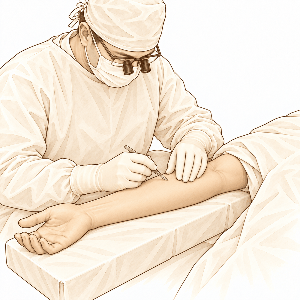
Se trata de una liberación nerviosa en el antebrazo. Su cirujano realiza una incisión en la parte anterior del antebrazo, cerca del codo, para llegar al nervio. La incisión se hace lo más pequeña posible. A través de ella, su cirujano localiza los puntos donde un tejido tenso está comprimiendo el nervio y libera ese tejido, dejando libre el nervio en todo su recorrido. Si una banda de tejido o un vaso sanguíneo anómalo presiona el nervio, también se extirpa.
La incisión se cierra con puntos de sutura y se cubre con un apósito. Se irá a casa con el apósito intacto.
La operación en sí es un trabajo concreto y minucioso en un espacio reducido. Liberar el nervio en el punto donde atraviesa el músculo del antebrazo requiere una disección más profunda que algunas otras liberaciones nerviosas, por lo que su cirujano se toma su tiempo para proteger el nervio y las estructuras que lo rodean.
Después de la operación
Despertará en la sala de recuperación, con las enfermeras vigilándole mientras la anestesia va desapareciendo. Es posible que el brazo se note pesado o entumecido durante un tiempo. Esto es esperable y se resuelve a medida que el medicamento pierde efecto. Recibirá analgésicos para mantenerle cómodo, y la mano y el antebrazo irán envueltos en un vendaje blando. Podrá moverse poco después de despertar, y alguien debe acompañarlo durante las primeras 24 horas. Su equipo le indicará si podrá irse a casa el mismo día o si deberá permanecer una noche en el hospital. Dejamos el vendaje puesto durante unos 10 días; por favor, no lo retire antes de ese plazo a menos que se lo indiquemos. Lo cambiaremos o lo quitaremos cuando venga a la consulta.
Recuperación
Durante los primeros días, la mano y el antebrazo pueden estar doloridos e hinchados. La zona alrededor de la incisión puede doler, y los dedos pueden notarse rígidos. El reposo, mantener la mano elevada sobre una almohada y los analgésicos ayudan a aliviarlo. Las molestias suelen ir disminuyendo con cada día que pasa.
Se irá a casa con un vendaje blando en el brazo. Lo dejamos puesto durante unos 10 días y lo cambiamos o lo quitamos cuando venga a la consulta. Una vez retirado, comienza la terapia de mano. Su terapeuta de mano, Ruby Doolan de Extend Rehabilitation, le guiará en los ejercicios y le confeccionará cualquier férula que necesite. Estos ejercicios mantienen los dedos en movimiento y evitan la rigidez mientras el nervio se calma.
En casa puede hacer la mayoría de las tareas cotidianas ligeras durante las primeras dos semanas aproximadamente, usando la mano según se lo permita la comodidad. Evite levantar peso, agarrar con fuerza o cualquier cosa que fuerce el antebrazo hasta que su terapeuta le dé el visto bueno. Duerma en la postura que le resulte cómoda; algunas personas prefieren apoyar el brazo sobre almohadas a su lado.
La recuperación sigue una pauta más que un calendario fijo. Primero se calman el dolor y la hinchazón. Después vuelven el movimiento y la fuerza de agarre. La fuerza y la sensibilidad de la mano siguen mejorando durante muchos meses, y el resultado final llega cuando el dolor y la fuerza dejan de cambiar. Cada persona se cura a su propio ritmo, así que su cronograma puede ser diferente; su cirujano y su terapeuta le guiarán durante todo el proceso.
Qué puede salir mal
La mayoría de los pacientes evolucionan bien, pero en ocasiones pueden surgir problemas. Su cirujano y el equipo lo vigilarán de cerca para detectar cualquier anomalía a tiempo.
Como esta liberación se realiza en la profundidad del músculo del antebrazo, la operación es más compleja que algunas otras liberaciones nerviosas. Su cirujano pone especial cuidado en proteger el nervio y el tejido que lo rodea.
El nervio que se libera se encuentra cerca de otros nervios que dan sensibilidad y movimiento a la mano. En raras ocasiones, uno de ellos puede irritarse durante la cirugía. Podría notar un entumecimiento, hormigueo o debilidad nuevos en parte de la mano o del antebrazo una vez que el bloqueo nervioso haya perdido efecto, lo que tarda unas 24 horas. Cierto entumecimiento y debilidad durante las primeras 24 horas son esperables por el propio bloqueo. Si dura más allá de ese plazo, llame a la clínica.
La herida también puede dar problemas. Llame a la clínica ese mismo día si tiene fiebre, un enrojecimiento que se extiende desde la incisión, o salida de líquido o pus por ella. Un dolor que sigue empeorando a pesar de sus analgésicos también requiere una llamada ese mismo día.
A veces se forma un bulto firme y sensible cerca de la herida en los días posteriores a la cirugía. Se trata de una acumulación de sangre bajo la piel. Si aumenta de tamaño, duele cada vez más o la piel que lo cubre se ve tensa y brillante, llame a la clínica.
Si la liberación se hace a través de una pequeña incisión mínimamente invasiva en lugar de una abierta, en ocasiones no se mantiene y puede ser necesaria otra operación. Su cirujano hablará de ello con usted al planificar su tratamiento.
La recuperación tras una cirugía de nervio suele ser lenta, y el nervio puede seguir mejorando durante muchos meses. Lo bien que se recupere depende en parte de cuánto tiempo estuvo comprimido antes de la cirugía. Si la presión ha estado presente durante mucho tiempo, es posible que algunos cambios en la mano no se reviertan del todo. Su cirujano hablará con usted sobre lo que cabe esperar en su caso.
En la tabla de complicaciones de esta página se detallan las tasas habituales, por si desea conocer los datos específicos.
¿Cuándo deben llamarnos?
La mayoría de los problemas aparecen en los primeros días. Algunos requieren una llamada rápida y otros, atención urgente.
Acuda al servicio de urgencias si la pantorrilla se le hincha o le duele, o si le falta el aire o tiene dolor en el pecho. Pueden ser signos de un coágulo de sangre. Acuda también si los dedos, la mano o el pulgar se ponen pálidos, fríos, blancos, azulados u oscuros, ya que esto indica un problema de riego sanguíneo.
Llame a la clínica ese mismo día si tiene fiebre, un enrojecimiento que se extiende desde la incisión, o salida de líquido o pus por ella. Un dolor que sigue empeorando a pesar de sus analgésicos también requiere una llamada ese mismo día.
Llame a la clínica si no siente el brazo, la mano o los dedos, o no puede moverlos, una vez que el bloqueo nervioso haya perdido efecto. El bloqueo tarda unas 24 horas en perder efecto, y el entumecimiento y la debilidad durante ese tiempo son esperables. Si dura más allá de ese plazo, llámenos.
Si no puede comunicarse con la clínica fuera del horario de atención o en fin de semana, acuda al servicio de urgencias más cercano.
Evidence & references
This is the clinical evidence summary written for health professionals. It is technical, and it lists the research this page was built from. You do not need to read it to understand your treatment or to make a decision about it.
Overview
- Surgical decompression can be considered for pronator syndrome when activities of daily living require frequent strong pronation and electromyography confirms compression [1].
- Other medical causes should be considered in anterior interosseous nerve syndrome with pure motor loss [1].
- Pronator syndrome can be successfully treated nonoperatively or surgically when the correct diagnosis is made [2].
- Cases of brachial plexus neuritis-induced anterior interosseous nerve palsy should be managed conservatively [3].
- Surgical decompression may be performed for specific instances of direct trauma causing anterior interosseous nerve palsy [3].
- Patients presenting with paresis in incomplete anterior interosseous nerve syndrome should be observed, as most will improve spontaneously without surgery [5].
- Bilateral pronator syndrome can be caused by an anomalous tendinous origin of the ulnar belly of the pronator teres muscle [6].
- Damage to the innervation of the pronator quadratus muscle risks impairment of the initiation of hand pronation [7].
- A patient with a very large neurilemmoma of the anterior interosseous nerve had no complaints and full flexion of the interphalangeal joint of the thumb and distal interphalangeal joint of the index finger one year after surgery [8].
- Supercharged end-to-side anterior interosseous nerve to ulnar motor nerve transfer followed by multimodal hand therapy provides measurable improvements in neurophysiology and function [9].
- Engagement in hand therapy and outcomes for supercharged end-to-side anterior interosseous nerve to ulnar motor nerve transfer appear to be mediated by comorbid physical and psychosocial health [9].
- Median nerve compression neuropathy by the lacertus fibrosus represents a subgroup of median nerve neuropathies where the lacertus fibrosus is the sole cause of compression [10].
- Patients who underwent cubital tunnel release and anterior interosseous nerve supercharge end-to-side transfer together had a significantly smaller cubital tunnel volume and cross-sectional area [11].
- Compression of the median nerve at the elbow is a pathology that is often unrecognized and can be isolated or associated with carpal tunnel syndrome [12].
- A patient with a nerve tumour causing anterior interosseous nerve palsy was able to return to work 3 months after surgery with increased pinch force and active interphalangeal joint motion of the thumb [4].
Anatomy & Pathophysiology
AIN Anatomy and Compression Sites
- Near its site of origin, the anterior interosseous nerve is vulnerable to compression from tendinous attachments or accessory muscles [13].
- Specific structures that can compress the AIN include tendinous origins of the deep head of pronator teres and of flexor digitorum superficialis to the middle finger [13].
- Accessory structures described as causes of AIN compression include the attachment of flexor digitorum superficialis to flexor pollicis longus, an accessory head of the flexor pollicis longus muscle (Gantzer's muscle), a tendinous origin of the muscle variant palmaris profundus, and a double lacertus fibrosis [13].
- Fractures at or around the elbow joint, direct injury to the nerve, and compression from plaster casts are recognized causes of anterior interosseous nerve syndrome [13].
- Vascular causes of anterior interosseous nerve syndrome have been described by Spinner [13].
- The AIN bifurcated at 3 cm (average) distal to the intercondylar line of the humerus in a report by Gunther and DiPasquale [15].
- Constrictions associated with AIN palsy occur in the median nerve trunk of the upper arm, a location where the cause cannot be explained by a mechanical factor alone [15].
- Structural factors, including nerve fascicles, must be considered before the onset of AIN palsy when constrictions occur in the median nerve trunk of the upper arm [15].
- In patients with nontraumatic AIN palsy, the relationship between hemangiomas and intraneural topography matched the AIN in specific cases, suggesting the hemangioma as the cause of the palsy [15].
- Intraepineurial constriction of nerve fascicles can be seen in pronator syndrome, similar to findings in AIN syndrome [15].
Pronator Syndrome Anatomy and Pathology
- The pronator quadratus muscle is of importance for the initiation of hand pronation [7].
- Damage to the innervation of the pronator quadratus muscle risks impairment of hand pronation initiation [7].
- Median nerve compression at the elbow by the lacertus fibrosus is a pathology that can be isolated or associated with carpal tunnel syndrome [12].
- Median nerve compression at the elbow by the lacertus fibrosus is a pathology often unrecognized where loss of muscle strength in the median nerve territory should evoke the diagnosis [17].
- Patients representing a subgroup of median nerve neuropathies may have the lacertus fibrosus as the sole cause of compression [10].
- Ultrasound examination findings may not lead to a diagnosis of pronator syndrome (LS) because median nerve compression in the forearm is often mild and may not produce positive US findings [16].
Clinical Presentation and Diagnostic Features
- Anterior interosseous nerve syndrome is characterized by weakness of pinch between the thumb and index finger [13].
- In AIN syndrome, the interphalangeal joint of the thumb and the distal interphalangeal joint of the index finger are hyperextended in the pinch position [13].
- Weakness of the pronator quadratus muscle can be tested with the forearm flexed to neutralize 75% of pronator teres action [13].
- Patients with AIN syndrome may complain of vague pain in the proximal forearm aggravated by exercise [13].
- Pronator syndrome in violinists and anterior interosseous nerve syndrome with pure motor loss are distinct clinical observations [1].
- If activities of daily living require frequent strong pronation and electromyography confirms compression, surgical decompression can be considered for pronator syndrome [1].
- Other medical causes should be considered in AIN syndrome rather than immediate surgical decompression based solely on activity demands [1].
Elbow Anatomy Context
- The flexor-pronator mass consists of the pronator teres, flexor carpi radialis, palmaris longus, flexor carpi ulnaris, and flexor digitorum superficialis [23].
- The medial epicondyle forms the attachment site for the origins of the flexor pronator mass [22].
- The ligament of Struthers is a variant anatomy arising from the supracondylar process to attach to the medial epicondyle and is a potential site of median nerve compression [23].
- The normal range of forearm pronation and supination is 75° and 85° respectively [19].
- The normal range of forearm pronosupination is 80 to 85 degrees in each direction [28].
- A functional arc for forearm rotation is 50 degrees [28].
Classification
- Pronator syndrome is characterized by pain in the proximal forearm that is aggravated by exercise [13].
- In anterior interosseous nerve syndrome, the interphalangeal joint of the thumb and the distal interphalangeal joint of the index finger are hyperextended during pinch [13].
- Anterior interosseous nerve syndrome can be caused by compression from tendinous attachments or accessory muscles near the site of nerve origin [13].
- Tendinous origins of the deep head of pronator teres and flexor digitorum superficialis to the middle finger are described causes of anterior interosseous nerve compression [13].
- Accessory structures causing anterior interosseous nerve compression include the attachment of flexor digitorum superficialis to flexor pollicis longus [13].
- An accessory head of the flexor pollicis longus muscle (Gantzer's muscle) is a described cause of anterior interosseous nerve compression [13].
- A tendinous origin of the muscle variant palmaris profundus is a described cause of anterior interosseous nerve compression [13].
- A double lacertus fibrosis is a described cause of anterior interosseous nerve compression [13].
- Fractures at or around the elbow joint and direct injury to the nerve are causes of anterior interosseous nerve syndrome [13].
- Compression from plaster casts is a recognized cause of anterior interosseous nerve syndrome [13].
- Vascular causes have been described for anterior interosseous nerve syndrome [13].
- Pronator syndrome can be caused by an anomalous tendinous origin of the ulnar belly of the pronator teres muscle [6].
- Median nerve compression at the elbow by the lacertus fibrosus is a pathology that is often unrecognized [12].
- Median nerve compression at the elbow by the lacertus fibrosus can be isolated or associated with carpal tunnel syndrome [12].
- Patients with median nerve compression at the elbow by the lacertus fibrosus represent a subgroup of median nerve neuropathies where the lacertus fibrosus is the sole cause of compression [10].
- Loss of muscle strength in the median nerve territory should prompt consideration of median nerve compression at the elbow [17].
- Nontraumatic anterior interosseous nerve palsy can be associated with palsy of the pronator teres, flexor carpi radialis, and/or palmaris longus muscles [15].
- Intraepineurial constriction of nerve fascicles can be observed in pronator syndrome [15].
- The cause of hourglass constrictions associated with anterior interosseous nerve palsy in the median nerve trunk of the upper arm cannot be explained by a mechanical factor alone [15].
- Structural factors, including nerve fascicles, must be considered before the onset of anterior interosseous nerve palsy when hourglass constrictions are present in the upper arm median nerve trunk [15].
- The anterior interosseous nerve bifurcates at an average of 3 cm distal to the intercondylar line of the humerus [15].
- Clinical signs and symptoms of spontaneous anterior interosseous nerve palsy with hourglass-like fascicular constriction are similar to those described for isolated neuritis [14].
- Ultrasound examination findings may not lead to a diagnosis of pronator syndrome because median nerve compression in the forearm is often mild [16].
Clinical Presentation
- AIN syndrome is characterized by weakness of pinch between the thumb and index finger [13].
- Patients with AIN syndrome may complain of vague pain in the proximal forearm that is aggravated by exercise [13].
- Pronator syndrome is associated with activities of daily living that require frequent strong pronation [1].
- Electromyography findings are used to confirm the presence of compression in pronator syndrome [1].
- Brachial plexus neuritis-induced anterior interosseous nerve palsy presents with clinical signs and symptoms similar to those described for isolated neuritis [14].
- Nontraumatic AIN palsy may be accompanied by palsy of the pronator teres, flexor carpi radialis, and/or palmaris longus muscles [15].
- Clinical findings in nontraumatic AIN palsy may be unassociated with high median nerve injury [15].
- Ultrasound examination findings for median nerve compression in the forearm may not lead to a diagnosis of pronator syndrome because the compression is often mild [16].
Investigations
Clinical Presentation and Physical Examination
- In anterior interosseous nerve syndrome, the interphalangeal joint of the thumb and the distal interphalangeal joint of the index finger are hyperextended in the pinch position [13].
- Patients with anterior interosseous nerve syndrome may complain of vague pain in the proximal forearm that is aggravated by exercise [13].
- Median nerve compression at the elbow is a pathology that is often unrecognized, where loss of muscle strength in the median nerve territory should evoke the diagnosis [17].
- Median nerve compression at the elbow can be isolated or associated with carpal tunnel syndrome [12].
- Patients with nontraumatic anterior interosseous nerve palsy may present with palsy of the pronator teres, flexor carpi radialis, and/or palmaris longus muscles [15].
Electrodiagnostic Studies
- Electromyography findings can confirm the presence of compression in pronator syndrome [1].
- Electromyography/nerve conduction velocity studies should be performed if any question about neurologic dysfunction exists [36].
Imaging
- Ultrasonographic examination can be challenging for diagnosing median nerve compression in the forearm, as findings may not lead to diagnosis because compression is often mild and may not produce positive US findings [16].
- Plain radiographs remain the hallmark and the best screening test for elbow evaluation [19].
- CT is helpful when assessing for malunion architecture and the location and pattern of osteophytes and/or loose bodies [36].
- MRI can be used to evaluate ligaments and tendons, but it is rarely indicated for elbow stiffness [36].
- MRI may be most helpful in evaluating associated injuries including partial or complete tears of the medial collateral ligament in valgus extension overload syndrome [38].
Treatment
Non-Operative Management
- Patients presenting with paresis of the anterior interosseous nerve should be observed, as most will improve spontaneously without surgery [5].
- When the correct diagnosis is made, pronator syndrome can be successfully treated nonoperatively [2].
Operative Management
- Surgical decompression can be considered for pronator syndrome if activities of daily living require frequent strong pronation and electromyography confirms the presence of compression [1].
- When the correct diagnosis is made, pronator syndrome can be successfully treated surgically if necessary [2].
- One year after surgery for a very large neurilemmoma of the anterior interosseous nerve, the patient had no complaints and full flexion of the interphalangeal joint of the thumb and distal interphalangeal joint of the index finger [8].
- Engagement in hand therapy and outcomes after supercharged end-to-side anterior interosseous nerve to ulnar motor nerve transfer appear to be mediated by comorbid physical and psychosocial health [9].
Anatomical Considerations in Treatment
- Tendinous origins of the deep head of pronator teres and of flexor digitorum superficialis to the middle finger are described as causes of anterior interosseous nerve compression [13].
- Accessory structures described as causes of anterior interosseous nerve compression include the attachment of flexor digitorum superficialis to flexor pollicis longus, an accessory head of the flexor pollicis longus muscle (Gantzer's muscle), a tendinous origin of the muscle variant palmaris profundus, and a double lacertus fibrosis [13].
- Compression from plaster casts has been recognized as a cause of anterior interosseous nerve syndrome [13].
- The median nerve compression neuropathy by the lacertus fibrosus represents a subgroup of median nerve neuropathies where the lacertus fibrosus is the sole cause of compression [10].
Complications
- Surgical decompression of the pronator quadratus muscle risks damage to its innervation, which is important for the initiation of hand pronation [7].
- Compression of the anterior interosseous nerve can result from tendinous attachments or accessory muscles, including the deep head of pronator teres and flexor digitorum superficialis to the middle finger [13].
- Accessory structures that can cause anterior interosseous nerve compression include the attachment of flexor digitorum superficialis to flexor pollicis longus, an accessory head of the flexor pollicis longus muscle (Gantzer's muscle), a tendinous origin of the palmaris profundus muscle variant, and a double lacertus fibrosis [13].
- The cause of hourglass constrictions associated with anterior interosseous nerve palsy in the median nerve trunk of the upper arm cannot be explained by a mechanical factor alone and requires consideration of structural factors including nerve fascicles [15].
Recovery
- Cases of brachial plexus neuritis-induced anterior interosseus nerve palsy should be managed conservatively [3].
- Surgical decompression for pronator syndrome can be considered if activities of daily living require frequent strong pronation and electromyography confirms the presence of compression [1].
- When the correct diagnosis is made, pronator syndrome can be successfully treated nonoperatively or surgically, if necessary [2].
Key Evidence
- [L5] If the activities of daily living of a patient require frequent strong pronation, and if the electromyography findings confirm the presence of compression, surgical decompression can be considered against the background of pronator syndrome, whereas other medical causes should be considered in AIN syndrome. [1] (10.1177/17531934221080018)
- [L5] When the correct diagnosis is made, pronator syndrome can be successfully treated nonoperatively or surgically, if necessary. [2] (10.1016/s0278-5919(05)70267-2)
- [L5] Cases of brachial plexus neuritis-induced anterior interosseus nerve palsy should be managed conservatively, while surgical decompression may be performed for specific instances of direct trauma. [3] (10.1016/s0363-5023(97)80025-1)
- [L5] The patient was able to return to work 3 months after surgery with increased pinch force and active interphalangeal joint motion of the thumb. [4] (10.1177/1753193412454801)
- [L4] Patients presenting with paresis should be observed, as most will improve spontaneously without surgery. [5] (10.1016/s0363-5023(85)80240-9)
- [L5] We report the case of a patient with bilateral pronator syndrome caused by an anomalous tendinous origin of the ulnar belly of the pronator teres muscle. [6] (10.1016/0363-5023(93)90373-b)
- [L5] This risks damage of the innervation of this muscle of importance for initiation of hand pronation. [7] (10.1016/j.jhsb.2005.06.017)
- [L5] One year later, the patient has no complaints and has full flexion of the interphalangeal joint of the thumb and D.I.P. joint of the index finger. [8] (10.1016/0266-7681(91)90141-a)
- [L4] SETS AIN to ulnar motor nerve followed by multimodal hand therapy provides measurable improvements in neurophysiology and function, although engagement in hand therapy and outcomes appear to be mediated by comorbid physical and psychosocial health. [9] (10.1016/j.jht.2020.03.021)
- [L4] The authors conclude that these patients represent a subgroup of median nerve neuropathies where the lacertus fibrosus is the sole cause of compression. [10] (10.1016/s0363-5023(86)80015-6)
- [L4] Patients who underwent CuTR and AIN-SETS together had a significantly smaller cubital tunnel volume (CTV) and cross-sectional area (CSA). [11] (10.1016/j.otsr.2024.103982)
- [L4] La compression du nerf médian au coude est une pathologie souvent méconnue qui peut être isolée ou associée à un syndrome du canal carpien. [12] (10.1016/j.hansur.2018.10.219)
- [L5] [13] (10.1016/s0266-7681(05)80231-1)
- [L4] The clinical signs and symptoms of these patients were similar to those described for isolated neuritis. [14] (10.1016/s0363-5023(96)80114-6)
- [L4] [15] (10.1053/jhsu.2003.50021)
- [L5] US examination can be challenging and its findings may not lead to diagnosis of LS, as median nerve compression in the forearm is often mild and may not produce positive US findings. [16] (10.1177/17531934251370272)
- [L4] La compression du nerf médian au coude est une pathologie souvent méconnue où une perte de la force musculaire dans le territoire du nerf médian doit faire évoquer le diagnostic. [17] (10.1016/j.hansur.2019.10.044)
References
[1] Two clinical observations: pronator syndrome in violinists and anterior interosseous nerve syndrome with pure motor loss. Journal of Hand Surgery (European Volume). 2022. DOI: 10.1177/17531934221080018
[2] PRONATOR SYNDROME. Clinics in Sports Medicine. 2001. DOI: 10.1016/s0278-5919(05)70267-2
[3] Brachial neuritis presenting as anterior interosseous nerve compression—Implications for diagnosis and treatment: A case report. The Journal of Hand Surgery. 1997. DOI: 10.1016/s0363-5023(97)80025-1
[4] Nerve tumour as a rare cause of anterior interosseous nerve palsy. Journal of Hand Surgery (European Volume). 2012. DOI: 10.1177/1753193412454801
[5] The incomplete anterior interosseous nerve syndrome. The Journal of Hand Surgery. 1985. DOI: 10.1016/s0363-5023(85)80240-9
[6] Bilateral pronator syndrome associated with anomalous heads of the pronator teres muscle: A case report. The Journal of Hand Surgery. 1993. DOI: 10.1016/0363-5023(93)90373-b
[7] An Anatomical Study of the Anterior Interosseous Nerve and its Innervation of the Pronator Quadratus Muscle. Journal of Hand Surgery. 2005. DOI: 10.1016/j.jhsb.2005.06.017
[8] A Very Large Neurilemmoma of the Anterior Interosseous Nerve. Journal of Hand Surgery. 1991. DOI: 10.1016/0266-7681(91)90141-a
[9] Postoperative management and rehabilitation after the supercharged end-to-side anterior interosseous nerve to ulnar motor nerve transfer: A report of 3 cases. Journal of Hand Therapy. 2021. DOI: 10.1016/j.jht.2020.03.021
[10] Median nerve compression neuropathy by the lacertus fibrosus: Report of three cases. The Journal of Hand Surgery. 1986. DOI: 10.1016/s0363-5023(86)80015-6
[11] Morphological characteristics of the cubital tunnel as indication for anterior interosseous nerve supercharge end-to-side transfer in treating advanced cubital tunnel syndrome. Orthopaedics & Traumatology: Surgery & Research. 2025. DOI: 10.1016/j.otsr.2024.103982
[12] Diagnostic et traitement chirurgical de la compression du nerf médian au coude par section isolée du lacertus fibrosus. À propos de 13 cas. Hand Surgery and Rehabilitation. 2018. DOI: 10.1016/j.hansur.2018.10.219
[13] An Anomaly of the Median Artery Associated with the Anterior Interosseous Nerve Syndrome. Journal of Hand Surgery. 1992. DOI: 10.1016/s0266-7681(05)80231-1
[14] Spontaneous anterior interosseous nerve palsy with hourglass-like fascicular constriction within the main trunk of the median nerve. The Journal of Hand Surgery. 1996. DOI: 10.1016/s0363-5023(96)80114-6
[15] Fascicular torsion in the median nerve within the distal third of the upper arm: Three cases of nontraumatic anterior interosseous nerve palsy. The Journal of Hand Surgery. 2003. DOI: 10.1053/jhsu.2003.50021
[16] Re: Fang J, Zhang LQ, Tang JB. Incidence of local tenderness at the lacertus fibrosus in healthy people. Journal of Hand Surgery (European Volume). 2025. DOI: 10.1177/17531934251370272
[17] Compression du nerf médian au coude par le lacertus fibrosus : à propos de 34 cas. Hand Surgery and Rehabilitation. 2019. DOI: 10.1016/j.hansur.2019.10.044
[19] Orthopaedic Knowledge Update 13 Ebook Without Multimedia. Anatomy, Biomechanics, Physical Examination, and Imaging of the Elbow > Summary and Conclusions.
[22] Orthopaedic Knowledge Update 13 Ebook Without Multimedia. Anatomy, Biomechanics, Physical Examination, and Imaging of the Elbow > Anatomy > Bony Anatomy.
[23] Miller S Review Of Orthopaedics. SECTION 16 PATELLAR TRACKING IN TOTAL KNEE ARTHROPLASTY > TABLE 2.3 Shoulder Spaces.
[28] Miller S Review Of Orthopaedics. ELBOW.
[36] Aaos Comprehensive Orthopaedic Review 3. Elbow Stiffness* > IV. Evaluation.
[38] Aaos Comprehensive Orthopaedic Review 3. Elbow Injuries in the Athlete* > III. Valgus Extension Overload Syndrome and Posterior Impingement.