Patients › Elbow
Fractura del olécrano
Olecranon fractures — patterns, non-operative care, and tension-band or plate fixation.
Qué está sintiendo
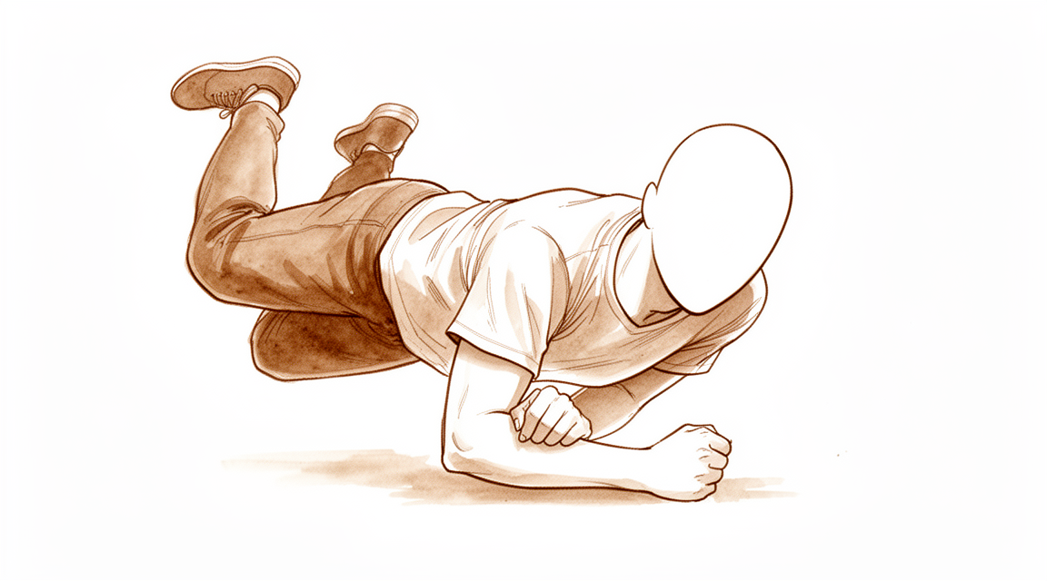
Una fractura del olécrano es una rotura de la punta ósea situada en la parte posterior del codo. Suele producirse de una de dos maneras. En las personas más jóvenes tiende a deberse a una caída fuerte o a un golpe directo en el codo. En las personas mayores suele deberse a una simple caída desde su propia altura. Un estiramiento brusco y forzado del brazo también puede arrancar un fragmento de hueso de la punta del codo.
Normalmente sentirá dolor justo en la parte posterior del codo, y suele doler en un punto concreto más que en toda la zona. La hinchazón sobre la parte posterior del codo es habitual y puede ser bastante marcada. Dado que esa punta ósea se encuentra justo debajo de la piel, a veces se puede palpar el borde de la fractura a través de la piel. Si la piel se abrió en la caída, la herida suele estar en la parte posterior del codo, donde golpeó contra el suelo. Puede resultar difícil usar el brazo, y estirarlo contra algo, como al impulsarse para levantarse de una silla, puede notarse débil o resultar imposible.
En los primeros días, el dolor suele empeorar cuando intenta mover el codo y puede alterarle el sueño. Las tareas cotidianas que doblan el codo o le someten a carga, como levantar una tetera, vestirse o apoyarse en el antebrazo, normalmente le dolerán. La hinchazón y los moratones disminuyen durante las primeras dos semanas, y el dolor se va aliviando poco a poco a medida que el hueso empieza a consolidar.
Algunas situaciones requieren atención urgente. Acuda a un servicio de urgencias ese mismo día si la piel sobre el codo está abierta, si el hueso queda a la vista o si el brazo presenta una deformidad evidente. Lo mismo se aplica si la mano o los dedos se ponen pálidos, fríos, blancos o azulados, o si pierde de repente la sensibilidad o el movimiento del brazo después de la lesión. Si los síntomas no se calman, empeoran a lo largo de semanas o le despiertan por la noche, consulte a su médico de cabecera o solicite una evaluación con un especialista. Si no puede contactar con la clínica, acuda al servicio de urgencias más cercano.
¿Qué ocurre realmente?
La punta ósea de la parte posterior del codo se llama olécrano. Es el extremo superior de uno de los huesos del antebrazo y funciona como el tope de una bisagra. Cuando estira el brazo, ese tope se desliza en un surco situado en el extremo del hueso del brazo. La fractura es importante por lo que tiene unido: el tendón del tríceps, el músculo de la parte posterior del brazo, se ancla justo en ese punto. Ese tendón es lo que le permite estirar el codo contra una carga.
Imagínelo como una puerta con un muelle fuerte que tira de ella para cerrarla. El olécrano es el soporte que sujeta ese muelle. Cuando el hueso se rompe, el muelle sigue tirando y arrastra el fragmento roto hacia arriba y hacia fuera, dejando un hueco en la superficie articular. Por eso el brazo no funciona con normalidad en este momento: el músculo sigue siendo fuerte, pero tira de un fragmento de hueso suelto en lugar de mover el antebrazo. Estirar el brazo contra resistencia se vuelve débil o imposible.
El hueso se cura soldándose de nuevo, y un fragmento desprendido que conserva su tendón unido puede volver a fijarse a medida que el hueso se suelda. Pero los fragmentos deben estar en la posición correcta para que esto funcione bien. Cuando la fractura no se ha desplazado, basta con mantener los fragmentos inmóviles mientras se sueldan. Cuando los fragmentos se han separado, o cuando la superficie articular se ha roto en varios fragmentos, el codo puede volverse inestable, y esas fracturas normalmente requieren cirugía para mantenerlos en su sitio mientras se curan.
Los cirujanos clasifican estas fracturas en grados según cuánto se han desplazado los fragmentos y si la articulación del codo se mantiene estable. Una fractura de grado uno no se ha desplazado. Una de grado dos se ha desplazado, pero la articulación sigue funcionando. Una de grado tres significa que la propia articulación del codo es inestable. Los grados desplazado e inestable son los que con más frecuencia requieren cirugía.
La mayoría de estas fracturas sanan bien, aunque es frecuente una ligera pérdida de la capacidad de estirar el codo por completo.
¿Qué podemos hacer al respecto?
El Dr. Kieran Hirpara, cirujano de extremidades superiores en el Mater Private Hospital Rockhampton, adapta el tratamiento a la lesión específica de cada paciente. Algunas de estas fracturas se curan sin operación y otras requieren cirugía poco después de la lesión, por lo que es importante una evaluación rápida. Por lo general, los pacientes son derivados a nuestra clínica por su médico de cabecera; si un fisioterapeuta le ha recomendado que nos consulte, igualmente necesitará una derivación de su médico de cabecera para poder acceder al reembolso de Medicare. En la clínica, tomamos el historial clínico, examinamos el brazo y solicitamos pruebas de imagen cuando son necesarias.
Si la fractura no se ha desplazado, o solo se ha desplazado un poco, y el codo está estable, podemos recomendar mantenerlo inmóvil mientras se cura. Eso supone llevar una férula o una escayola durante un tiempo, con controles mediante radiografías repetidas para asegurarnos de que los fragmentos permanecen en su sitio. Esta opción es habitual en pacientes mayores y en personas que usan el brazo para tareas cotidianas y no para trabajos pesados. Así se evita una operación y los riesgos que conlleva. La contrapartida es que puede que al final el codo no llegue a estirarse por completo, y a algunas personas les cuesta aceptarlo. La movilidad se recupera por etapas con su terapeuta de mano, que también le fabricará cualquier férula que necesite.
Si los fragmentos se han separado, o la articulación del codo está inestable, normalmente recomendaremos cirugía desde el principio. El objetivo es mantener los fragmentos en la posición correcta para que el hueso pueda soldarse y usted pueda empezar a moverse pronto. La forma de fijar el hueso depende del patrón de la fractura, y ningún método es adecuado para todas las fracturas. A veces ambas opciones son realmente posibles, por ejemplo en un paciente mayor con una fractura desplazada. Hablaremos de lo que es importante para usted: cuánto dolor está dispuesto a soportar y si le molestaría perder los últimos grados de extensión.
Sea cual sea la opción elegida, las primeras semanas son parecidas. El alivio del dolor le mantiene cómodo mientras la hinchazón disminuye. La lesión debe protegerse mientras el hueso se suelda, y le indicaremos lo que puede y lo que no puede hacer con el brazo. Su terapeuta de mano guiará sus movimientos en el momento adecuado, con suavidad al principio y aumentando a medida que el hueso se cura. La mayoría de estas fracturas sanan bien, aunque es frecuente una ligera pérdida de la capacidad de estirar el codo por completo. El material metálico que queda justo debajo de la piel en la parte posterior del codo puede resultar molesto, y algunas personas necesitan más adelante una pequeña operación para retirarlo.
Qué esperar
La mayoría de estas fracturas sanan bien. Es frecuente una ligera pérdida de la capacidad de estirar el codo por completo, y la mayoría de las personas conservan un buen uso del brazo. Los resultados a largo plazo tras la cirugía de fracturas complejas son buenos, incluso cuando en radiografías posteriores aparece cierta artrosis por desgaste en la articulación.
Si su fractura se mantiene inmóvil con una férula o una escayola, el hueso se suelda a lo largo de semanas, no de días. Se le harán radiografías repetidas para asegurarnos de que los fragmentos permanecen donde deben. Esta opción es adecuada para pacientes mayores y para personas que usan el brazo para tareas cotidianas y no para trabajos pesados. En ese grupo, los resultados son satisfactorios tanto a corto como a largo plazo. La contrapartida es que puede que al final el codo no llegue a estirarse por completo.
Si se somete a cirugía, el hueso se mantiene en su sitio con material metálico mientras se suelda. El movimiento empieza pronto y avanza por etapas con su terapeuta de mano. Tras esta lesión es de esperar cierta pérdida de la capacidad de estirar el codo por completo, sea cual sea la opción elegida. En un seguimiento prolongado de personas operadas por una fractura desplazada, el 96 % tuvo resultados buenos o excelentes hasta 25 años después de la lesión.
La recuperación es un regreso gradual a sus actividades habituales. Primero vuelven las tareas diarias ligeras y, después, levantar peso y un uso más exigente a medida que el hueso se fortalece. Cuándo podrá conducir, trabajar o hacer deporte depende de su trabajo, de su brazo y de cómo se cure el hueso, y le explicaremos cada etapa.
Conviene conocer las posibles complicaciones. El material metálico queda justo debajo de la piel en la parte posterior del codo y puede resultar tan molesto que algunas personas deciden que se les retire más adelante. Es un problema recurrente tras esta cirugía. Otros problemas, como que el hueso consolide en una mala posición o que el codo quede rígido, son menos frecuentes. Si sus síntomas no se calman, empeoran a lo largo de semanas o le despiertan por la noche, consulte a su médico de cabecera o solicite una evaluación con un especialista. Si no puede contactar con la clínica, acuda al servicio de urgencias más cercano.
¿Cuándo consultar a un médico?
Busque atención urgente si el codo presenta una deformidad evidente, si la piel sobre la lesión está abierta o si la mano o los dedos se ponen pálidos, fríos, blancos o azulados. La pérdida repentina de sensibilidad o de movimiento del brazo después de la lesión también requiere acudir a un servicio de urgencias ese mismo día. Estos signos significan que la lesión debe revisarse de inmediato.
Para todo lo demás, empiece por su médico de cabecera. Puede examinarle el brazo y solicitar radiografías, que normalmente son todo lo que se necesita para ver este tipo de fractura. Si su médico de cabecera considera que debe hacerse cargo un especialista, le derivará para una evaluación.
Vuelva a consultar, o solicite una evaluación con un especialista, si el dolor no se calma o si la hinchazón, el movimiento o el uso del brazo no mejoran semana a semana a medida que el hueso se cura. Los síntomas que empeoran a lo largo de semanas, le despiertan por la noche o le impiden trabajar o usar el brazo también merecen una nueva revisión. Si no puede contactar con la clínica, acuda al servicio de urgencias más cercano.
En mayor profundidad
Advanced reading: the deeper science (optional)
Esta sección profundiza más de lo necesario para que usted tome decisiones sobre su propio tratamiento. La fractura del olécrano merece una lectura adicional debido a un hallazgo que invierte la lógica habitual en la cirugía de fracturas: en pacientes de edad avanzada, una fractura desplazada del olécrano puede dejarse sin fijar y, aun así, el codo funciona correctamente.
La fractura desplazada que no requiere corrección
Una fractura en el olécrano separa el tríceps del antebrazo; por ello, la lógica habitual indica que debe reinsertarse para que el codo pueda estirarse contra resistencia. Sin embargo, en pacientes mayores de 70 años esta lógica no se aplica. Una revisión sistemática demostró que las fracturas desplazadas del olécrano en este grupo pueden tratarse eficazmente sin cirugía, logrando alta satisfacción y un rango de movimiento funcional [1].
Lo que se “paga” como costo es medible: una pérdida de extensión, la imposibilidad de estirar completamente el codo y una disminución de la fuerza en dicha extensión. En cambio, lo que se evita es una herida sobre un hueso subcutáneo en piel delgada y frágil, así como el uso de material metálico bajo dicha piel. Dado que la presencia de implantes es la causa más frecuente de una segunda intervención tras la fijación del olécrano, evitarlos elimina por completo la complicación más habitual.
Para una persona mayor cuyo brazo se utiliza para tareas cotidianas y no para empujes fuertes, un codo que no alcanza los últimos grados de extensión supone una molestia menor que cualquier complicación derivada de una herida. Para un paciente más joven o con mayores exigencias funcionales, no es así; por eso este hallazgo es específico de la edad y no de carácter general.
En los casos en que se emplea la fijación, la elección del implante varía
Para el patrón común de fracturas desplazadas pero sencillas, compiten dos tipos de implantes: una estructura de banda de tensión hecha con alambres y una placa. En 827 pacientes con fracturas de tipo II según la clasificación de Mayo, la fijación con placa demostró mayor eficacia y seguridad que el uso de bandas de tensión con alambres; los autores señalan, no obstante, que se incluyeron pocos estudios y que aún se requieren ensayos aleatorizados de mayor calidad [2]. Una revisión de 2025 realizada con 472 pacientes, que compara técnicas modernas de fijación con el método tradicional de bandas de tensión, llega a la misma conclusión [3].
Este es uno de los pocos casos en este sitio donde la comparación entre técnicas arroja resultados claros en lugar de dejar la cuestión en el aire. No obstante, conviene tomar estos hallazgos con cautela, dado que los propios autores advierten sobre la limitada base de evidencia disponible.
Se desarrolla artritis, pero en la mayoría de los casos no supone un problema
Los hallazgos imagenológicos a largo plazo pueden resultar alarmantes si se comunican sin contexto. Tras fracturas aisladas del olécrano, la incidencia media de artrosis postraumática fue del 19 % con un seguimiento medio de 41 meses, y los resultados finales reportados por los pacientes variaron entre buenos y excelentes, independientemente del tipo de fractura o de la presencia de artritis [4].
Ambas partes de esa oración son esenciales. La aparición de artritis en radiografías de seguimiento tras esta lesión es frecuente; sin embargo, en este período de tiempo, dicha artritis apenas influye en la percepción que los pacientes tienen de la funcionalidad de su codo. Un hallazgo radiológico no equivale automáticamente a un problema clínico, y por sí solo no debería ser motivo para iniciar intervenciones adicionales.
Referencias
[1] Alvara CA, Biedron G, Dunn JC. Tratamiento no quirúrgico de las fracturas del olécrano en pacientes ancianos: una revisión sistemática. Hand (N Y). 2020;17(4):734-9. https://doi.org/10.1177/1558944720944261
[2] Jia Y, Liu A, Guo T, Chen J, Yu W, Zhai J. Eficacia y seguridad del alambre de banda de tensión frente a la placa en fracturas del olécrano de tipo Mayo II: una revisión sistemática y metaanálisis. J Orthop Surg Res. 2022;17(1). https://doi.org/10.1186/s13018-022-03262-7
[3] Wang C, Li C. Técnicas modernas de fijación frente al método tradicional de alambre de banda de tensión para fracturas del olécrano: una revisión sistemática y metaanálisis. J Orthop Surg Res. 2025;20(1). https://doi.org/10.1186/s13018-025-06061-y
[4] Wiersma JP, de Klerk HH, Priester-Vink S, Doornberg JN, Bhasyam AR, van den Bekerom MP. Incidencia de artrosis postraumática en fracturas del olécrano y el papel del tipo de fractura: una revisión sistemática. J Shoulder Elbow Surg. 2026. https://doi.org/10.1016/j.jse.2026.02.024
Evidence & references
This is the clinical evidence summary written for health professionals. It is technical, and it lists the research this page was built from. You do not need to read it to understand your treatment or to make a decision about it.
Overview
General Outcomes and Complications
- A majority of olecranon fractures heal uneventfully with good or excellent results, with a small loss of motion expected [11].
- 96% of individuals with a closed displaced fracture of the olecranon treated by open reduction and internal fixation had excellent or good outcomes up to 25 years after the fracture [4].
- Surgical treatment of olecranon fractures is associated with a high rate of complications [18].
- Patients undergoing revisions beyond implant removal after olecranon fracture fixation had poorer functional outcomes [18].
- No significant differences in functional outcomes or secondary operations were found with respect to fracture type, gender, or surgical method for olecranon fractures [18].
- The incidence of post-traumatic osteoarthritis following isolated olecranon fractures is 19% at a median follow-up of 41 months [22].
Operative Management
- No single technique is suitable for the management of all olecranon fractures [21].
- Low-profile double-plate osteosynthesis is a safe and effective alternative treatment for olecranon fractures with excellent subjective and objective clinical outcome measures [1].
- Plating of the olecranon leads to predictable union, though the most common complication was lack of full extension in 39% of patients [16].
- Both Kirschner wire tension band and anatomical locking plate procedures effectively treat Mayo type II olecranon fractures [9].
- Current randomized evidence suggests that tension band wiring and plate fixation may yield similar functional outcomes for displaced olecranon fractures, based on a limited number of randomized controlled trials [24].
- Tension band wiring remains an effective treatment for appropriately selected olecranon fractures and outperformed plate osteosynthesis in one cohort [40].
- The timing of fixation of displaced olecranon fractures does not significantly increase the rate of early complications or reoperation [15].
- Excision of the olecranon fragment with repair of the triceps mechanism is supported as a preferred method of treatment due to significantly less morbidity compared with internal fixation [65].
Non-Operative Management
- Close radiographic follow-up is recommended for nonsurgically treated olecranon fractures in children [2].
- Some isolated displaced (>2 mm) olecranon fractures in elderly patients can be successfully treated nonoperatively [6].
- Satisfactory short-term and long-term outcomes were found following the nonoperative management of isolated displaced olecranon fractures in older, lower-demand patients [13].
- Displaced olecranon fractures in patients older than 70 years may be effectively managed with nonoperative measures to produce high satisfaction and functional range of motion [23].
- Nonoperative management of olecranon fractures in elderly patients offers comparable functional outcomes to surgical intervention while significantly reducing exposure to potential complications and healthcare costs [41].
- Nonoperative treatment is a reasonable option for displaced stable olecranon fractures in elderly patients [29].
Elderly Population Specifics
- Olecranon fractures in the elderly have higher than expected 1-year mortality rates [5].
- Patients with olecranon fractures have essentially similar demographic characteristics compared to patients with distal radius fractures [10].
- The literature on the treatment of olecranon fractures in elderly patients is limited [7].
Anatomy & Pathophysiology
Bony Anatomy
- The olecranon and the coronoid process form the greater sigmoid notch, which articulates with the trochlea of the distal humerus [37].
- The intrinsic anatomy of the ulnohumeral articulation allows flexion/extension movement of the elbow joint and provides stability for the elbow [37].
- The olecranon serves as the insertion for the triceps tendon, which blends with the periosteum of the proximal ulna [37].
- The exposed, subcutaneous position of the olecranon renders it vulnerable to direct trauma and violent muscular contractions from the triceps [37].
- In children, the olecranon is predominantly cartilage, particularly in younger children, which reduces the chance of fracture occurring with a direct blow [48].
- In children, the thick periosteum and relatively thin metaphyseal cortex of the olecranon predispose it to minimally displaced greenstick fractures [48].
- Radiographic measurements of normal elbows guide the surgeon to obtain a more reliable length of the olecranon and maintain articular geometry during fixation [69].
Mechanisms of Injury
- Olecranon fractures can result from a direct blow, a fall on an outstretched hand with the elbow in flexion, or high-energy trauma associated with radial head fractures or elbow dislocation [37].
- Sudden and violent triceps muscle contraction can produce an avulsion fracture of varying size of the olecranon tip [37].
- A bimodal distribution of olecranon fractures is seen in young patients with high-energy trauma and elderly patients with low-energy trauma such as a fall from standing [37].
- Olecranon fractures typically occur as low-energy falls from standing in older patients or high-energy injury mechanisms in younger patients [38].
- The bone typically fails from a direct blow to the elbow or as a tensile force from the pull of the triceps tendon [38].
- Regardless of the mechanism of injury, the triceps acts as the main deforming mechanism pulling the fractured fragment proximally, creating a gap at the articular surface, and an incompetent extensor mechanism [38].
- In children, olecranon fractures are usually the result of a hyperextension injury [48].
- In children, flexion injuries are usually caused by a fall on an outstretched hand with the elbow flexed, resulting from a strong eccentric contraction of the triceps pulling the olecranon over the fulcrum of the distal humerus [48].
- In children, shear injuries result from a force to the proximal ulna just anterior to the humeral condyles, causing the olecranon to fracture through metaphyseal bone with anterior displacement of the distal fragment [48].
- Olecranon fractures commonly occur with a direct blow, generally resulting in comminuted fractures, or as an avulsion injury with triceps contracture [54].
- Contraction of the triceps often results in transverse or short oblique types of olecranon fractures [54].
- In children, hyperextension injuries are frequently associated with other elbow injuries, where the direction of the associated coronal plane force determines the corresponding injuries [48].
- In children, a valgus hyperextension force may produce an associated radial neck or medial epicondyle fracture [48].
- In children, a varus hyperextension injury may be associated with lateral dislocation of the radial head or a Bado type III Monteggia lesion [48].
- In children, flexion injuries are generally transverse (perpendicular to the axis of the ulna), displaced posteriorly rather than anteriorly, and rarely associated with other injuries [48].
Associated Injuries
- Olecranon fractures typically occur in isolation; however, given the subcutaneous location of the injury, there is a risk to soft tissues as well as risk of the fracture being open [38].
- In an epidemiologic study of olecranon fractures, 22% of patients had injuries to the ipsilateral limb and 6.4% of fractures were open [38].
- In children, olecranon fractures are associated with other elbow injuries (usually the medial epicondyle) in 20% to 50% of cases [48].
Classification
- The Mayo classification is the most common classification used for olecranon fractures, based on the amount of fracture displacement, the presence of comminution, and stability of the ulnohumeral joint [38].
- In the Mayo classification, Type I fractures are nondisplaced, Type II fractures are displaced, and Type III fractures have an unstable ulnohumeral joint [38].
- In the Mayo classification, modifier A indicates simple (noncomminuted) fractures and modifier B indicates comminuted fractures [38].
- The Mayo classification is simple to use, can help guide fracture management, and has good interobserver reliability [38].
- Displaced olecranon fracture lines entered along the medial side of the trochlear notch and exited at the base of the coronoid, while minimally displaced fractures entered and exited the trochlear notch at the base of the coronoid [38].
- The Schatzker and Colton classification attempted to classify olecranon fractures based on different fracture pattern morphology; however, its application was limited [38].
- A proposed fragment-specific classification system for complex olecranon fractures is anatomically based and considers the deforming forces from ligaments and tendons [64].
- In a study of 2462 fractures, the distribution included a larger share of comminuted central fractures (29%) compared to a single-center report from Scotland [8].
- In a study of 2462 fractures, an almost threefold larger share of distal olecranon fractures was observed compared to the 6.2% reported in a Scottish study of 64 olecranon fractures [8].
- In a study of 2462 fractures, the distribution differed from a single-center report from Scotland where 74% of the fractures were simple central fractures and 81% were central fractures [8].
Classification
Classification Systems and Reproducibility
- The Mayo classification is the most commonly used classification system in clinical practice for olecranon fractures [43].
- The Mayo classification consists of 3 types with a modifier to indicate comminution [43].
- Type I fractures in the Mayo classification are nondisplaced [43].
- Type II fractures in the Mayo classification are displaced [43].
- Type III fractures in the Mayo classification have accompanying injuries [43].
- The Mayo classification was designed to simplify categorization of olecranon fractures but does not achieve this goal due to poor reproducibility [27].
- Multiple classification systems for olecranon fractures exist, including AO, Mayo, Schatzker and Colton, but none are widely accepted or provide direct and reliable advice on operative strategies [88].
- The low reproducibility rates of existing olecranon fracture classification systems raise questions about their use in clinical and research contexts [88].
- The Colton classification had the best intra- and inter-observer agreement among the Mayo, AO/OTA, and Colton & Schatzker classifications [111].
- The AO classification subsumes olecranon fractures to proximal forearm injuries [88].
- Schatzker and Colton divide olecranon fractures into groups by quantity of fragments and fracture lines [88].
Fracture Morphology and Distribution
- Quantitative 3-dimensional computed tomography analysis further clarified the fracture morphology of Mayo type I, II, and III fractures [62].
- In a study of 2462 fractures from the Swedish Fracture Register, 29% of olecranon fractures were comminuted central fractures [8].
- The share of distal olecranon fractures in the Swedish Fracture Register was almost threefold larger than the 6.2% reported in a Scottish study of 64 olecranon fractures [8].
- In a study of 64 olecranon fractures, the most frequent injury according to the AO fracture classification was the 21-B1.1 type [94].
- In a study of 64 olecranon fractures, a simple isolated displaced olecranon fracture (Mayo type 2A) was the most common according to the Mayo classification [94].
- In a study of 44 patients treated with tension band wiring, Type IIA fractures were the most common pattern, accounting for 91% of cases [107].
- In a study of 57 patients treated with a shape-memory alloy concentrator, the Mayo classification system was used to distinguish fracture displacement, presence of comminution, and ulnohumeral stability [60].
- Mayo type III fractures are categorized into anterior and posterior olecranon fracture-dislocations based on the radio-ulnar relationship [106].
- Articular impaction is a common feature of geriatric olecranon fractures [32].
Pediatric and Adolescent Considerations
- Fracture of the olecranon in a child is often only part of a complex injury to the bone and soft tissues around the elbow [30].
- The olecranon apophyseal ossification system, when combined with age and sex, successfully predicts peak height velocity within a year in 90% of cases [93].
Clinical Presentation
Mechanisms and Epidemiology
- The triceps acts as the main deforming mechanism pulling the fractured fragment proximally, creating a gap at the articular surface, and an incompetent extensor mechanism [38].
- The incidence of olecranon fractures increased by 29% over a 20-year study period in Denmark [12].
- In a Swedish study of 2462 fractures, 29% were comminuted central fractures and the share of distal olecranon fractures was almost threefold larger than in a Scottish study [8].
- In a Swedish study, high-energy injury mechanisms accounted for 5.7% of female and 11.6% of male patients overall, while low-energy mechanisms accounted for 87.4% of female and 77.8% of male patients overall [8].
- Fractures of the ipsilateral olecranon associated with the radial neck are not as rare as previously reported [42].
Physical Examination
- Pain is usually localized to the posterior part of the elbow [37].
- Given the subcutaneous location of the olecranon, the fracture itself may be palpable [37].
- Extensive posterior swelling is typical [37].
- A careful examination of the integrity of the extensor mechanism with gravity eliminated can aid surgical decision making [37].
- If present, open wounds are typically posterior and result from the direct impact of the posterior surface of the elbow against an unyielding structure [37].
- The affected extremity from shoulder to hand should be thoroughly examined for any soft tissue compromise [38].
- There is a low suspicion for open fractures given the subcutaneous nature of the olecranon [38].
- The elbow will often present with a large fluid collection subcutaneously over the olecranon [38].
- Examination of the extensor mechanism both with and without gravity eliminated should be performed to assess competency [38].
Imaging
- Plain radiographs are usually sufficient for isolated fractures of the olecranon [37].
- A true lateral radiograph is necessary to accurately identify the plane of the fracture and the number of fracture fragments [37].
- The examiner should assess for fracture comminution and impaction on radiographs [37].
- In more complex cases, CT may help delineate the comminution or impaction better, but this is not routinely required [37].
- Anteroposterior and lateral radiographs of the elbow are required for olecranon fractures [38].
- If there is concern for a radial head fracture, a radiocapitellar view of the elbow can be obtained [38].
- In the setting of an elbow dislocation, post-reduction radiographs should be obtained [38].
- Computed tomography (CT) can be used for preoperative planning for comminuted fractures of the olecranon if there is an associated radial head or coronoid fracture, but this is not routinely utilized [38].
Classification
- The Mayo classification is based on the amount of fracture displacement, the presence of comminution, and stability of the ulnohumeral joint [38].
- The modifier A (simple) and B (comminuted) in the Mayo classification indicate the presence of comminution [38].
- The Schatzker and Colton classification attempted to classify olecranon fractures based on different fracture pattern morphology, but its application was limited [38].
Prognosis and Outcomes
- A majority of olecranon fractures heal uneventfully with good or excellent results with a small loss of motion to be expected [11].
- Primary elbow instability and fracture morphology are prognostic factors for elbow function and development of arthrosis after operative treatment of olecranon fractures [45].
- The median incidence of post-traumatic osteoarthritis following isolated olecranon fractures is 19% at a median follow-up of 41 months [22].
- Patients aged 50 years or more with olecranon fracture should be evaluated for osteoporosis and fracture risks for secondary prevention of subsequent fractures [36].
Investigations
Clinical Examination
- The history may help distinguish a triceps avulsion from an actual direct blow to the elbow [37].
- Extensive posterior swelling is typical of olecranon fractures [37].
- Open wounds, if present, are typically posterior and result from the direct impact of the posterior surface of the elbow against an unyielding structure [37].
- The affected extremity from shoulder to hand should be thoroughly examined to identify associated ipsilateral injuries [38].
- The arm should be examined for soft tissue compromise, with a low suspicion for open fractures maintained given the subcutaneous nature of the olecranon [38].
Imaging
- In more complex cases, CT may help delineate comminution or impaction better, though this is not routinely required [37].
- Quantitative analysis of olecranon fractures via CT further clarified fracture morphology of Mayo type I, II, and III fractures [62].
Classification
- The distribution of olecranon fractures in a Swedish register study showed a larger share of comminuted central fractures (29%) compared to a single-center report from Scotland [8].
- The Swedish register study observed an almost threefold larger share of distal olecranon fractures than the 6.2% reported in a Scottish study of 64 olecranon fractures [8].
Pediatric Specifics
- Olecranon fractures are associated with other elbow injuries (usually the medial epicondyle) in 20% to 50% of pediatric cases [48].
- In younger children, the olecranon is predominantly cartilage, which reduces the chance of a fracture occurring with a direct blow [48].
- The thick periosteum and relatively thin metaphyseal cortex of the pediatric olecranon predispose it to minimally displaced greenstick fractures [48].
Treatment
General Principles and Indications
- Conventional indications for surgical treatment of olecranon fractures include open fractures, displaced fractures, and fractures causing disruption to the extensor mechanism affecting the insertion of the triceps muscle into the olecranon [28].
- The aims of treatment for all olecranon fractures are restoration of function and stability to the elbow joint to allow early mobilization, while minimizing associated complications [113].
- Fixation must be secure enough to permit early motion to avoid significant stiffness of the elbow joint [20].
- Good results overall are to be expected after surgical management of olecranon fractures [43].
Non-Operative Management
- Mayo type I stable undisplaced fractures can be managed effectively with nonoperative management [113].
- Some authors suggest splinting the elbow in 45° to 90° of flexion for three to four weeks followed by supervised mobilization for stable undisplaced fractures [113].
- Early active mobilization is recommended where pain allows, and time in a cast should be minimized to reduce stiffness [113].
- The acceptable degree of fracture displacement for nonoperative management is commonly quoted as < 2 mm of articular displacement on the lateral radiograph [113].
- Aggregate data support the non-operative treatment of isolated undisplaced olecranon fractures in children with good results [33].
- Nonoperative management of isolated displaced olecranon fractures in older, lower-demand patients yields satisfactory short-term and long-term outcomes [13].
Operative Management: Techniques and Outcomes
- There are four common methods in the surgical management of olecranon fractures: tension band wiring, plate fixation, intramedullary fixation, and excision of the proximal fractured fragment with re-attachment of triceps tendon [28].
- Both operative procedures (Kirschner wire tension band and anatomical locking plate) effectively treat Mayo type II olecranon fractures [9].
- Current randomized evidence suggests that tension band wiring (TBW) and plate fixation (PF) may yield similar functional outcomes for displaced olecranon fractures, based on a limited number of RCTs [24].
- Among active patients with a simple isolated, displaced fracture of the olecranon, no difference was found between TBW and plate fixation in patient-reported outcome at 1 year following surgery [35].
- Plate fixation has better efficacy and safety for Mayo II olecranon fractures compared to tension band wiring [51].
- TBW remains an effective treatment for appropriately selected olecranon fractures and outperformed plate osteosynthesis in one cohort [40].
- Internal fixation by cable pin system (CPS) is associated with a shorter healing time, fewer complications, and better function than TBW [34].
- Low-profile double-plate osteosynthesis is a safe and effective alternative treatment of olecranon fractures with excellent subjective and objective clinical outcome measures [1].
- Low-profile double-plate osteosynthesis for treating olecranon fractures resulted in good clinical outcomes [85].
- Suture tension band fixation reduces hardware complications in olecranon fractures [3].
- In all patients treated with percutaneously placed K-wires and absorbable tension-band sutures for displaced olecranon fractures, fracture united within 6 weeks and full painless range of movement was regained within 6 months [47].
- Fractures through an ununited olecranon apophysis are successfully treated with plate and screw fixation with bone grafting [71].
- The Nickel-Titanium olecranon memory connector (OMC) can be an effective alternative to treat olecranon fractures [77].
- The study supports the rationale for excision of the fragment of the olecranon with repair of the triceps mechanism as the preferred method of treatment due to significantly less morbidity compared with internal fixation [65].
- Functional results after at least 2 years were equally good with tension band and screwing techniques, but there was a considerably higher rate of separation at the fracture after screwing than after inserting a tension band [46].
Complications and Risk Factors
- Patients undergoing revisions beyond implant removal had poorer functional outcomes after olecranon fracture fixation [18].
- No significant differences in functional outcomes or secondary operations were found with respect to fracture type, gender, or surgical method for olecranon fracture fixation [18].
- Patients who have operative fixation of a fracture of the olecranon can be counseled that most patients keep their implants, that only 3% experience implant migration, and that technical factors such as the type or configuration of an implant seem less important than personal factors in determining who requests a second surgery for implant removal [25].
- Risk factors associated with a poorer outcome following operative treatment of displaced olecranon fractures are patient factors, fracture morphology, and associated elbow instability or fractures [113].
Complications
General Outcomes and Mortality
- ORIF for olecranon fractures demonstrates low short-term complication rates [101].
- Specific comorbidities such as smoking and ascites were associated with increased risk of short-term complications following ORIF for olecranon fractures [101].
Hardware and Fixation Complications
- Symptomatic hardware requiring removal is a recurring and frequent problem in olecranon fracture management [43].
- The most common complication following plating of the olecranon was lack of full extension in 39% of patients [16].
- Only 3% of patients who undergo operative fixation of an olecranon fracture experience implant migration [25].
- Technical factors such as the type or configuration of an implant seem less important than personal factors in determining who requests a second surgery for implant removal [25].
- Internal fixation by cable pin system (CPS) is associated with fewer complications than tension band wiring (TBW) for olecranon fractures [34].
- Suture tension band fixation reduces hardware complications in olecranon fractures compared to traditional techniques [3].
Long-Term and Specific Complications
- Articular impaction in olecranon fractures is associated with worse postoperative outcomes [67].
- Inadequate reduction of articular impaction contributes to increased step-off and poorer function in olecranon fractures [67].
Recovery
General Outcomes
- Articular impaction in olecranon fractures is associated with worse postoperative outcomes, with inadequate impaction reduction contributing to increased step-off and poorer function [67].
Operative Recovery
- Surgical treatment of olecranon fractures is associated with a high rate of complications, and patients undergoing revisions beyond implant removal had poorer functional outcomes [18].
- No significant differences in functional outcomes or secondary operations were found with respect to fracture type, gender, or surgical method following open reduction and fixation of olecranon fractures [18].
- Both tension band wiring and plate fixation yield excellent or good clinical outcomes with minimal loss of physical capacity, little pain, and disability in the majority of patients with simple and comminuted displaced olecranon fractures [68].
- Among active patients with a simple isolated, displaced fracture of the olecranon, no difference was found between tension band wiring and plate fixation in the patient-reported outcome at 1 year following surgery [35].
- Internal fixation by cable pin system is an effective method for olecranon fracture and is associated with a shorter healing time, fewer complications, and better function than tension band wiring [34].
- Tension band wiring remains an effective treatment for appropriately selected olecranon fractures and in one cohort outperformed plate osteosynthesis [40].
- Both Kirschner wire tension band combined with anatomical locking plate and standard operative procedures effectively treat Mayo type II olecranon fractures [9].
Non-Operative Recovery
- Close radiographic follow-up for nonsurgically treated olecranon fractures is recommended due to the risk of further displacement after initial nonsurgical treatment in children [2].
- All 10 cases of olecranon fractures in children treated with a sling were free of symptoms and had a full range of elbow movement at 3 weeks [17].
Elderly-Specific Outcomes
Key Evidence
- [L3] Low-profile double-plate osteosynthesis is a safe and effective alternative treatment of olecranon fractures with excellent subjective and objective clinical outcome measures. [1] (10.1016/j.otsr.2019.08.019)
- [L4] Close radiographic follow-up for nonsurgically treated olecranon fractures is recommended. [2] (10.5435/jaaos-d-25-00821)
- [L3] Future studies should focus on the long-term outcomes of this technique, as compared to traditional and more established techniques to treat olecranon fractures. [3] (10.1016/j.jseint.2026.101734)
- [L3] 96% of individuals with a closed displaced fracture of the olecranon treated by open reduction and internal fixation had excellent or good outcomes up to 25 years after the fracture. [4] (10.1067/mse.2002.124548)
- [L3] Olecranon fractures in the elderly have higher than expected 1 year mortality rates. [5] (10.1177/1758573221994860)
- [L4] Some isolated displaced (>2 mm) olecranon fractures in elderly patients can be successfully treated nonoperatively. [6] (10.1016/j.ocl.2016.08.011)
- [L4] The literature on the treatment of olecranon fractures in elderly patients is limited. [7] (10.1007/s11678-018-0488-7)
- [L4] [8] (10.1007/s00068-021-01765-2)
- [L3] Both operative procedures effectively treat Mayo type II olecranon fractures. [9] (10.1186/s12891-025-08843-1)
- [L3] Patients with olecranon fractures have essentially similar demographic characteristics compared to patients with distal radius fractures. [10] (10.1177/17585732221124301)
- [L4] A majority of olecranon fractures heal uneventfully with good/excellent results with a small loss of motion to be expected. [11] (10.1016/j.hcl.2015.07.003)
- [L3] The incidence of olecranon fractures increased by 29% over the 20-year study period. [12] (10.1186/s13018-025-05970-2)
- [L4] We found satisfactory short-term and long-term outcomes following the nonoperative management of isolated displaced olecranon fractures in older, lower-demand patients. [13] (10.2106/jbjs.l.01137)
- [L3] The timing of fixation of displaced olecranon fractures does not significantly increase the rate of early complications or reoperation. [15] (10.1016/j.jhsg.2023.09.002)
- [L3] Plating of the olecranon leads to predictable union, though the most common complication was lack of full extension in 39% of patients. [16] (10.1016/j.injury.2016.04.015)
- [L4] All 10 cases treated with a sling were free of symptoms and had a full range of elbow movement at 3 weeks. [17] (10.1016/0020-1383(75)90056-x)
- [L4] Surgical treatment of olecranon fractures is associated with a high rate of complications, and patients undergoing revisions beyond implant removal had poorer functional outcomes; however, no significant differences in functional outcomes or secondary operations were found with respect to fracture type, gender, or surgical method. [18] (10.1016/j.xrrt.2025.08.004)
- [L5] Fixation must be secure enough to permit early motion to avoid significant stiffness of the elbow joint. [20] (10.5435/00124635-200007000-00007)
- [Paper] No one technique is suitable for the management of all olecranon fractures. [21] (10.1016/j.injury.2008.12.013)
- [L4] This review identified a median OA incidence of 19% at a median follow-up of 41 months following isolated olecranon fractures. [22] (10.1016/j.jse.2026.02.024)
- [L4] Displaced olecranon fractures in patients older than 70 years may be effectively managed with nonoperative measures to produce high satisfaction and functional range of motion. [23] (10.1177/1558944720944261)
- [L1] Current randomized evidence suggests that TBW and PF may yield similar functional outcomes for displaced olecranon fractures; however, these findings are based on a limited number of RCTs, with several secondary outcomes derived from only 2 studies. [24] (10.1016/j.xrrt.2026.100817)
- [L3] Patients who have operative fixation of a fracture of the olecranon can be counseled that most patients keep their implants, that only 3% experience implant migration, and that technical factors such as the type or configuration of an implant seem less important than personal factors in determining who requests a second surgery for implant removal. [25] (10.1007/s11999-015-4488-2)
- [L5] The Mayo classification was designed to simplify categorization of olecranon fractures but does not achieve this goal due to poor reproducibility. [27] (10.1097/corr.0000000000000614)
- [L1] [28] (10.1002/14651858.cd010144.pub2)
- [L1] This supports nonoperative treatment as a reasonable option for displaced stable olecranon fractures in elderly patients. [29] (10.2106/jbjs.24.00655)
- [L4] Fracture of the olecranon in a child is often only part of a complex injury to the bone and soft tissues around the elbow. [30] (10.1016/0020-1383(80)90009-1)
- [L4] Articular impaction is a common feature of geriatric olecranon fractures. [32] (10.5435/jaaos-d-20-01293)
- [L4] Aggregate data support the non-operative treatment of isolated undisplaced olecranon fractures with good results, and support the operative treatment of fractures displaced ≥4 mm. [33] (10.1302/2058-5241.5.190082)
- [L1] Internal fixation by CPS is an effective method for olecranon fracture and is associated with a shorter healing time, fewer complications and better function than TBW. [34] (10.1177/147323001204000324)
- [L1] Among active patients with a simple isolated, displaced fracture of the olecranon, no difference was found between TBW and plate fixation in the patient-reported outcome at 1 year following surgery. [35] (10.2106/jbjs.16.00773)
- [L4] Our results suggest that patients aged 50 years or more with olecranon fracture should be evaluated for osteoporosis and fracture risks for secondary prevention of subsequent fractures. [36] (10.11005/jbm.2017.24.3.175)
- [L4] TBW remains an effective treatment for appropriately selected olecranon fractures and in this cohort outperformed plate osteosynthesis. [40] (10.1007/s00590-015-1724-0)
- [L5] Nonoperative management of olecranon fractures in elderly patients offers comparable functional outcomes to surgical intervention while significantly reducing exposure to potential complications and healthcare costs. [41] (10.1016/j.injury.2025.112579)
- [L3] Fractures of the ipsilateral olecranon associated with the radial neck are not so rare as previously reported. [42] (10.1186/s13018-021-02373-x)
- [L4] [43] (10.1016/j.jhsa.2012.12.036)
- [L3] Primary elbow instability and fracture morphology are prognostic factors for elbow function and development of arthrosis after operative treatment of olecranon fractures. [45] (10.1016/j.injury.2003.12.002)
- [L4] [46] (10.1016/0020-1383(87)90386-x)
- [L4] In all patients, fracture was united within 6 weeks and all patients regained full painless range of movement of elbow within 6 months. [47] (10.1097/bte.0b013e318254642f)
- [L1] Plate has better efficacy and safety for Mayo II olecranon fractures. [51] (10.1186/s13018-022-03262-7)
- [L4] [60] (10.1186/s13018-020-01982-2)
- [L4] Quantitative analysis of olecranon fractures further clarified fracture morphology of Mayo type I, II, and III fractures. [62] (10.1016/j.jse.2015.10.002)
- [L4] This proposed classification system is anatomically based and considers the deforming forces from ligaments and tendons. [64] (10.1016/j.jse.2023.12.021)
- [L3] The study supports the rationale for excision of the fragment of the olecranon with repair of the triceps mechanism as the preferred method of treatment of fractures of the olecranon due to significantly less morbidity compared with internal fixation. [65] (10.2106/00004623-198163050-00005)
- [L3] Articular impaction in olecranon fractures is associated with worse postoperative outcomes, with inadequate impaction reduction contributing to increased step-off and poorer function. [67] (10.1302/0301-620x.108b1.bjj-2025-0444.r1)
- [L3] Both TBW and plate fixation yield excellent/good clinical outcomes with minimal loss of physical capacity, little pain and disability in the majority of patients with simple and comminuted displaced olecranon fractures. [68] (10.1007/s00402-014-2021-9)
- [L4] The authors conclude that these measurements guide the surgeon to obtain a more reliable length of the olecranon and maintain articular geometry during fixation. [69] (10.1002/ca.20431)
- [L5] Fractures through an ununited olecranon apophysis are successfully treated with plate and screw fixation with bone grafting. [71] (10.1142/s2424835520720017)
- [L2] The study showed that OMC could be an effective alternative to treat olecranon fractures. [77] (10.1007/s00264-013-1878-5)
- [L3] Low-profile double-plate osteosynthesis for treating olecranon fractures resulted in good clinical outcomes. [85] (10.1016/j.jse.2020.01.091)
- [L4] [88] (10.1016/j.jor.2019.09.017)
- [L3] When combined with age and sex, the olecranon system successfully predicts PHV within a year in 90% of cases, establishing a single lateral view of the olecranon as a simple alternative to more complex grading systems. [93] (10.2106/jbjs.20.01856)
- [L3] [94] (10.1016/j.injury.2011.10.017)
- [L4] ORIF for olecranon fractures demonstrates low short-term complication rates; however, specific comorbidities such as smoking and ascites were associated with increased risk. [101] (10.1016/j.jseint.2025.06.017)
- [Paper] [106] (10.1007/s00402-016-2593-7)
- [L4] [107] (10.1016/j.jhsa.2013.05.012)
- [L4] [111] (10.1016/j.injury.2024.111496)
- [L4] [113] (10.1302/0301-620x.105b2.bjj-2022-0703.r1)
References
[1] Clinical evaluation of double-plate osteosynthesis for olecranon fractures: A retrospective case-control study. Orthopaedics & Traumatology: Surgery & Research. 2019. DOI: 10.1016/j.otsr.2019.08.019
[2] Further Displacement After Initial Nonsurgical Treatment of Minimally Displaced Olecranon Fractures in Children. Journal of the American Academy of Orthopaedic Surgeons. 2026. DOI: 10.5435/jaaos-d-25-00821
[3] Suture tension band fixation reduces hardware complications in olecranon fractures: a comparative study. JSES International. 2026. DOI: 10.1016/j.jseint.2026.101734
[4] Comparison of tension-band and figure-of-eight wiring techniques for treatment of olecranon fractures. Journal of Shoulder and Elbow Surgery. 2002. DOI: 10.1067/mse.2002.124548
[5] Complications and mortality associated with olecranon fractures in the elderly: a retrospective cohort comparison from a large level one trauma centre. Shoulder & Elbow. 2021. DOI: 10.1177/1758573221994860
[6] Controversies in Fractures of the Proximal Ulna. Orthopedic Clinics of North America. 2017. DOI: 10.1016/j.ocl.2016.08.011
[7] Nonoperative treatment of olecranon fractures in the elderly—a systematic review. Obere Extremität. 2018. DOI: 10.1007/s11678-018-0488-7
[8] Epidemiology, classification and treatment of olecranon fractures in adults: an observational study on 2462 fractures from the Swedish Fracture Register. European Journal of Trauma and Emergency Surgery. 2021. DOI: 10.1007/s00068-021-01765-2
[9] Efficacy evaluation of Kirschner wire tension band combined with anatomical locking plate in the treatment of Mayo type II olecranon fractures. BMC Musculoskeletal Disorders. 2025. DOI: 10.1186/s12891-025-08843-1
[10] Mortality and subsequent fractures of patients with olecranon fractures compared to other upper extremity osteoporotic fractures. Shoulder & Elbow. 2022. DOI: 10.1177/17585732221124301
[11] Olecranon Fractures. Hand Clinics. 2015. DOI: 10.1016/j.hcl.2015.07.003
[12] Epidemiology and Treatment of Olecranon Fractures: a nationwide register-based analysis of 27,880 cases in Denmark from 1999 to 2018. Journal of Orthopaedic Surgery and Research. 2025. DOI: 10.1186/s13018-025-05970-2
[13] Nonoperative Management of Displaced Olecranon Fractures in Low-Demand Elderly Patients. Journal of Bone and Joint Surgery. 2014. DOI: 10.2106/jbjs.l.01137
[15] Timing of Olecranon Fracture Fixation Does Not Affect Early Complication or Reoperation Rates. Journal of Hand Surgery Global Online. 2024. DOI: 10.1016/j.jhsg.2023.09.002
[16] Outcomes after plating of olecranon fractures: A multicenter evaluation. Injury. 2016. DOI: 10.1016/j.injury.2016.04.015
[17] Olecranon fractures in children. Injury. 1975. DOI: 10.1016/0020-1383(75)90056-x
[18] Risk factors for complications and poor function after open reduction and fixation of olecranon fractures. JSES Reviews, Reports, and Techniques. 2025. DOI: 10.1016/j.xrrt.2025.08.004
[20] Olecranon Fractures: Treatment Options. Journal of the American Academy of Orthopaedic Surgeons. 2000. DOI: 10.5435/00124635-200007000-00007
[21] Olecranon fractures. Injury. 2009. DOI: 10.1016/j.injury.2008.12.013
[22] Incidence of Post-traumatic Osteoarthritis in Olecranon Fractures and the Role of Instability and Comminution in its Development: A Systematic Review. Journal of Shoulder and Elbow Surgery. 2026. DOI: 10.1016/j.jse.2026.02.024
[23] Nonoperative Management of Olecranon Fractures in Elderly Patients: A Systematic Review. HAND. 2020. DOI: 10.1177/1558944720944261
[24] Comparative efficacy and safety of tension band wiring versus plate fixation for olecranon fractures: a meta-analysis and trial sequential analysis of randomized clinical trials. JSES Reviews, Reports, and Techniques. 2026. DOI: 10.1016/j.xrrt.2026.100817
[25] Factors Associated With Reoperation After Fixation of Displaced Olecranon Fractures. Clinical Orthopaedics & Related Research. 2016. DOI: 10.1007/s11999-015-4488-2
[27] Classifications in Brief: Mayo Classification of Olecranon Fractures. Clinical Orthopaedics & Related Research. 2018. DOI: 10.1097/corr.0000000000000614
[28] Surgical interventions for treating fractures of the olecranon in adults. Cochrane Database of Systematic Reviews. 2014. DOI: 10.1002/14651858.cd010144.pub2
[29] Surgery for Olecranon Fractures in the Elderly (SOFIE). Journal of Bone and Joint Surgery. 2025. DOI: 10.2106/jbjs.24.00655
[30] Fractures of the olecranon in children. Injury. 1980. DOI: 10.1016/0020-1383(80)90009-1
[32] Incidence and Management of Articular Impaction in Geriatric Olecranon Fractures. Journal of the American Academy of Orthopaedic Surgeons. 2021. DOI: 10.5435/jaaos-d-20-01293
[33] Paediatric olecranon fractures: a systematic review. EFORT Open Reviews. 2020. DOI: 10.1302/2058-5241.5.190082
[34] Randomized Prospective Study of Olecranon Fracture Fixation: Cable Pin System versus Tension Band Wiring. Journal of International Medical Research. 2040. DOI: 10.1177/147323001204000324
[35] Plate Versus Tension-Band Wire Fixation for Olecranon Fractures. Journal of Bone and Joint Surgery. 2017. DOI: 10.2106/jbjs.16.00773
[36] Olecranon Fractures Have Features of Osteoporotic Fracture. Journal of Bone Metabolism. 2017. DOI: 10.11005/jbm.2017.24.3.175
[37] Aaos Comprehensive Orthopaedic Review 3. Fractures of the Elbow > II. Olecranon Fractures.
[38] Rockwood And Green S Fractures In Adults. 40: Fractures of the Proximal Forearm: Olecranon, Proximal Radius, and Radial Head > Assessment of Olecranon Fractures.
[40] Outcome after olecranon fracture repair: Does construct type matter?. European Journal of Orthopaedic Surgery & Traumatology. 2015. DOI: 10.1007/s00590-015-1724-0
[41] How should displaced olecranon fractures in the elderly be treated - nonoperatively or operatively?. Injury. 2025. DOI: 10.1016/j.injury.2025.112579
[42] Management strategies of pediatric ipsilateral olecranon with associated radial neck fractures based on multicenter experience. Journal of Orthopaedic Surgery and Research. 2021. DOI: 10.1186/s13018-021-02373-x
[43] Olecranon Fractures. The Journal of Hand Surgery. 2013. DOI: 10.1016/j.jhsa.2012.12.036
[45] Olecranon fractures in adults: factors influencing outcome. Injury. 2004. DOI: 10.1016/j.injury.2003.12.002
[46] The complications of surgical treatment of displaced fractures of the olecranon. Injury. 1987. DOI: 10.1016/0020-1383(87)90386-x
[47] Outcome of Open Reduction and Internal Fixation With Percutaneously Placed K-wires and Absorbable Tension-band Sutures for Displaced Olecranon Fractures in Children. Techniques in Shoulder & Elbow Surgery. 2012. DOI: 10.1097/bte.0b013e318254642f
[48] Tachdjian S Pediatric Orthopaedics From The Texas Scottish Rite Hospital For Children E Book. Pigmented Villonodular Synovitis and Giant Cell Tumor of the Tendon Sheath > Olecranon Fractures.
[51] Efficacy and safety of tension band wire versus plate for Mayo II olecranon fractures: a systematic review and meta-analysis. Journal of Orthopaedic Surgery and Research. 2022. DOI: 10.1186/s13018-022-03262-7
[54] A Lange Medical Book Current Diagnosis Treatment In Orthopedics Fifth Edition. 2Musculoskeletal Trauma Surgery > OLECRANON FRACTURES (ICD-9:813.01).
[60] Application of a novel shape-memory alloy concentrator in displaced olecranon fractures: a report of the technique and mid-term clinical results. Journal of Orthopaedic Surgery and Research. 2020. DOI: 10.1186/s13018-020-01982-2
[62] Quantitative 3-dimensional computed tomography analysis of olecranon fractures. Journal of Shoulder and Elbow Surgery. 2016. DOI: 10.1016/j.jse.2015.10.002
[64] A novel fragment specific classification of complex olecranon fractures: 3-dimensional model design, radiological validation, and proposed surgical algorithm. Journal of Shoulder and Elbow Surgery. 2024. DOI: 10.1016/j.jse.2023.12.021
[65] Operative treatment of olecranon fractures. Excision or open reduction with internal fixation.. The Journal of Bone & Joint Surgery. 1981. DOI: 10.2106/00004623-198163050-00005
[67] Articular impaction of olecranon fracture is associated with poor postoperative clinical outcome. The Bone & Joint Journal. 2026. DOI: 10.1302/0301-620x.108b1.bjj-2025-0444.r1
[68] Simple and comminuted displaced olecranon fractures: a clinical comparison between tension band wiring and plate fixation techniques. Archives of Orthopaedic and Trauma Surgery. 2014. DOI: 10.1007/s00402-014-2021-9
[69] Radiographic measurements of normal elbows: Clinical relevance to olecranon fractures. Clinical Anatomy. 2006. DOI: 10.1002/ca.20431
[71] Olecranon Fracture through Persistent Olecranon Apophysis in a 21-Year-Old Male: A Case Report and Systematic Review of the Literature. The Journal of Hand Surgery (Asian-Pacific Volume). 2020. DOI: 10.1142/s2424835520720017
[77] Design and application of Nickel-Titanium olecranon memory connector in treatment of olecranon fractures: a prospective randomized controlled trial. International Orthopaedics. 2013. DOI: 10.1007/s00264-013-1878-5
[85] Can low-profile double-plate osteosynthesis for olecranon fractures reduce implant removal? A retrospective multicenter study. Journal of Shoulder and Elbow Surgery. 2020. DOI: 10.1016/j.jse.2020.01.091
[88] Plate fixation and tension band wiring after isolated olecranon fracture comparison of outcome and complications. Journal of Orthopaedics. 2020. DOI: 10.1016/j.jor.2019.09.017
[93] The Relationship of Olecranon Apophyseal Ossification and Sanders Hand Scores with the Timing of Peak Height Velocity in Adolescents. Journal of Bone and Joint Surgery. 2021. DOI: 10.2106/jbjs.20.01856
[94] The epidemiology of fractures of the proximal ulna. Injury. 2012. DOI: 10.1016/j.injury.2011.10.017
[101] Short-term complications of open reduction and internal fixation of olecranon fractures: a national database study. JSES International. 2025. DOI: 10.1016/j.jseint.2025.06.017
[106] Fracture line distribution of olecranon fractures. Archives of Orthopaedic and Trauma Surgery. 2016. DOI: 10.1007/s00402-016-2593-7
[107] Use of Kirschner Wires With Eyelets for Tension Band Wiring of Olecranon Fractures. The Journal of Hand Surgery. 2013. DOI: 10.1016/j.jhsa.2013.05.012
[111] Olecranon fractures: An old fixation device for a new surgical technique. Injury. 2024. DOI: 10.1016/j.injury.2024.111496
[113] Olecranon fractures. The Bone & Joint Journal. 2023. DOI: 10.1302/0301-620x.105b2.bjj-2022-0703.r1