Patients › Shoulder
Fractura de clavícula
Clavicle fractures — when conservative management is fine and when fixation is indicated.
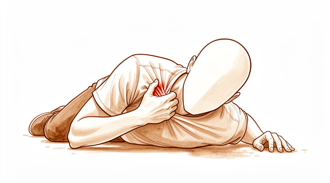
Qué está sintiendo
La fractura de clavícula suele producirse por una caída sobre el hombro o un golpe directo en esa zona. El deporte, el ciclismo y los accidentes de tráfico son causas frecuentes. La clavícula se encuentra justo debajo de la piel, en la parte delantera del hombro, por lo que la fractura a menudo se nota como un bulto o un cambio de forma. Si el hueso se desplaza, un músculo del cuello tira de uno de los fragmentos hacia arriba, mientras que el peso del brazo tira del otro fragmento hacia abajo. Esto puede dejar un bulto visible a lo largo del hueso.
El dolor aparece de inmediato y suele ser agudo. Es posible que sienta náuseas o falta de aire durante unos minutos. El hombro puede verse hinchado y amoratado, y su forma puede parecer distinta a la del otro lado. Probablemente no querrá mover el brazo, y cosas sencillas como vestirse, levantar una bolsa o dormir sobre ese lado resultan dolorosas. La mayoría de las fracturas se producen en la parte media del hueso. Alrededor del 80 % se producen ahí, con alrededor del 15 % en el extremo externo, cerca del hombro, y el 5 % en el extremo interno, cerca del esternón.
En los primeros días, el dolor es peor con el movimiento y por la noche. Encontrar una postura cómoda para dormir suele ser lo más difícil. Durante las semanas siguientes, el dolor disminuye a medida que el hueso empieza a soldarse. El bulto en el lugar donde consolidó el hueso puede quedarse, pero por lo general molesta menos con el tiempo.
Algunos signos de alarma requieren actuar con rapidez. Acuda a un servicio de urgencias el mismo día si la piel sobre la fractura está abierta o se ve el hueso, si la mano o los dedos se ponen pálidos, fríos, blancos o azulados, o si pierde de repente la sensibilidad o el movimiento del brazo después de la lesión. Son poco frecuentes, pero hay que revisarlos de inmediato. Consulte a su médico de cabecera o solicite una evaluación por un especialista si el dolor no disminuye, empeora con el paso de las semanas, le despierta por la noche o le impide trabajar o usar el brazo.
¿Qué ocurre realmente?
La clavícula es un puntal delgado que une el esternón con el omóplato. Es la estructura que mantiene el hombro separado del pecho, por lo que, cuando se rompe, todo el hombro pierde su soporte. La fractura suele producirse donde el hueso es más estrecho y tiene menos músculo alrededor, y por eso la mayoría de las fracturas se producen en la parte media.
Una vez que el hueso se rompe, los músculos toman el control y separan los fragmentos. Un músculo del cuello eleva el fragmento interno, mientras que el peso del brazo arrastra el fragmento externo hacia abajo y hacia dentro, hacia el pecho. Piense en el mástil de una tienda de campaña que se ha partido por la mitad: las dos mitades se abren hacia los lados y la lona se hunde. Por eso el hombro se ve caído y con bultos, y por eso el brazo todavía no funciona con normalidad. El hueso no puede mantener el hombro estable hasta que se haya vuelto a soldar.
El hueso consolida formando hueso nuevo a través de la fractura, un poco como el pegamento que fragua en una unión. Esto lleva semanas, y los fragmentos tienen que permanecer quietos mientras ocurre. Si los fragmentos quedan separados o superpuestos, el hueso puede soldarse en una posición acortada y angulada. Eso deja un bulto permanente y puede acortar ligeramente el hombro. La mayoría de las personas conservan igualmente una buena función, pero a algunas les molesta la forma o el roce de la piel sobre el bulto.
La ubicación de la fractura cambia la situación. Las fracturas de la parte media suelen soldarse bien con un cabestrillo y tiempo. Las fracturas del extremo externo, cerca del hombro, son distintas: el fragmento roto es pequeño y los ligamentos que lo sujetan suelen estar desgarrados, por lo que es más probable que ese fragmento no llegue a consolidar en absoluto. Las fracturas del extremo interno, cerca del esternón, son poco frecuentes y suelen deberse a accidentes más graves.
Su cirujano valorará cuánto se han desplazado los fragmentos y le explicará si el reposo por sí solo o la cirugía le ofrece más posibilidades de lograr un hueso recto y consolidado.
Qué podemos hacer al respecto
El Dr. Kieran Hirpara, cirujano de miembro superior en el Mater Private Hospital Rockhampton, adapta el tratamiento a la lesión específica que usted presenta. La opción adecuada depende de dónde se rompió el hueso, de cuánto se han desplazado los fragmentos y de lo que necesita hacer con el brazo. Algunas fracturas consolidan bien sin operación y otras necesitan cirugía poco después de la lesión, por lo que es importante una valoración rápida. Por lo general, los pacientes son derivados a nuestra clínica por su médico de cabecera; si un fisioterapeuta le ha sugerido que nos consulte, igualmente necesitará una derivación de su médico de cabecera para poder acceder al reembolso de Medicare.
Muchas fracturas se tratan sin cirugía. Si los fragmentos están cerca uno del otro, sostenemos el brazo con un cabestrillo mientras el dolor disminuye, lo que suele llevar de 1 a 3 semanas. Una vez que el dolor cede, le animamos a empezar a mover el hombro, y su fisioterapeuta le guía mientras el hueso se suelda. Un cabestrillo sencillo es más cómodo que un vendaje en ocho, que causa más dolor y no mantiene el hueso en una mejor posición. Las fracturas del extremo interno, cerca del esternón, también se tratan casi siempre de esta forma, con un cabestrillo para mayor comodidad y sin mover el hombro durante 2 a 6 semanas. Los deportes de contacto deben esperar al menos 2 a 3 meses para que el hueso consolide por completo.
Se recomienda la cirugía desde el principio cuando la fractura es grave. Si el hueso se ha acortado 2 cm o más, se ha desplazado todo su grosor o se ha roto en varios fragmentos, una operación mantiene los fragmentos en su posición normal mientras consolidan. También es adecuada en algunas lesiones con daño de nervios o vasos sanguíneos, y en personas que necesitan volver a usar el brazo rápidamente. En la operación se utiliza una placa moldeada para adaptarse a la clavícula, fijada con tornillos. La cirugía hace más probable que el hueso consolide y le permite usar el brazo antes. La decisión es realmente compartida: algunas fracturas pueden consolidar sin cirugía, pero el hueso puede soldarse más corto o angulado y dejar un bulto permanente.
Ambos caminos comparten las mismas primeras semanas. Los analgésicos le mantienen cómodo, y encontrar la manera de dormir suele ser lo más difícil. Protege el brazo mientras el hueso se suelda y, después, recupera la fuerza y el movimiento con su fisioterapeuta en el momento adecuado.
Qué esperar
La mayoría de las fracturas de clavícula sanan bien. Con un cabestrillo y tiempo, el hueso se suelda a través de la fractura a lo largo de varias semanas, y el dolor disminuye a medida que lo hace. Su fisioterapeuta le guía para volver a mover el hombro una vez que cede el dolor inicial, y después llega la recuperación de la fuerza. Las tareas cotidianas como vestirse y levantar objetos vuelven poco a poco a medida que el hueso se suelda. Los deportes de contacto deben esperar al menos 2 a 3 meses para que el hueso consolide por completo. El noventa y cinco por ciento de las fracturas de clavícula consolidan sin problemas.
Algunas cosas pueden retrasar o complicar la consolidación. Si el hueso se ha acortado más de 2 cm, hay más probabilidad de que no llegue a soldarse en absoluto, o de que se suelde en una posición acortada que deje dolor y debilidad al mover el hombro. Una fractura en el extremo externo del hueso tiene más probabilidades de no consolidar que una en la parte media. El hueso consolidado también puede quedar angulado o acortado y dejar un bulto permanente. En los niños, el hueso suele remodelarse por sí solo y la forma se normaliza. En los adultos, normalmente hay que aceptar un ligero cambio de forma, salvo que el bulto sea muy visible o roce la piel.
La cirugía mantiene los fragmentos quietos en su posición normal mientras consolidan, por lo que es más probable que el hueso se suelde y usted puede usar el brazo antes. Las personas operadas suelen volver al trabajo antes que las que mantienen el brazo en reposo. La principal contrapartida es que la cirugía tiene sus propios riesgos. La placa o los tornillos pueden irritar la piel sobre la clavícula, y algunas personas notan entumecimiento alrededor de la cicatriz. Un pequeño número de personas necesita otra operación para retirar el material una vez que el hueso ha consolidado. En conjunto, unas 8 de cada 100 personas operadas de la clavícula tienen una complicación en los primeros 30 días.
Sea cual sea el camino que elija, el hombro puede notarse rígido y débil durante semanas mientras lo recupera. Si el dolor no disminuye, empeora con el paso de las semanas, le despierta por la noche o le impide trabajar o usar el brazo, consulte a su médico de cabecera o solicite una evaluación por un especialista.
¿Cuándo consultar a un profesional?
La mayoría de las fracturas de clavícula se valoran poco después de la lesión, normalmente en un servicio de urgencias o por su médico de cabecera. Busque atención urgente si la piel sobre la fractura está abierta, si la mano o los dedos se ponen pálidos, fríos, blancos o azulados, o si pierde de repente la sensibilidad o el movimiento del brazo después de la lesión. Estos signos requieren acudir a un servicio de urgencias el mismo día. Una deformidad evidente a lo largo de la clavícula también requiere una valoración rápida.
Una vez que le hayan atendido, la consolidación lleva semanas. Consulte a su médico de cabecera o solicite una evaluación por un especialista si el dolor no disminuye, empeora con el paso de las semanas, le despierta por la noche o le impide trabajar o usar el brazo. Lo mismo se aplica si la hinchazón, el movimiento o la fuerza no mejoran semana a semana a medida que el hueso se suelda. Si no puede contactar con la clínica fuera del horario de atención o en fin de semana, acuda al servicio de urgencias más cercano.
En mayor profundidad
Advanced reading: the deeper science (optional)
Esta sección profundiza más de lo necesario para que usted tome sus propias decisiones de tratamiento. La fractura de clavícula merece una lectura adicional, ya que la cirugía garantiza, sin duda, la consolidación ósea; sin embargo, la evidencia de que mejora el funcionamiento del hombro es considerablemente menor.
La cirugía favorece la consolidación ósea; la diferencia funcional es menor
Se han realizado numerosas comparaciones, y los resultados son consistentes si se interpretan con atención. En un total de 1,760 pacientes, el tratamiento quirúrgico de las fracturas del tercio medio de la clavícula produjo menos casos de no consolidación, menos maluniones y un retorno al trabajo más rápido [1].
Sin embargo, el resultado que más interesa a la mayoría de los pacientes presenta una diferencia menor. En 1,965 pacientes, la cirugía aumentó la probabilidad de consolidación ósea al cabo de un año, pero no elevó los índices funcionales en magnitudes que los pacientes considerarían clínicamente relevantes [2].
Ambas afirmaciones son ciertas al mismo tiempo; la distinción entre ellas es precisamente lo que determina la decisión terapéutica. La cirugía favorece una consolidación ósea más fiable y permite volver al trabajo antes. No obstante, no se ha demostrado que mejore la función del hombro al año de la intervención.
¿Qué factor determina el riesgo de no consolidación?
Si la consolidación ósea es el principal beneficio esperado, entonces la pregunta lógica es: ¿cuál es la probabilidad de que la fractura no se una sin intervención quirúrgica? Pues ese es el riesgo que la cirugía busca eliminar.
Uno de esos factores es cuantificable y modificable. El hábito de fumar implica un cociente de riesgo de 3,68 para la no consolidación en fracturas desplazadas de la tercera media de la clavícula tratadas de forma conservadora; los autores recomiendan informar a los pacientes al respecto y ofrecerles apoyo para dejar de fumar [3].
Un aumento cercano al cuádruple en el riesgo es suficiente para alterar el cálculo del riesgo. En el caso de un fumador con una fractura desplazada, la opción no quirúrgica implica un riesgo considerablemente mayor que en un no fumador con la misma lesión; además, dejar de fumar constituye una intervención inmediata y sin necesidad de incisión alguna.
La fractura en el extremo externo se comporta de manera distinta
Las fracturas en el extremo externo de la clavícula, cerca del hombro, constituyen un problema aparte: el fragmento óseo es pequeño y los ligamentos que normalmente lo sujetan suelen estar dañados, por lo que las tasas de no unión son más elevadas.
Al comparar distintos métodos de fijación en 2,284 pacientes, las placas con gancho arrojaron puntuaciones Constant-Murley significativamente más bajas, así como tasas de complicaciones y revisiones quirúrgicas más altas que la fijación coracoclavicular; no obstante, no hubo diferencias en la tasa de unión ósea. Por otro lado, los pacientes tratados de forma no quirúrgica presentaron buenos resultados funcionales [4].
De esto se desprenden dos conclusiones. En cuanto a los tratamientos quirúrgicos, la placa con gancho rinde peor, lo cual coincide con lo que indica la literatura sobre la misma prótesis en relación con la articulación acromioclavicular. Además, el tratamiento no quirúrgico de estas fracturas permite obtener buenos resultados funcionales a pesar de la mayor tasa de no unión; esto sugiere que dicha no unión suele ser más tolerable de lo que el término implica.
¿Cuánto cuesta realmente una consolidación anómala?
Dado que la ventaja funcional de la cirugía es mínima, vale la pena saber qué implica aceptar una consolidación anómala. Una clavícula que se ha soldado de forma más corta y con inclinación genera un bulto visible y acorta ligeramente el cinturón escapular. La mayoría de las personas se adaptan a ello sin sufrir ninguna pérdida funcional apreciable.
El cambio estético es permanente y real; para algunas personas, eso basta como motivo para optar por la cirugía. Se trata de un motivo legítimo para someterse al procedimiento, aunque distinto del objetivo de lograr un mejor funcionamiento del hombro.
Referencias
[1] Smeeing DP, van der Ven DJ, Hietbrink F, Timmers TK, van Heijl M, Kruyt MC, et al. Tratamiento quirúrgico versus no quirúrgico de las fracturas del tercio medio de la clavícula en pacientes de 16 años o más: revisión sistemática, metanálisis y comparación de ensayos controlados aleatorizados y estudios observacionales. Am J Sports Med. 2016;45(8):1937-45. https://doi.org/10.1177/0363546516673615
[2] Axelrod DE, Ekhtiari S, Bozzo A, Bhandari M, Johal H. ¿Cuál es la mejor evidencia para el manejo de las fracturas desplazadas del tercio medio de la clavícula? Una revisión sistemática y metanálisis de red de 22 ensayos controlados aleatorizados. Clin Orthop Relat Res. 2019;478(2):392-402. https://doi.org/10.1097/CORR.0000000000000986
[3] Dietrich G, Terrier A, Favre M, Elmers J, Stockton L, Soppelsa D, et al. Influencia del tabaquismo en la consolidación de las fracturas desplazadas del tercio medio de la clavícula tratadas de forma conservadora: revisión sistemática y metanálisis. Bone Joint J. 2023;105-B(7):801-7. https://doi.org/10.1302/0301-620X.105B7.BJJ-2022-1336.R1
[4] Uittenbogaard SJ, van Es LJ, den Haan C, van Deurzen DF, van den Bekerom MP. Resultados, tasa de consolidación y complicaciones tras el tratamiento quirúrgico y no quirúrgico de las fracturas distales de la clavícula de tipo II según Neer: revisión sistemática y metanálisis. Am J Sports Med. 2021;51(2):534-44. https://doi.org/10.1177/03635465211053336
Evidence & references
This is the clinical evidence summary written for health professionals. It is technical, and it lists the research this page was built from. You do not need to read it to understand your treatment or to make a decision about it.
Overview
General Principles
- Specific treatment of clavicle fractures should be individualized based on fracture characteristics and patient expectations rather than broadly applied [20].
- The most common complications following clavicle fractures, whether treated operatively or non-operatively, are non-unions and malunions [22].
Non-Operative Management
- Most patients with clavicle fractures have an excellent outcome using conservative management [5].
- Initial nonsurgical management of clavicle fractures may be reasonable because patients had similar functional outcomes even when surgery was delayed [2].
- Displaced midshaft clavicular fractures with the intent of achieving 'good' outcome must be managed non-operatively [66].
- Close follow-up of nonoperatively treated clavicle fractures is warranted [3].
- If patients with medial clavicle fractures can survive the initial trauma, there is every reason to expect good clinical and functional outcomes, regardless of whether surgical or nonsurgical management is chosen [1].
- Most mid-shaft clavicle fractures can be treated effectively by non-operative means [71].
- Current evidence suggests that the majority of clavicular fractures in adolescents can and should be treated nonoperatively [39].
- Nonoperative treatment of adolescent clavicle fractures demonstrated lower complication rates and similar satisfaction and functional outcomes compared to operative treatment [15].
- Surgery demonstrated no benefit in patient-reported quality of life, satisfaction, shoulder-specific function, or prevention of complications after completely displaced clavicle shaft fractures in adolescents at 2 years after injury [26].
Operative Management
- A select group of patients with completely displaced fractures, shortening of 2 cm or more, or specific indications benefit from surgical fixation which has been shown to result in improved outcomes compared with non-operative measures [71].
- Complication rates following surgical clavicle fracture care averaged 8.1% [9].
- Operative treatment of displaced medial clavicle fractures provides an excellent long-term functional outcome [16].
- Clavicle fixation is a safe and effective procedure in the pediatric population with a lack of serious complications [34].
- Operative treatment with plate and screw application has consistently good outcomes with a low complication rate in selected cases of adolescent clavicular fractures [39].
- Excellent functional outcomes continue to be observed several years after clavicle fracture fixation [52].
- No difference in reoperation rates between plate types or location could be detected following open reduction and plate fixation of displaced mid-shaft clavicle fractures [52].
- While most distal clavicle fracture fixation methods can achieve stable union, there is a highly variable complication profile associated with each fixation method [30].
- In the unit described, there is no clearly favoured method of internal fixation of lateral clavicle fractures [53].
- When indicated, operative management of displaced midshaft clavicle fractures with dual-plating is cost-effective compared to single-plating [183].
- Patient selection for surgery may influence functional outcome after midshaft clavicle fracture [166].
- There is an increasing trend toward stabilization and fixation of markedly displaced midshaft clavicle fractures in adolescents due to concerns about symptomatic malunion and poor functional outcomes with nonsurgical management, though definitive indications for fixation in this population remain unclear [160].
- Although ORIF of displaced midshaft clavicle fractures remains controversial in the adolescent population, there may be additional circumstances beyond absolute indications for surgical intervention that warrant ORIF at initial presentation [89].
- The optimal management of type II distal clavicle fractures remains controversial, with many authors advocating surgical intervention due to a nonunion rate that has generally averaged 30% [86].
Special Populations
- The authors recommend a more prolonged surveillance period in children with recurrent fractures of the clavicle [12].
Anatomy & Pathophysiology
Bony Anatomy
- The clavicle is the only long bone to ossify by intramembranous ossification [31].
- The clavicle is one of the first bones to ossify, beginning from two primary ossification centers (medial and lateral) by 5 to 6 weeks of gestation [99].
- By 7 to 8 weeks of gestation, the clavicle has already assumed its overall contour and "S" shape [99].
- The clavicle is one of the last bones to complete ossification [99].
- The lateral epiphysis of the clavicle forms and fuses at around 18 to 19 years of age [99].
- The medial epiphysis is the last in the body to ossify, at the age of 18 to 20 years, and the last to complete ossification, at the age of 23 to 25 years [99].
- Most growth (80%) of the clavicle occurs from the medial physis [99].
- The clavicle is relatively straight when anteriorly viewed, whereas in the transverse plane, it resembles an italic S [101].
- The greater radius of curvature occurs at the medial curve of the clavicle, which is anteriorly convex [101].
- The smaller lateral curve of the clavicle is posteriorly convex [101].
- The bone is somewhat rounded in its midsection and medially and relatively flat laterally [101].
- The medial end of the clavicle has a 30% incidence of a rhomboid fossa on its inferior surface where the costoclavicular ligaments insert [101].
- The medial end of the clavicle has a 2.5% incidence of an actual articular surface facing inferiorly toward the first rib [101].
- The middle portion of the clavicle contains the subclavian groove where the subclavius muscle has a fleshy insertion [101].
- The lateral portion of the clavicle has the coracoclavicular process when present [101].
- The conoid ligament attaches to the clavicle at the conoid tubercle [101].
- The trapezoid ligament attaches at the trapezoid line, which lies in an anteroposterior direction just lateral to the conoid tubercle [101].
- The distance from the lateral edge of the clavicle to the medial edge of the conoid tubercle is approximately 45 mm in male and female specimens [101].
- The distance from the lateral edge of the clavicle to the center of the trapezoid tuberosity is approximately 25 mm in male and female specimens [101].
- The primary blood supply to the clavicle is periosteal; there is no nutrient blood supply [31].
- The clavicle is subcutaneous, and its muscular envelope includes the platysma, pectoralis major, deltoid, and some of the strap muscles of the neck [31].
- The clavicle forms a unique S-shaped curve on the axial view [31].
- The distal clavicle is flat in the AP plane [31].
Muscular Anatomy
- The pectoralis major muscle originates from the clavicular shaft anteroinferiorly [106].
- The sternocleidomastoid originates superiorly on the medial clavicle [106].
- The pectoralis origin merges with the origin of the anterior deltoid laterally [106].
- The trapezius insertion blends superiorly with the deltoid origin at the lateral margin [106].
- The medial clavicular fragment is elevated by the unopposed pull of the sternocleidomastoid muscle after fracture [106].
- The distal clavicular fragment is held inferiorly by the deltoid and medially by the pectoralis major after fracture [106].
- The undersurface of the clavicle is the insertion site of the subclavius muscle [106].
- The subclavius muscle serves as a soft tissue buffer in the subclavicular space superior to the brachial plexus and subclavian vessels [106].
- The platysma usually envelopes the anterior and superior aspects of the clavicle and runs in the subcutaneous tissues [106].
- The deltoid originates on the anterior portion of the inner surface of the lateral curve of the clavicle [101].
- The pectoralis major originates from the anterior portion of the medial two-thirds of the clavicle [101].
- The sternocleidomastoid largely originates on the posterior portion of the middle third of the clavicle [101].
- The sternohyoid to a small extent originates on the clavicle, just medial to the origin of the sternocleidomastoid [101].
- The trapezius inserts on the posterosuperior surface of the distal end of the clavicle [101].
Ligaments and Joints
- The clavicle serves as the primary stabilizer between the axial skeleton (via the sternoclavicular joint) and the appendicular skeleton (via the acromioclavicular joint) [31].
- The coracoclavicular ligaments consist of the conoid (medial) and trapezoid (lateral) components [31].
- The coracoclavicular ligaments are the primary stabilizers to superior (vertical) translation of the distal clavicle [31].
- The Superior Shoulder Suspensory Complex (SSSC) is a bone–soft-tissue ring that provides a stable connection of the glenoid and scapula to the clavicle [31].
- The SSSC is composed of four bony landmarks: distal clavicle, acromion, coracoid process, and glenoid neck [31].
- The SSSC includes the supporting ligamentous complexes of the AC joint and the CC ligaments [31].
- The shoulder is a closed chain mechanism and constitutes the combined function of 4 joints: the sternoclavicular, the acromioclavicular, the scapulothoracic, and the glenohumeral joint [4].
- Two of the four shoulder joints are articulations of the clavicle [4].
- Clavicle malunion affects the whole shoulder girdle [4].
Neurovascular Anatomy
- The supraclavicular nerves originate from cervical roots C3 and C4 and exit from a common trunk behind the posterior border of the sternocleidomastoid muscle [115].
- There are typically three major branches of the supraclavicular nerves (anterior, middle, and posterior) that cross the clavicle superficially from medial to lateral [115].
- The subclavian vein runs directly below the subclavius muscle and above the first rib [115].
- The subclavian artery and the brachial plexus lie more posteriorly than the subclavian vein, separated from the vein and clavicle by the additional layer of the scalenus anterior muscle medially [115].
- The brachial plexus is closest to the clavicle in its midportion [115].
- In the middle third of the clavicle, the subclavian artery is a mean of 17 mm from the clavicle [115].
- In the middle third of the clavicle, the subclavian vein is a mean of 13 mm from the clavicle [115].
- In the middle third of the clavicle, the subclavian vessels are located at an approximate angle of 60 degrees to the horizontal, posterior-inferior to the clavicle [115].
- Laterally, the subclavian artery is a mean of 63 mm from the clavicle [115].
- Laterally, the subclavian vein is a mean of 76 mm from the clavicle [115].
- The subclavian vessels are closest to the clavicle at the medial end, with the vein directly apposed to the posterior cortex of the medial clavicle in some cases [115].
- The mean distance of the neurovascular bundle from the posterior border of the clavicle is 9.2 mm [83].
Pathophysiology and Mechanism of Injury
- A direct blow on the point of the shoulder is the commonest reported mechanism of injury that produces a midshaft fracture of the clavicle [103].
- Most (85%) clavicle fractures occur in the midshaft of the bone where the bone is narrowest and enveloping soft tissue structures are most scarce [103].
- The direction of the initial deforming force, and both gravitational and muscular forces on the clavicle result in the typical deformity seen after fracture [103].
- After a midshaft fracture, the distal fragment is translated inferiorly, anteriorly, and medially (shortened), and rotated anteriorly [103].
- The typical deformity of middle-third fractures is caused by a medial fragment pulled superiorly by the sternocleidomastoid muscle, with the weight of gravity pulling downward on the lateral fragment [31].
- A fall on the shoulder or the outstretched hand may fracture the clavicle [14].
- In a clavicle fracture, the lateral fragment is pulled down by the weight of the arm, while the medial fragment is held up by the sternomastoid muscle [14].
- The most common mechanism of injury in clavicle fractures is a direct blow to the shoulder, whether following a fall or because of direct trauma [63].
- A fall on an outstretched hand can result in a clavicle fracture, though this mechanism was initially thought to be the most common cause [63].
- Clavicle fractures are rarely open, despite being caused by high-energy trauma [63].
- The force of the traumatic impact follows the curve of the clavicle and disperses on reaching the lateral curve in Group I (middle third) fractures [80].
- The proximal and distal segments of the clavicle are mechanically secured by ligamentous structures and muscular attachments, whereas the central segment is relatively free [80].
- In Type II distal clavicle fractures, the outer fragment is pulled downward and forward by the weight of the arm when the patient is erect [80].
- The pectoralis major, pectoralis minor, and latissimus dorsi draw the distal segment downward and medially, thereby causing overriding in Type II distal clavicle fractures [80].
- The scapula might rotate the distal segment as the arm is moved in Type II distal clavicle fractures [80].
- The trapezius muscle attaches to the entire outer two-thirds of the clavicle, whereas the sternocleidomastoid muscle attaches to the medial third, acting to draw the clavicular segment superiorly and posteriorly [80].
- Medialization of a clavicular fracture more than 20 mm is associated with a measurable decrease in functional outcome [31].
- The observed association of glenohumeral internal rotation deficit with operative treatment of mid-shaft clavicle fractures may be related to a tendency toward scapular protraction [11].
- Clavicle fractures do not increase the occurrence of later subacromial pain syndrome, and results do not suggest protraction of the scapula as a major risk factor for the development of subacromial pain syndrome [6].
Classification
General Epidemiology and Mechanism
- Clavicle fractures have an incidence of 30 per 100,000 and represent 2.6%-4% of all fractures [55].
- Fractures of the clavicle shaft account for 69% of all clavicle fractures [55].
- Lateral clavicle fractures account for 28% of all clavicle fractures [55].
- Medial clavicle fractures account for 3% of all clavicle fractures [55].
- The most common mechanism of injury is a direct blow to the shoulder, whether following a fall or because of direct trauma [63].
- A fall on an outstretched hand can result in a clavicle fracture, a mechanism initially thought to be the most common cause [63].
- The classical injury mechanisms are a simple fall on the shoulder (31%), road traffic accidents (27%), and sports (23%) [55].
- Medial clavicle fractures occur more often as part of a high-energy trauma or trauma with multiple injuries [55].
- Most clavicle fractures result from a fall onto the shoulder or a direct blow, with the majority occurring in males and associated with vehicular trauma, falls, or sporting injuries [8].
Allman Classification
- The Allman classification divides the clavicle into thirds [169].
- Type I fractures involve the middle third of the clavicle [63].
- Type II fractures involve the lateral third of the clavicle [63].
- Type III fractures involve the medial third of the clavicle [63].
- Group I fractures, or fractures of the middle third, are the most common fractures seen in adults and children [80].
- Group I fractures occur at the point at which the clavicle changes to a flattened cross section from a prismatic cross section [80].
- Group I fractures account for 80% of clavicle fractures [80].
- Group II fractures account for 12% to 15% of all clavicle fractures [80].
Neer Classification (Distal Clavicle)
- The Neer classification separates distal third clavicle fractures into five main types [169].
- Type I distal clavicle fractures are distal to the coracoclavicular ligaments, within 2 cm of the acromioclavicular joint without intra-articular extension, and are minimally displaced [169].
- Type IIA distal clavicle fractures are medial to the conoid ligament, and the proximal fragment is displaced [169].
- Type IIB distal clavicle fractures occur between the conoid and trapezoid ligaments [169].
- Type II distal clavicle fractures are medial to the coracoclavicular ligaments and are usually unstable [63].
- Type II distal clavicle fractures require surgical management [63].
- Type III distal clavicle fractures are intra-articular fractures through the acromioclavicular joint with intact coracoclavicular ligaments [63].
- Type III distal clavicle fractures are usually stable but can result in the development of acromioclavicular joint arthritis [63].
- Type IV distal clavicle fractures involve disruption of the clavicular periosteal sleeve in pediatric patients [63].
- In Type IV distal clavicle fractures, the epiphysis and physis remain with the acromioclavicular joint and displacement occurs at the junction of the metaphysis and physis [63].
- Type V distal clavicle fractures involve a small, inferior cortical bone fragment remaining attached to the coracoclavicular ligaments [63].
- In Type V distal clavicle fractures, the proximal and distal fragments of the clavicle fracture are not connected to the coracoid process [63].
- Type V distal clavicle fractures are rare and generally require surgical intervention for reduction and stabilization [63].
- The modified Neer classification remains the predominantly cited classification system for distal clavicle fractures [104].
- The intra- and interobserver reliability of the modified Neer classification has been demonstrated to be inconsistent [104].
- Inconsistent reliability of the modified Neer classification can lead to incorrect treatment choices and misclassifications in research [104].
Craig Classification
- The Craig classification was introduced in 1990 based on variable fracture patterns within the three broad groups of Allman’s classification [80].
- In Craig Group II Type I fractures, the ligaments remain intact to hold the fragments together and prevent rotation, tilting, or significant fracture displacement [80].
- Craig Group II Type I fractures are interligamentous fractures occurring between the conoid and trapezoid or between the coracoclavicular and acromioclavicular ligaments [80].
- Craig Group II Type I fractures are the most common by a ratio of 4:1 [80].
- In Craig Group II Type II fractures, the coracoclavicular ligaments are detached from the medial segment [80].
- In Craig Group II Type IIA fractures, both the conoid and trapezoid ligaments are on the distal fragment [80].
- In Craig Group II Type IIB fractures, the conoid ligament is ruptured while the trapezoid ligament remains attached to the distal segment [80].
- There is no functional difference between Craig Group II Type IIA and Type IIB fractures [80].
- The high rate of nonunion in Craig Group II Type II fractures may be secondary to excessive motion at the fracture site [80].
- Craig Group II Type III fractures involve the articular surface of the acromioclavicular joint alone [80].
- Craig Group II Type III fractures are characterized by a break in the articular surface without a ligamentous injury [80].
- Craig Group II Type IV fractures occur in children with ligaments intact to the periosteum and displacement of the proximal fragment [80].
- Craig Group II Type V fractures are comminuted, with ligaments attached neither proximally nor distally, but to an inferior, comminuted fragment [80].
- The Craig classification best predicted nonunion or delayed union of lateral third clavicle fractures [63].
Robinson/Edinburgh Classification
- The Edinburgh classification was proposed in 1998, dividing clavicular fractures by anatomic location into type I (medial third), type II (middle third), and type III (lateral third) [63].
- In the Edinburgh classification, Subgroup A indicates displacement less than 100% and subgroup B indicates displacement more than 100% [63].
- In the Edinburgh classification, Subgroup 1 indicates no articular involvement and subgroup 2 indicates intra-articular extension of the fracture [63].
- In the Edinburgh classification, Type II fractures are subdivided into subgroup 1 (simple or wedge-type fractures) and subgroup 2 (comminuted or segmented fractures) [63].
- The Robinson classification had the best prognostic potential for middle third clavicle fractures [63].
- The Edinburgh classification has the highest correlation with prognosis for the middle-third fractures of the clavicle [41].
Pediatric Lateral Clavicle Classification
- The most widely used classification scheme for injuries involving the lateral end of the clavicle and acromioclavicular joint in children is based on a similar classification for adults [60].
- In children, displacement of the lateral clavicle occurs through the periosteal sleeve rather than through the coracoclavicular ligaments [60].
- Most injuries to the lateral end of the clavicle in the immature skeleton are fractures involving the metaphyseal or physeal regions (Salter-Harris type I or II fractures) [60].
- The lateral epiphysis of the clavicle does not ossify until the age of 18 or 19 years [60].
- Pediatric lateral clavicle injuries are classified into six types [60].
- Type I pediatric lateral clavicle injuries are caused by low-energy trauma, with mild strain of the acromioclavicular ligaments and no disruption of the periosteal tube [60].
- In Type II pediatric lateral clavicle injuries, the acromioclavicular ligaments are completely disrupted, with partial damage to the superolateral aspect of the periosteal sleeve [60].
- Type II pediatric lateral clavicle injuries result in mild instability of the lateral end of the clavicle [60].
- In Type III pediatric lateral clavicle injuries, complete disruption of the acromioclavicular ligaments occurs in addition to greater disruption of the periosteal sleeve [60].
- Type III pediatric lateral clavicle injuries result in gross instability of the distal end of the clavicle [60].
- In Type III pediatric lateral clavicle injuries, superior displacement of the lateral clavicle is seen on AP radiographs, with the coracoid–clavicle interval increased by 25% to 100% compared to the uninjured contralateral side [60].
- In Type IV pediatric lateral clavicle injuries, the soft tissue disruption is similar to that in type III injuries, but the lateral clavicle displaces posteriorly and can pierce the trapezius muscle and/or fascia [60].
- In Type V pediatric lateral clavicle injuries, the superior aspect of the periosteal sleeve is completely disrupted, resulting in displacement of the distal clavicle through the trapezial fascia into the subcutaneous tissues [60].
- In Type V pediatric lateral clavicle injuries, the deltoid and trapezius muscles may be detached from the clavicle [60].
- In Type V pediatric lateral clavicle injuries, the coracoid-clavicle interval is increased by 100% or more compared to that of the contralateral side [60].
- Type VI pediatric lateral clavicle injuries are rare and occur when the distal clavicle displaces inferiorly, with the distal end displaced inferior to the coracoid process [60].
New Classification Systems
- A new simple classification system for lateral clavicle fractures showed substantial inter- and intraobserver reliability [100].
- A new classification system for distal clavicle fractures demonstrated moderate interobserver and substantial intraobserver reliability [107].
- The interrater agreement of the modified Neer classification system for lateral clavicle fractures was fair [120].
- Additional 3D CT did not improve the overall level of interrater or intrarater agreement of the modified Neer classification system or associated treatment choice [120].
Clinical Presentation
Mechanism and Epidemiology
- Most clavicle fractures result from a fall onto the shoulder or a direct blow [8].
- Clavicle fractures typically occur because of a direct blow to the shoulder, often in the setting of a fall [21].
- Clavicle fractures most commonly affect young, active, working individuals, with most occurring in male individuals younger than 30 years [21].
- The majority of clavicle fractures occur in males and are associated with vehicular trauma, falls, or sporting injuries [8].
- Clavicle fractures account for 2.6 to 4% of all fractures in adults [21].
- Clavicle fractures account for 3.8% of all fractures and 35.0% to 45.0% of all shoulder girdle injuries [31].
- Midshaft fractures make up approximately 80% of all clavicle fractures [21].
- Approximately 15% of clavicular fractures are distal third, 80% are middle third, and 5% are medial third [31].
- In adolescents, contact sport is the most common mechanism of injury for lateral-end clavicle fractures, followed by cycling, falls, road traffic accidents, and direct blows [24].
- Adolescent clavicle fractures occurred more commonly in male patients during sports, secondary to a direct blow to the shoulder, and on the nondominant side [121].
- Bilateral clavicle fractures are extremely rare and associated with polytrauma [45].
Physical Examination and Deformity
- When displacement occurs in clavicle fractures, the medial fragment typically displaces posterosuperiorly because of the pull of the sternocleidomastoid muscle, and the lateral fragment typically displaces inferomedially because of the pull of the pectoralis major and the weight of the arm [21].
- The displacement pattern described above often results in what is described as a “Z” deformity [21].
- A fall on the shoulder or the outstretched hand may fracture the clavicle; the lateral fragment is pulled down by the weight of the arm, while the medial fragment is held up by the sternomastoid muscle [14].
- The fracture is often displaced, producing a lump along the ‘collar-bone’ [14].
- Fractures of the outer third are easily mistaken for acromioclavicular injuries [14].
- A distal neurovascular examination is important because of the proximity of the brachial plexus and the subclavian vessels to the zone of injury [31].
- Tenting of the skin should be evaluated carefully because it can be a sign of impending open fracture [31].
- Clinicians must carefully examine patients with isolated clavicle fractures for concomitant injuries to the ipsilateral shoulder girdle, particularly in the context of compression mechanisms [37].
Imaging
- Upright and supine radiographs, including an AP view of the clavicle and a 15° cephalad tilt view, should be obtained to define displacement when the patient is upright [31].
- A bilateral panoramic view of both shoulders should be obtained to measure clavicular shortening [31].
- CT is the most accurate modality for determining fracture shortening and morphology but is not typically obtained [31].
- X-rays show that the fracture is usually in the middle third of the bone and the lateral fragment lies below the medial [14].
- Outer-third injuries need special views to define any fracture [14].
- Lateral clavicle fractures can be well visualized with AP radiographs using the Zanca view, which helps delineate the fracture by removing the overlap of the thoracic cage [156].
- For medial clavicle fractures, CT scanning is the procedure of choice [156].
- Fractures of the medial clavicle, especially those involving the SC joint, are notoriously difficult to accurately assess with plain radiographs [164].
- CT scanning is the radiographic procedure of choice when the anatomy of a medial clavicle fracture is unclear [164].
- CT scanning can help distinguish between a medial epiphyseal fracture and true SC dislocations [164].
- Fractures of the medial end of the clavicle can be difficult to diagnose on plain X-ray due to the superimposed cervical and thoracic spine [131].
Classification
- The Allman classification defines fractures of the proximal (medial), middle (midshaft), and distal (lateral) thirds of the clavicle [31].
- Neer classified lateral-third fractures based on the integrity of the CC ligament complex and the involvement of the AC joint [31].
- Medial third fractures are classified according to the displacement and involvement of the SC joint [31].
- The Robinson classification divides the clavicle into thirds and adds variables that are of proven diagnostic value, including intra-articular extension, displacement, and comminution [164].
- The AO/OTA Fracture and Dislocation Classification Compendium designates the clavicle as segment 15 and divides it into medial metaphyseal, diaphyseal, and lateral metaphyseal fractures [164].
Complications
- Vascular and neurological complications are rare in clavicle fractures [14].
- Although rare, limb or life-threatening neurovascular complications from clavicle surgery have potentially devastating implications for young, active patients who typically undergo surgical management of these fractures [17].
- Despite deformity, damage to the lung or vessels beneath the clavicle is very rare [14].
- Clavicle nonunions in adolescents are rare, occurring mostly in older males with displaced fractures and prior ipsilateral fractures [81].
Investigations
Imaging Protocols and Views
- Simple anteroposterior (AP) radiographs are usually sufficient to establish the diagnosis of a clavicle fracture [61].
- The diagnosis may also be made from a single AP chest radiograph, which may be the only available film in an urgent trauma setting [61].
- A chest radiograph can be used to evaluate the deformity of the involved clavicle relative to the normal side and to look for associated skeletal injuries such as rib, glenoid, and scapular fractures [61].
- A measurement of length can be made on the chest radiograph comparing the injured to the uninjured side [61].
- Shortening of 2 cm or more represents a relative indication for primary fixation [61].
- To best delineate a clavicular fracture, a radiograph should be taken in the upright position where gravity will demonstrate maximal deformity [61].
- Ideally, the radiographic beam for the AP radiograph of the clavicle should be angled 20 degrees superiorly to eliminate the overlap of the thoracic cage and show the clavicle in profile [61].
- If the torso is internally rotated a similar 20 degrees, the scapula and shoulder girdle are placed parallel to the cassette for a true AP film [61].
- An upright chest radiograph should be obtained to evaluate midshaft clavicle fracture displacement, as it represents the physiologic stress across the fracture when considering nonoperative management [196].
Advanced Imaging and Measurement Limitations
- CT scanning of midshaft clavicular fractures is rarely performed in the clinical setting [61].
- CT can demonstrate the complex three-dimensional deformity that affects the shoulder girdle with these injuries, including significant scapular angulation and protraction [61].
- CT is useful for evaluating fractures of the medial third of the clavicle and the remainder of the shoulder girdle, such as the glenoid neck in cases of a “floating shoulder” [61].
- CT is the most accurate modality for determining fracture shortening and morphology but not typically obtained [31].
- When clavicle shortening is considered in the decision to pursue operative management, the use of plain radiograph-based measurements is not recommended [200].
- Standard plain unilateral radiographs of the clavicle are insufficient to reliably determine the degree of shortening of clavicle fractures and the need for surgery among shoulder/sports medicine fellowship–trained orthopaedic surgeons [202].
Clinical Examination
Follow-up and Prognostic Imaging
- Delayed assessment at 6 weeks following displaced midshaft clavicle fracture enables an accurate prediction of patients who are likely to have union with nonoperative management [48].
- Once clavicle fractures are healed, further radiographic imaging does not provide any notable information [7].
- For patients undergoing ORIF of isolated clavicular fractures, obtaining a postoperative chest radiograph may be an unnecessary practice, especially given their low sensitivity [208].
Treatment
Non-Operative Management
- Nonsurgical management has been the preferred initial mode of treatment for most clavicle fractures [49].
- Historically, most clavicle fractures have been managed nonoperatively [47].
- In a study year, 8.6% of clavicle fractures were managed by primary surgery, with the highest prevalence in OTA type B diaphyseal fractures at 10.4% [47].
- The majority of operatively treated clavicle fractures in a Swedish study were OTA type B diaphyseal fractures [47].
- Accurate closed reduction is neither possible nor essential for undisplaced middle-third fractures [14].
- For undisplaced middle-third fractures, supporting the arm in a sling until pain subsides (usually 1–3 weeks) is the primary requirement [14].
- Active shoulder exercises should be encouraged after pain subsides in patients treated with a sling, particularly in older patients [14].
- Comparative studies have shown no advantage of the figure-of-eight bandage over a simple sling for nonoperative management [47].
- A simple sling causes less discomfort and fewer complications than a figure-of-eight bandage [47].
- The figure-of-eight bandage is associated with higher pain scores compared to a simple sling [47].
- There is no difference in the amount of clavicular shortening between patients treated with a sling versus a figure-of-eight bandage [47].
- A sling is usually maintained for 2 weeks before physical therapy is started in nonoperative management [47].
- Nondisplaced clavicle fractures continue to be treated conservatively with a simple sling until the fracture is healed according to radiographs and clinical assessment [128].
- Nonoperative care for displaced midshaft fractures typically consists of a standard arm sling for 6 weeks with physical therapy for strengthening once healing is evident [68].
- Radiographic malunion was universal in the non-operative group of a multicenter randomized clinical trial for completely displaced middle third fractures [68].
- The non-union rate was 14% in the non-operative group of a multicenter randomized clinical trial for completely displaced middle third fractures [68].
- Complications, including non-union and symptomatic malunion, were more frequent in the non-operative group compared to the operative group in a multicenter randomized clinical trial [68].
- There is a direct relationship in the non-operative group between increased displacement and worse DASH score [68].
- The management of medial third clavicle fractures is usually nonsurgical, with satisfactory outcomes and low nonunion rates of 4% to 8% [177].
- Treatment for medial third clavicle fractures consists of an arm sling for comfort, with shoulder immobilization for 2 to 6 weeks [177].
- Shoulder range of motion is started as soon as pain subsides or becomes tolerable in medial third clavicle fractures [177].
- Contact sports should be avoided for at least 2 to 3 months to allow complete fracture healing and shoulder rehabilitation in medial third clavicle fractures [177].
- Nondisplaced distal clavicle fractures can be treated with nonoperative management (Neer types I, III, and IV) [78].
- Nondisplaced fractures of the distal end of the clavicle are best treated with a sling [87].
- The earliest return to sports activity for nondisplaced distal clavicle fractures is generally around 2 months after the injury [87].
- Nonsurgical and surgical management provide similar results for distal clavicle fractures [40].
- Nonoperative management of displaced distal clavicle fractures results in higher nonunion rates, but shoulder function remains excellent, and risk of complications and delayed surgery are low [29].
- Nonsurgical treatment should be considered as an option to surgery for fully displaced lateral fractures of the clavicle more often [134].
- Nonoperative management of adolescent mid-shaft clavicle fractures results in excellent functional outcomes at long-term follow-up [18].
- Teenage patients with completely displaced clavicle fractures can expect excellent radiographic and clinical outcomes 5 years post-injury if treated non-operatively [27].
- Comparably excellent outcomes of severe clavicle fractures in adolescent athletes can be achieved with non-operative treatment [110].
- Proportional shortening of 8% is not associated with impaired function or patient dissatisfaction in nonoperatively treated clavicle fractures [47].
- Proportional shortening of 8% did not significantly correlate with the DASH score, Constant score, or SF-12 score at any time during follow-up in nonoperatively treated patients [47].
- There was no correlation between proportional shortening of 8% and patient satisfaction in nonoperatively treated patients [47].
Operative Management
- There is a growing trend towards internal fixation of acute clavicular fractures associated with severe displacement, fragmentation or shortening [14].
- Specific contoured locking plates are available for the treatment of displaced middle-third fractures [14].
- An advantage of internal fixation for displaced middle-third fractures is that the patient can mobilize the arm and return to work and independence more quickly [14].
- Outer-third fractures are quite troublesome and may need open reduction and internal fixation [14].
- Current evidence-based indications for surgical fixation of midshaft clavicular fractures include radiographic shortening of greater than or equal to 2 cm [149].
- Current evidence-based indications for surgical fixation of midshaft clavicular fractures include displacement in a caudal/cephalad plane by equal to or greater than 100% or one bone width [149].
- Current evidence-based indications for surgical fixation of midshaft clavicular fractures include significant comminution [149].
- Additional indications for surgical intervention include open fractures, fractures with underlying neurovascular injury, and fractures that result in significant scapular malposition or winging [149].
- Fracture-specific indications for operative treatment of midshaft clavicle fractures include displacement >2 cm [28].
- Fracture-specific indications for operative treatment of midshaft clavicle fractures include shortening >2 cm [28].
- Fracture-specific indications for operative treatment of midshaft clavicle fractures include increasing comminution (>3 fragments) [28].
- Fracture-specific indications for operative treatment of midshaft clavicle fractures include segmental fractures [28].
- Fracture-specific indications for operative treatment of midshaft clavicle fractures include open fractures [28].
- Fracture-specific indications for operative treatment of midshaft clavicle fractures include impending open fractures with soft tissue compromise [28].
- Fracture-specific indications for operative treatment of midshaft clavicle fractures include obvious clinical deformity [28].
- Fracture-specific indications for operative treatment of midshaft clavicle fractures include scapular malposition and winging on initial examination [28].
- Associated injuries that are indications for operative treatment include vascular injury requiring repair [28].
- Associated injuries that are indications for operative treatment include progressive neurologic deficit [28].
- Associated injuries that are indications for operative treatment include ipsilateral upper extremity injuries/fractures [28].
- Associated injuries that are indications for operative treatment include multiple ipsilateral upper rib fractures [28].
- Associated injuries that are indications for operative treatment include "floating shoulder" [28].
- Associated injuries that are indications for operative treatment include bilateral clavicle fractures [28].
- Patient factors that are indications for operative treatment include polytrauma with requirement for early upper extremity weight-bearing/arm use [28].
- Patient factors that are indications for operative treatment include patient motivation for rapid return of function (e.g., elite sports or the self-employed professional) [28].
- Operative repair should be reserved for medically well, physically active patients who stand to benefit the most from a rapid restoration of normal anatomy and stable fixation [28].
- Most mid-shaft clavicle fractures can be treated effectively by non-operative means, but a select group of patients with completely displaced fractures, shortening of 2 cm or more, or specific indications benefit from surgical fixation [71].
- Surgical fixation has been shown to result in improved outcomes compared with non-operative measures for a select group of patients with completely displaced fractures or shortening of 2 cm or more [71].
- For healthy, active adults, midshaft clavicular fractures should undergo consideration for surgical stabilization if significantly displaced (2 cm of shortening, 100% displacement or significant comminution) [67].
- The use of a pre-contoured plate facilitates surgical care of clavicular fractures, reducing hardware prominence and secondary surgical procedures [67].
- Dual plating may be used as an adjuvant in highly comminuted cases [67].
- Dual plating with 2.4- or 2.7-mm plates may additionally have a benefit of decreased hardware prominence [67].
- Parameters for the management of distal and medial clavicle fractures remain unclear and should be assessed on a case-by-case basis [67].
- Fracture displacement and the development of nonunion may not, in isolation, affect outcome especially in the elderly for distal and medial clavicle fractures [67].
- Floating shoulder injuries should be carefully assessed on the basis of component injuries individually because surgical management has not been shown to result in improved clinical outcomes [67].
- Multiple meta-analyses of existing randomized trials have favored surgical management because it provides significantly lower rates of nonunion, malunion, and complications excluding planned hardware removals [149].
- Earlier functional return compared with nonsurgical management of displaced midshaft clavicular fractures has been identified in multiple meta-analyses [149].
- Fixation of displaced fractures of the clavicle can be accomplished by either open plating or intramedullary pinning [149].
- A meta-analysis of five randomized controlled trials demonstrated that intramedullary fixation resulted in fewer complications such as prominent hardware and superficial infection compared to plating [149].
- Constant scores were similar between intramedullary fixation (93.8) and plating (89.3) in a meta-analysis of five randomized controlled trials [149].
- Intramedullary fixation has been shown to be inferior to plating in terms of rotational control and axial rigidity [149].
- Intramedullary fixation may not be the best choice in comminuted fracture patterns requiring increased rotatory control and axial stability [149].
- There is a higher rate of removal of intramedullary pins or nails compared to plates [149].
- A 2013 cadaver study has shown no significant difference in distance from screw tips to neurovascular structures between anterior-inferior plating and superior plating [149].
- Hardware prominence has been shown to be no different between anterior-inferior plating and superior plating [149].
- A study reported on revision surgery rates in midshaft clavicular fractures undergoing surgical fixation and found no association between revision surgery and plate position [149].
- The overall rate of revision surgery for hardware irritation in midshaft clavicular fractures undergoing surgical fixation was 17% [149].
- The development of precontoured clavicular plates has resulted in a significant reduction in hardware prominence compared with the prior standard use of straight compression or reconstruction plates [149].
- Precontoured 2.7-mm plates have demonstrated biomechanical superiority to reconstruction plates in both load to failure and bending failure stiffness [149].
- Dual plating of the clavicle, usually superiorly and anteriorly, has shown similar outcomes to single plating [149].
- Biomechanically, dual mini-fragment plating was not significantly different from single superior or anterior plate in terms of axial, bending, torsional stiffness, or bending load to failure in a cadaver study of simulated butterfly fracture [149].
- Dual plating may be useful in cases of complex comminution or fragment containment where a bridging technique is required [149].
- Hardware prominence and secondary surgery for hardware removal has been suggested as a potential benefit of dual plating over single plating [149].
- Operative treatment leads to improved short-term functional outcomes compared with conservative treatment [141].
- Operative treatment leads to increased patient satisfaction compared with conservative treatment [141].
- Operative treatment leads to an earlier return to sports compared with conservative treatment [141].
- Operative treatment leads to lower rates of non-union compared with conservative treatment [141].
- In terms of cost-effectiveness, operative treatment seems to be advantageous compared with conservative treatment [141].
- Operative treatment is associated with an increased risk of complications compared with conservative treatment [141].
- Operative treatment is associated with an increased risk of re-operations compared with conservative treatment [141].
- Long-term shoulder functional outcomes are similar between operative and conservative treatment [141].
- The optimal treatment strategy should be one tailor-made to the patient and his/her specific needs and expectations by utilizing a shared decision-making model [141].
- Specific treatment of clavicle fractures should not be broadly applied but rather should be individualized based on fracture characteristics and patient expectations [20].
- A targeted approach to the management of mid-shaft clavicle fractures is needed, with simple fractures treated nonoperatively and complex displaced fractures considered for surgery to prevent non-union [126].
- The plate is very effective and safe for the management of medial clavicle fractures [112].
- Fractures that are significantly displaced may warrant operative repair, especially if there is posterior displacement of the shaft fragment in medial clavicle fractures [147].
- The primary technical difficulty with medial clavicle fractures is the fixation in the medial fragment [147].
- The subclavian vessels are in close proximity to the bone medially during medial clavicle fracture fixation [147].
- Definitive plate fixation can be performed in a variety of ways for medial clavicle fractures [147].
- If the medial fragment is large enough, standard plate and screw fixation can be performed for medial clavicle fractures [147].
- A plate with an expanded end section may augment multiple screw purchase in medial clavicle fractures [147].
- The significant expansion of the medial clavicle allows for placement of longer (22 to 24 mm) cancellous screws [147].
- If there is insufficient purchase, the plate can be extended across the joint onto the sternum for medial clavicle fractures [147].
- A construct extended across the joint onto the sternum will eventually loosen due to motion at the SC joint but will typically stabilize the fracture long enough (3 months) for union to occur [147].
- The plate should be removed at the point of union when extended across the joint onto the sternum [147].
- Rarely, fixation with a hook plate intrasternally or retrosternally may be required for medial clavicle fractures [147].
- Cardiovascular support should be available in the event of inadvertent injury to the vascular structures found retrosternally when using a hook plate intrasternally or retrosternally [147].
- Fixation of the fracture using smooth wires or pins alone is contraindicated for medial clavicle fractures due to the potential for migration and visceral injury [147].
- Surgical management of medial third clavicle fractures is usually reserved for fractures associated with injury to the mediastinal structures secondary to fracture displacement [177].
- These fractures should be reduced fairly emergently, with an attempt at closed reduction [177].
- Open reduction and internal fixation (ORIF) is sometimes necessary to maintain reduction of an unstable medial third clavicle fracture [177].
- Several techniques have been used for ORIF of displaced medial third clavicle fractures including wire fixation, plate-and-screw constructs, or interosseous sutures [177].
- A recent study published the outcomes following surgical management of medial third clavicle fractures in 27 patients, with 24 acute fractures and 3 nonunions [177].
- Surgical treatment consisted of transosseous sutures in 8 cases, and plate and screws fixation in 19 cases in a study of 27 medial third clavicle fractures [177].
- The authors reported 100% union rate and excellent DASH functional outcome scores in a study of 27 medial third clavicle fractures [177].
- In our unit there is no clearly favoured method of internal fixation of lateral clavicle fractures [53].
- Evidence-based indications for acute surgical intervention in lateral third clavicle fractures are lacking [162].
- Current expert opinion on surgical indications for distal clavicle fractures includes severely displaced fractures, high-demand patients or failure of nonsurgical care [162].
- Most distal clavicle injuries, particularly in middle-aged or elderly populations, should be managed nonsurgically [162].
- Initial nonsurgical management with delayed surgery for patients who develop a symptomatic nonunion appears to be a viable treatment strategy for distal clavicle fractures [162].
- A recent
Complications
General and Non-Operative
- Vascular and neurological complications are rare [14].
- Damage to the lung or vessels beneath the clavicle is very rare despite deformity [14].
- Non-union sometimes occurs in middle-third fractures and is treated by bone graft and plating [14].
- Malunion is inevitable in displaced fractures; in children, the bone is soon remodelled, but in adults the slight deformity has to be accepted unless there is a very unsightly bump with skin irritation [14].
- Treating displaced middle-third fractures with shortening of more than 2 cm by simple splintage incurs a risk of symptomatic malunion, mainly pain and lack of power during shoulder movements, and an increased incidence of non-union [14].
- The non-union rate was 14% in the non-operative group for completely displaced middle-third fractures [68].
- Complications, including non-union and symptomatic malunion, were more frequent in the non-operative group compared to the operative group for completely displaced middle-third fractures [68].
- Shortening of greater than 2 cm has been reported as a risk factor for nonunion and malunion [152].
- Final shortening of greater than 2 cm was associated with a nonunion rate of 15% and subsequent poor outcomes [152].
- Robinson and colleagues noted a nonunion rate of 21% for fractures with greater than 2 cm of shortening [152].
- Nowak and colleagues reported that 46% of patients with clavicle fractures had residual symptoms, especially those with no bony contact and those with comminuted fractures [152].
- Of 252 clavicle fractures, 31% of patients with completely displaced midshaft fractures had residual symptoms [152].
- The risk factors for nonunion after nonoperative treatment of midshaft clavicle fractures are multifactorial [84].
- Several patients became symptomatic for fractures with a larger proportional change in clavicular length or displacement during long-term conservative treatment [54].
- Close follow-up of nonoperatively treated clavicle fractures is warranted due to the potential for progressive displacement in the peri-injury period [3].
Operative
- The overall 30-day complication rate following open reduction and internal fixation (ORIF) for clavicle fractures was 1.9% [57].
- Surgical treatment of midshaft clavicle fractures leads to a slightly higher incidence of complications compared with the nonoperative approach [70].
- Although rare, limb or life-threatening neurovascular complications from clavicle surgery have potentially devastating implications for young, active patients [17].
- The percentage of patients reporting implant irritation was 70% for plate fixation and 66% for intramedullary nail fixation, with no significant difference between the two groups [19].
- Intramedullary fixation was associated with a higher likelihood of implant removal (82%) compared to plate fixation (50%) [19].
- A limited incision approach for plating of acute midshaft clavicle fractures achieved a low complication rate comparable to the reported rate for standard incision techniques [62].
- In the Canadian Orthopaedic Trauma Association study, complications including non-union and symptomatic malunion were more frequent in the non-operative group than the operative group [68].
- The technique of clavicle pinning resulted in minimal complications, short hospital stay and excellent functional outcomes [124].
Associated Injuries and Specific Phenomena
- Ipsilateral clavicle fracture and acromioclavicular (AC) joint injury is much more common than traditionally believed, with an incidence of 6.8% overall [175].
- Patients who have sustained high-energy vehicular trauma are more likely to have associated injuries to the thoracic cage, including ipsilateral rib fractures, scapular and/or glenoid fractures, proximal humeral fractures, and hemo/pneumothoraces [50].
- Several studies from Level 1 trauma centers have noted a high mortality rate (20% to 34%) from associated chest and head traumas in polytrauma patients with clavicle fractures [50].
- In-hospital mortality rates for medial clavicle fractures have been reported to be as high as 20%, predominantly due to concomitant intra-cranial and intra-thoracic injuries [13].
- Medial clavicle fractures have a high proportion of patients who will die within 3 years of the injury [35].
- The observed association of glenohumeral internal rotation deficit with a tendency toward scapular protraction may provide insight into this phenomenon after operative treatment of mid-shaft clavicle fractures [11].
- Women reported higher rates of distal clavicle fracture in the postoperative period following acromioclavicular joint reconstruction, with a high fracture rate in the female cohort (90 days, 19.6%) [76].
Recovery
General Outcomes and Prognosis
- If patients with medial clavicle fractures survive the initial trauma, good clinical and functional outcomes are expected regardless of whether surgical or nonsurgical management is chosen [1].
- Medial clavicle fractures have favorable functional outcomes and pain relief at minimum 1-year follow-up among patients who survive the trauma, but a high proportion will die within 3 years of the injury [35].
- Clavicle fractures do not increase the occurrence of later subacromial pain syndrome, with results suggesting that protraction of the scapula is not a major risk factor for its development [6].
- The prognosis for obtaining bony union after infected clavicle fractures is poor, with only two of six patients achieving union [46].
Non-Operative Management
- Close follow-up of nonoperatively treated clavicle fractures is warranted due to displacement related to patient position and progressive displacement in the peri-injury period [3].
- Proportional shortening of 8% in nonoperatively treated displaced midshaft clavicular fractures is not associated with impaired function or patient dissatisfaction [47].
- Significant early improvements in fracture alignment were observed in a substantial percentage of adolescent patients with completely displaced clavicle fractures, indicating that the true final deformity is commonly less than that present at the time of injury [212].
- Clavicle fractures with associated ipsilateral rib fractures tend to demonstrate an increased amount of displacement on follow-up radiographs compared with those without ipsilateral rib fractures [96].
- A sling is suggested to be worn for about 2 weeks followed by a physical therapy regimen for nonoperative management, as comparative studies have shown no advantage of the figure-of-eight bandage over a simple sling [47].
- Low-intensity pulsed ultrasound (LIPUS) did not confirm acceleration of clinical healing time of fresh clavicle shaft fractures [210].
Operative Management
- Surgical treatment of midshaft clavicle fractures significantly reduces the nonunion rate and shortens the time to union as compared with the nonoperative approach, leading to better shoulder functional scores at short- and long-term follow-up despite a slightly higher incidence of complications [70].
- Surgical treatment led to a greater likelihood of union at 1 year of follow-up among adult patients with displaced mid-third clavicle fractures [213].
- Functional outcome is excellent following the treatment of both acute and non-united clavicle fractures, but recovery occurs earlier following acute treatment [33].
- The improvement in DASH and CSS scores seen with primary fixation of displaced clavicle fractures persists at 2 years but does not differ from values seen after 1 year of followup [138].
- Patients reported a good quality of life and functional outcome after plating for midshaft clavicular fractures [133].
- No difference in reoperation rates between plate types or location could be detected, and excellent functional outcomes continue to be observed several years after clavicle fracture fixation [52].
- A limited incision approach for plating of acute midshaft clavicle fractures achieved good functional and radiographic outcomes with a low complication rate comparable to the reported rate for standard incision techniques [62].
- Carefully indicated adolescent patients undergoing ORIF of clavicle fractures can return to play more quickly than previously thought [91].
- Over the past 5 years, nearly 50% of NFL players with a completely displaced middle-third clavicle fracture were treated successfully with acute surgical fixation without sequela and healed at an average of 8.8 weeks [94].
- Risk factors for nonunions after lateral clavicle fracture fixation include early mechanical stress, a lateral clavicular fragment larger than 3 cm, and time delay to surgery [93].
- The observed association of glenohumeral internal rotation deficit with a tendency toward scapular protraction may provide insight into this subclinical phenomenon after operative treatment of mid-shaft clavicle fractures [11].
Pediatric and Adolescent Specifics
Key Evidence
- [L5] If patients with medial clavicle fractures can survive the initial trauma, there is every reason to expect good clinical and functional outcomes, regardless of whether surgical or nonsurgical management is chosen. [1] (10.1097/corr.0000000000001916)
- [L3] Initial nonsurgical management of clavicle fractures may be reasonable because patients had similar functional outcomes even when surgery was delayed. [2] (10.5435/jaaos-d-16-00130)
- [L2] Close follow-up of nonoperatively treated clavicle fractures is warranted. [3] (10.1016/j.jse.2018.01.004)
- [L4] [4] (10.3109/17453674.2010.480939)
- [L3] Most patients with clavicle fractures have an excellent outcome using conservative management. [5] (10.1016/j.jse.2019.06.022)
- [L4] The results do not suggest protraction of the scapula as a major risk factor for the development of SAPS. [6] (10.1016/j.xrrt.2024.01.008)
- [L3] Once clavicle fractures are healed, further radiographic imaging does not provide any notable information. [7] (10.5435/jaaos-d-17-00598)
- [L3] Complication rates following surgical clavicle fracture care averaged 8.1%. [9] (10.1186/s12891-022-05075-5)
- [L4] The observed association with a tendency toward scapular protraction may provide insight into this phenomenon. [11] (10.1186/s13018-026-06856-7)
- [L5] The authors recommend a more prolonged surveillance period in children with recurrent fractures of the clavicle. [12] (10.1097/bpb.0000000000000231)
- [Paper] [13] (10.1016/j.injury.2016.06.011)
- [L2] Nonoperative treatment of adolescent clavicle fractures demonstrated lower complication rates and similar satisfaction and functional outcomes compared to operative treatment. [15] (10.1177/2325967119s00428)
- [L4] Operative treatment of displaced medial clavicle fractures provides an excellent long-term functional outcome. [16] (10.1007/s00068-018-1024-6)
- [L5] Although rare, limb or life-threatening neurovascular complications from clavicle surgery have potentially devastating implications for young, active patients who typically undergo surgical management of these fractures. [17] (10.1097/bte.0000000000000097)
- [L3] Nonoperative management of adolescent mid-shaft clavicle fractures results in excellent functional outcomes at long-term follow-up. [18] (10.1302/0301-620x.103b5.bjj-2020-1929.r1)
- [L5] Specific treatment of clavicle fractures should not be broadly applied but rather should be individualized based on fracture characteristics and patient expectations. [20] (10.1016/j.jse.2011.08.053)
- [L1] [21] (10.5435/jaaos-d-23-00472)
- [L4] [24] (10.1177/17585732221131922)
- [L2] Surgery demonstrated no benefit in patient-reported quality of life, satisfaction, shoulder-specific function, or prevention of complications after completely displaced clavicle shaft fractures in adolescents at 2 years after injury. [26] (10.1177/03635465221114420)
- [L2] Teenage patients with completely displaced clavicle fractures can expect excellent radiographic and clinical outcomes 5 years post-injury if treated non-operatively. [27] (10.1177/2325967123s00041)
- [L4] Nonoperative management of displaced distal clavicle fractures results in higher nonunion rates, but shoulder function remains excellent, and risk of complications and delayed surgery are low. [29] (10.1016/j.jse.2023.12.006)
- [L3] While most distal clavicle fracture fixation methods can achieve stable union, there is a highly variable complication profile associated with each fixation method. [30] (10.1016/j.otsr.2019.03.012)
- [L3] Functional outcome is excellent following the treatment of both acute and non-united clavicle fractures, but recovery occurs earlier following acute treatment. [33] (10.1016/j.otsr.2017.03.021)
- [L4] Clavicle fixation is a safe and effective procedure in the pediatric population with a lack of serious complications. [34] (10.1177/2325967119s00056)
- [L4] Medial clavicle fractures have favorable functional outcomes and pain relief at minimum 1-year follow-up among those patients who survive the trauma, but a high proportion will die within 3 years of the injury. [35] (10.1097/corr.0000000000001839)
- [L4] Clinicians must carefully examine patients with isolated clavicle fractures for concomitant injuries to the ipsilateral shoulder girdle, particularly in the context of compression mechanisms. [37] (10.1177/03635465000280062301)
- [L4] Current evidence suggests that the majority of clavicular fractures in adolescents can and should be treated nonoperatively, although operative treatment with plate and screw application has consistently good outcomes with a low complication rate in selected cases. [39] (10.2106/jbjs.22.01036)
- [L4] Nonsurgical and surgical management provide similar results for distal clavicle fractures. [40] (10.5435/00124635-201107000-00002)
- [L3] [41] (10.1016/j.jse.2014.09.037)
- [L4] Bilateral clavicle fractures are extremely rare and associated with polytrauma. [45] (10.1186/s12891-023-06228-w)
- [L4] The prognosis for obtaining bony union after infected clavicle fractures is poor, with only two of six patients achieving union. [46] (10.1097/01.blo.0000183088.60639.05)
- [L1] Delayed assessment at 6 weeks following displaced midshaft clavicle fracture enables an accurate prediction of patients who are likely to have union with nonoperative management. [48] (10.2106/jbjs.19.00955)
- [L4] No difference in reoperation rates between plate types or location could be detected, and excellent functional outcomes continue to be observed several years after clavicle fracture fixation. [52] (10.1016/j.injury.2014.04.032)
- [L4] In our unit there is no clearly favoured method of internal fixation of lateral clavicle fractures. [53] (10.1007/s00590-021-03173-z)
- [L4] Moreover, several patients became symptomatic for fractures with a larger proportional change in clavicular length or displacement. [54] (10.1186/s13018-023-04450-9)
- [L4] [55] (10.1007/s00068-019-01122-4)
- [L4] The overall 30-day complication rate following ORIF for clavicle fractures was 1.9 %, with 128 patients experiencing adverse events out of a cohort of 6753 patients. [57] (10.1016/j.injury.2024.111883)
- [L5] In this large cohort with long-term follow-up, a limited incision approach for plating of acute midshaft clavicle fractures achieved good functional and radiographic outcomes with a low complication rate comparable to the reported rate for standard incision techniques. [62] (10.1016/j.jse.2025.06.002)
- [L3] Displaced midshaft clavicular fractures with the intent of achieving 'good' outcome must be managed non-operatively. [66] (10.1016/j.injury.2020.10.019)
- [L1] Surgical treatment of midshaft clavicle fractures significantly reduces the nonunion rate and shortens the time to union as compared with the nonoperative approach and, despite a slightly higher incidence of complications, leads to better shoulder functional scores at short- and long-term follow-up. [70] (10.1177/0363546519826961)
- [L3] Women reported higher rates of distal clavicle fracture in the postoperative period, and the high fracture rate in the female cohort (90 days, 19.6%) highlights a potentially under-recognized clinical consideration. [76] (10.1002/ars2.70063)
- [Paper] Nondisplaced distal clavicle fractures can be treated with nonoperative management (Neer types I, III, and IV). [78] (10.2106/jbjs.rvw.25.00260)
- [L3] Clavicle nonunions in adolescents are rare, occurring mostly in older males with displaced fractures and prior ipsilateral fractures. [81] (10.1016/j.jse.2017.06.040)
- [L5] [83] (10.1016/s0020-1383(15)30035-8)
- [L3] The risk factors for nonunion after nonoperative treatment of midshaft clavicle fractures are multifactorial. [84] (10.1016/j.otsr.2014.11.018)
- [L5] The optimal management of type II distal clavicle fractures remains controversial, with many authors advocating surgical intervention due to a nonunion rate that has generally averaged 30%. [86] (10.1016/s0278-5919(02)00108-4)
- [Case_report] Although ORIF of displaced midshaft clavicle fractures remains controversial in the adolescent population, there may be additional circumstances beyond absolute indications for surgical intervention that warrant ORIF at initial presentation. [89] (10.1016/j.xrrt.2023.03.004)
- [L3] These data suggest that carefully indicated adolescent patients undergoing ORIF of clavicle fractures can RTP more quickly than previously thought. [91] (10.1177/23259671251374299)
- [L4] Risk factors for nonunions include early mechanical stress, a lateral clavicular fragment larger than 3 cm, and time delay to surgery. [93] (10.1007/s00402-018-3075-x)
- [L4] Over the past 5 years, nearly 50% of NFL players with a completely displaced middle-third clavicle fracture were treated successfully with acute surgical fixation without sequela and healed at an average of 8.8 weeks. [94] (10.1177/0363546510372795)
- [L2] Clavicle fractures with associated ipsilateral rib fractures tend to demonstrate an increased amount of displacement on follow-up radiographs compared with those without ipsilateral rib fractures. [96] (10.1097/bot.0000000000000758)
- [L4] The presented classification system as well as associated treatment algorithms for lateral clavicle fractures showed substantial inter- and intraobserver reliability. [100] (10.1016/j.jse.2025.04.021)
- [L5] The modified Neer classification remains the predominantly cited classification system for distal clavicle fractures, yet its intra- and interobserver reliability has been demonstrated to be inconsistent, which can lead to incorrect treatment choices and misclassifications in research. [104] (10.1097/corr.0000000000001456)
- [L3] The study demonstrated moderate interobserver and substantial intraobserver reliability of the new classification system and the associated treatment choice for distal clavicle fractures. [107] (10.1016/j.otsr.2018.05.015)
- [L2] Comparably excellent outcomes of severe clavicle fractures in adolescent athletes can be achieved with non-operative treatment. [110] (10.1177/2325967121s00214)
- [L5] We conclude that the plate is very effective and safe for the management of medial clavicle fractures. [112] (10.1097/bte.0000000000000006)
- [L3] The interrater agreement of the modified Neer classification system for lateral clavicle fractures was fair, and additional 3D CT did not improve the overall level of interrater or intrarater agreement of the classification system or associated treatment choice. [120] (10.1177/0363546515593949)
- [L4] Adolescent clavicle fractures occurred more commonly in male patients during sports, secondary to a direct blow to the shoulder, and on the nondominant side. [121] (10.1177/2325967120921344)
- [L4] The technique of clavicle pinning resulted in minimal complications, short hospital stay and excellent functional outcomes. [124] (10.4103/0973-6042.57895)
- [L5] A targeted approach to the management of mid-shaft clavicle fractures is needed, with simple fractures treated nonoperatively and complex displaced fractures considered for surgery to prevent non-union. [126] (10.1016/j.injury.2020.11.066)
- [L4] Nondisplaced clavicle fractures continue to be treated conservatively with a simple sling until the fracture is healed according to radiographs and clinical assessment. [128] (10.3810/psm.2011.09.1930)
- [L4] [131] (10.1177/1758573220923122)
- [L3] Patients reported a good quality of life and functional outcome after plating for midshaft clavicular fractures. [133] (10.1016/j.injury.2017.10.032)
- [L3] Nonsurgical treatment should be considered as an option to surgery for fully displaced lateral fractures of the clavicle more often. [134] (10.1016/j.injury.2024.111422)
- [L1] The improvement in DASH and CSS scores seen with primary fixation of displaced clavicle fractures persists at 2 years but does not differ from values seen after 1 year of followup. [138] (10.1007/s11999-011-1915-x)
- [L4] [141] (10.1302/2058-5241.3.170033)
- [Paper] [152] (10.1097/01.bte.0000187285.99726.0e)
- [L5] There is an increasing trend toward stabilization and fixation of markedly displaced midshaft clavicle fractures in adolescents due to concerns about symptomatic malunion and poor functional outcomes with nonsurgical management, though definitive indications for fixation in this population remain unclear. [160] (10.5435/00124635-201301000-00002)
- [L1] This review shows that patient selection for surgery may influence functional outcome after midshaft clavicle fracture. [166] (10.1177/1758573218777996)
- [L5] [169] (10.1007/s10140-018-1586-y)
- [L3] Ipsilateral clavicle fracture and AC joint injury is much more common than traditionally believed, with an incidence of 6.8% overall. [175] (10.1016/j.injury.2016.12.021)
- [L5] When indicated, operative management of displaced midshaft clavicle fractures with dual-plating is cost-effective compared to single-plating. [183] (10.1177/2325967123s00167)
- [L4] An upright chest radiograph should be obtained to evaluate midshaft clavicle fracture displacement, as it represents the physiologic stress across the fracture when considering nonoperative management. [196] (10.1097/bot.0000000000000727)
- [L4] When clavicle shortening is considered in the decision to pursue operative management, the use of plain radiograph-based measurements is not recommended. [200] (10.4055/cios.2016.8.4.367)
- [L3] Standard plain unilateral radiographs of the clavicle are insufficient to reliably determine the degree of shortening of clavicle fractures and the need for surgery among shoulder/sports medicine fellowship–trained orthopaedic surgeons. [202] (10.1177/0363546514523926)
- [L4] Therefore, for patients undergoing ORIF of isolated clavicular fractures obtaining a postoperative chest radiograph may be an unnecessary practice, especially given their low sensitivity. [208] (10.1016/j.jse.2018.09.016)
- [L1] Our findings did not confirm that LIPUS accelerates clinical healing time of fresh clavicle shaft fractures. [210] (10.1016/j.injury.2008.04.004)
- [L3] Significant early improvements in fracture alignment were observed in a substantial percentage of adolescent patients with completely displaced clavicle fractures, indicating that the true final deformity is commonly less than that present at the time of injury. [212] (10.1177/2325967121s00031)
- [L1] Surgical treatment led to a greater likelihood of union at 1 year of follow-up among adult patients with displaced mid-third clavicle fractures. [213] (10.1097/corr.0000000000000986)
References
[1] CORR Insights®: What Are the Functional Outcomes and Pain Scores after Medial Clavicle Fracture Treatment?. Clinical Orthopaedics & Related Research. 2021. DOI: 10.1097/corr.0000000000001916
[2] Factors Affecting Functional Outcomes After Clavicle Fracture. Journal of the American Academy of Orthopaedic Surgeons. 2016. DOI: 10.5435/jaaos-d-16-00130
[3] Displacement of diaphyseal clavicle fractures related to patient position and progressive displacement in the peri-injury period. Journal of Shoulder and Elbow Surgery. 2018. DOI: 10.1016/j.jse.2018.01.004
[4] Malunion after midshaft clavicle fractures in adults. Acta Orthopaedica. 2010. DOI: 10.3109/17453674.2010.480939
[5] Plate fixation of clavicle fractures: comparison between early and delayed surgery. Journal of Shoulder and Elbow Surgery. 2020. DOI: 10.1016/j.jse.2019.06.022
[6] Clavicle fractures do not increase the occurrence of later subacromial pain syndrome. A registry-based case-control study with 15-25 years of follow-up of 131.838 persons from the Danish National Patient Register. JSES Reviews, Reports, and Techniques. 2024. DOI: 10.1016/j.xrrt.2024.01.008
[7] Potential Economic Benefits of Limited Clinical and Radiographic Follow-up After Plate Fixation of Midshaft Clavicle Fractures. Journal of the American Academy of Orthopaedic Surgeons. 2019. DOI: 10.5435/jaaos-d-17-00598
[8] 7. Clavicle Fractures: Epidemiology, Clinical Evaluation, Imaging, and Classification. 2010.
[9] Surgical treatment, complications, reoperations, and healthcare costs among patients with clavicle fracture in England. BMC Musculoskeletal Disorders. 2022. DOI: 10.1186/s12891-022-05075-5
[11] Glenohumeral internal rotation deficit after operative treatment of mid-shaft clavicle fractures: a subclinical, scapula-related phenomenon. Journal of Orthopaedic Surgery and Research. 2026. DOI: 10.1186/s13018-026-06856-7
[12] Post-traumatic nonunion of a clavicle fracture in a 9-year-old child. Journal of Pediatric Orthopaedics B. 2016. DOI: 10.1097/bpb.0000000000000231
[13] Natural history of medial clavicle fractures. Injury. 2016. DOI: 10.1016/j.injury.2016.06.011
[14] Apley And Solomon S Concise System Of Orthopaedics And Trauma. FRACTURES OF THE CLAVICLE.
[15] Two-year Functional Outcomes Of Operative Vs. Non-operative Treatment Of Completely Displaced Midshaft Clavicle Fractures In Adolescents: Results From A Prospective, Multicenter, Level 2 Study. Orthopaedic Journal of Sports Medicine. 2019. DOI: 10.1177/2325967119s00428
[16] Displaced medial clavicle fractures: operative treatment with locking compression plate fixation. European Journal of Trauma and Emergency Surgery. 2018. DOI: 10.1007/s00068-018-1024-6
[17] Neurovascular Complications in Clavicle Fractures Plating: A Simple Procedure to Avoid Them. Techniques in Shoulder & Elbow Surgery. 2017. DOI: 10.1097/bte.0000000000000097
[18] Adolescent mid-shaft clavicular fracture displacement does not predict nonunion or inferior functional outcome at long-term follow-up. The Bone & Joint Journal. 2021. DOI: 10.1302/0301-620x.103b5.bjj-2020-1929.r1
[19] Orthopaedic Knowledge Update Trauma. Fracture of the Clavicle and Scapula > Annotated References.
[20] Treatment of clavicle fractures: current concepts review. Journal of Shoulder and Elbow Surgery. 2012. DOI: 10.1016/j.jse.2011.08.053
[21] American Academy of Orthopaedic Surgeons Clinical Practice Guideline Summary on the Treatment of Clavicle Fractures. Journal of the American Academy of Orthopaedic Surgeons. 2023. DOI: 10.5435/jaaos-d-23-00472
[22] 10. Complications of Clavicle Fractures—Diagnosis and Management. 2011.
[24] Adolescent displaced lateral-end clavicle fractures are not comparable to the adult variant. Epidemiology, fracture patterns and outcome of non-operative management. Shoulder & Elbow. 2022. DOI: 10.1177/17585732221131922
[26] Two-Year Functional Outcomes of Operative vs Nonoperative Treatment of Completely Displaced Midshaft Clavicle Fractures in Adolescents: Results From the Prospective Multicenter FACTS Study Group. The American Journal of Sports Medicine. 2022. DOI: 10.1177/03635465221114420
[27] Paper 15: 5 Year Radiographic and Functional Outcomes of Non-Operative Treatment of Completely Displaced Clavicle Fractures in Teenagers, A Prospective Study. Orthopaedic Journal of Sports Medicine. 2023. DOI: 10.1177/2325967123s00041
[28] Rockwood And Green S Fractures In Adults. 29: Principles of Nonunion and Bone Defect Treatment > Operative Treatment of Clavicle Fractures.
[29] Are displaced distal clavicle fractures associated with inferior clinical outcomes following nonoperative management? A systematic review. Journal of Shoulder and Elbow Surgery. 2024. DOI: 10.1016/j.jse.2023.12.006
[30] Complications after surgical treatment of distal clavicle fractures. Orthopaedics & Traumatology: Surgery & Research. 2019. DOI: 10.1016/j.otsr.2019.03.012
[31] Aaos Comprehensive Orthopaedic Review 3. Fractures of the Clavicle, Scapula, and Glenoid > I. Clavicular Fractures.
[33] Functional recovery following early mobilization after middle third clavicle osteosynthesis for acute fractures or nonunion: A case-control study. Orthopaedics & Traumatology: Surgery & Research. 2017. DOI: 10.1016/j.otsr.2017.03.021
[34] INCREASING VALUE: CAN MIDSHAFT CLAVICLE FRACTURES BE SURGICALLY TREATED AS AN OUTPATIENT?. Orthopaedic Journal of Sports Medicine. 2019. DOI: 10.1177/2325967119s00056
[35] What Are the Functional Outcomes and Pain Scores after Medial Clavicle Fracture Treatment?. Clinical Orthopaedics & Related Research. 2021. DOI: 10.1097/corr.0000000000001839
[37] Ipsilateral Clavicle Fracture, Sternoclavicular Joint Subluxation, and Long Thoracic Nerve Injury: An Unusual Constellation of Injuries Sustained during Wrestling. The American Journal of Sports Medicine. 2000. DOI: 10.1177/03635465000280062301
[39] Clavicular Fractures in the Adolescent. Journal of Bone and Joint Surgery. 2023. DOI: 10.2106/jbjs.22.01036
[40] Management of Distal Clavicle Fractures. Journal of the American Academy of Orthopaedic Surgeons. 2011. DOI: 10.5435/00124635-201107000-00002
[41] The long-term outcome of displaced mid-third clavicle fractures on scapular and shoulder function: variations between immediate surgery, delayed surgery, and nonsurgical management. Journal of Shoulder and Elbow Surgery. 2015. DOI: 10.1016/j.jse.2014.09.037
[45] Simultaneous bilateral traumatic clavicle fractures: incidence, characteristics, and surgical outcomes. BMC Musculoskeletal Disorders. 2023. DOI: 10.1186/s12891-023-06228-w
[46] Infection after Clavicle Fractures. Clinical Orthopaedics and Related Research. 2005. DOI: 10.1097/01.blo.0000183088.60639.05
[47] Rockwood And Green S Fractures In Adults. 9: Principles of Nonoperative Management of Fractures > Clavicle Fractures.
[48] Displaced Midshaft Clavicle Fracture Union Can Be Accurately Predicted with a Delayed Assessment at 6 Weeks Following Injury. Journal of Bone and Joint Surgery. 2020. DOI: 10.2106/jbjs.19.00955
[49] Orthopaedic Knowledge Update Sports Medicine 6. Disorders of the Acromioclavicular Joint, Sternoclavicular Joint, and Clavicle > Clavicle Fractures.
[50] Rockwood And Green S Fractures In Adults. 29: Principles of Nonunion and Bone Defect Treatment > Injuries Associated with Clavicle Fractures.
[52] Reoperation following open reduction and plate fixation of displaced mid-shaft clavicle fractures. Injury. 2014. DOI: 10.1016/j.injury.2014.04.032
[53] Operative management of lateral third clavicle fractures: a comparison of internal fixation methods. European Journal of Orthopaedic Surgery & Traumatology. 2021. DOI: 10.1007/s00590-021-03173-z
[54] Long-term conservative treatment outcomes for midshaft clavicle fractures: a 10-to-30-year follow-up. Journal of Orthopaedic Surgery and Research. 2023. DOI: 10.1186/s13018-023-04450-9
[55] Clavicle fractures in adults; current concepts. European Journal of Trauma and Emergency Surgery. 2019. DOI: 10.1007/s00068-019-01122-4
[57] Risk factors for adverse events after clavicle fracture open reduction and internal fixation: A NSQIP study. Injury. 2024. DOI: 10.1016/j.injury.2024.111883
[60] Rockwood And Matsen S The Shoulder. Fractures, Dislocations, and Acquired Problems of the Shoulder in Children > Lateral Clavicle Injuries.
[61] Rockwood And Green S Fractures In Adults. 29: Principles of Nonunion and Bone Defect Treatment > Imaging and Other Diagnostic Studies for Clavicle Fractures > Midshaft Fractures.
[62] Limited incision plating of midshaft clavicle fractures: a case series of 1,038 patients. Journal of Shoulder and Elbow Surgery. 2026. DOI: 10.1016/j.jse.2025.06.002
[63] Orthopaedic Knowledge Update Sports Medicine 6. Disorders of the Acromioclavicular Joint, Sternoclavicular Joint, and Clavicle > Clavicle Fractures > Mechanism of Injury and Classification.
[66] Displaced midshaft clavicle fractures in adults – is non-operative management enough?. Injury. 2021. DOI: 10.1016/j.injury.2020.10.019
[67] Orthopaedic Knowledge Update Trauma. Fracture of the Clavicle and Scapula > Summary.
[68] 8. Non-operative Management of Clavicle Fractures: Indications, Techniques, and Outcomes. n.d..
[70] Midshaft Clavicle Fractures: Surgery Provides Better Results as Compared With Nonoperative Treatment: A Meta-analysis. The American Journal of Sports Medicine. 2019. DOI: 10.1177/0363546519826961
[71] 9. Operative Management of Clavicle Fractures: Indications, Techniques, and Outcomes. 2010.
[76] Loss of Reduction and Clavicle Fractures Commonly Occur in Patients Undergoing Acromioclavicular Joint Reconstruction and Female Sex Is Associated With Higher Fracture Risk. Arthroscopy, Sports Medicine, and Rehabilitation. 2026. DOI: 10.1002/ars2.70063
[78] Treatment of Distal Clavicle Fractures in Adults. JBJS Reviews. 2026. DOI: 10.2106/jbjs.rvw.25.00260
[80] Rockwood And Matsen S The Shoulder. Shoulder and Elbow Specialty Clinic Workers’ Survey > CLASSIFICATION OF CLAVICLE FRACTURES > Craig’s Classification.
[81] Adolescent clavicle nonunions: potential risk factors and surgical management. Journal of Shoulder and Elbow Surgery. 2018. DOI: 10.1016/j.jse.2017.06.040
[83] Late recurrent peripheral upper limb ischemia after non-union of a clavicle fracture. Injury. 2015. DOI: 10.1016/s0020-1383(15)30035-8
[84] Intrinsic and extrinsic risk factors for nonunion after nonoperative treatment of midshaft clavicle fractures. Orthopaedics & Traumatology: Surgery & Research. 2015. DOI: 10.1016/j.otsr.2014.11.018
[86] Evaluation and treatment of distal clavicle fractures. Clinics in Sports Medicine. 2003. DOI: 10.1016/s0278-5919(02)00108-4
[87] Rockwood And Matsen S The Shoulder. Shoulder and Elbow Specialty Clinic Workers’ Survey > Distal Clavicle Fractures.
[89] Consequences of delayed surgical intervention of a displaced midshaft clavicle fracture: a case report. JSES Reviews, Reports, and Techniques. 2023. DOI: 10.1016/j.xrrt.2023.03.004
[91] Safety of Accelerated Return to Play After Anatomic Open Reduction and Internal Fixation of Adolescent Clavicle Fractures. Orthopaedic Journal of Sports Medicine. 2025. DOI: 10.1177/23259671251374299
[93] Which lateral clavicle fractures can be treated by an arthroscopic-assisted endobutton procedure? An analysis of risk factors. Archives of Orthopaedic and Trauma Surgery. 2018. DOI: 10.1007/s00402-018-3075-x
[94] Evolving Management of Middle-Third Clavicle Fractures in the National Football League. The American Journal of Sports Medicine. 2010. DOI: 10.1177/0363546510372795
[96] Association of Ipsilateral Rib Fractures With Displacement of Midshaft Clavicle Fractures. Journal of Orthopaedic Trauma. 2017. DOI: 10.1097/bot.0000000000000758
[99] Rockwood And Matsen S The Shoulder. Fractures, Dislocations, and Acquired Problems of the Shoulder in Children > FRACTURES OF THE CLAVICLE.
[100] Differentiating and treating lateral clavicle fractures: a new simple classification system. Journal of Shoulder and Elbow Surgery. 2026. DOI: 10.1016/j.jse.2025.04.021
[101] Rockwood And Matsen S The Shoulder. Developmental Anatomy of the Shoulder and Anatomy of the Glenohumeral Joint > Clavicle.
[103] Rockwood And Green S Fractures In Adults. 29: Principles of Nonunion and Bone Defect Treatment > Assessment of Clavicle Fractures.
[104] Classifications in Brief: The Modified Neer Classification for Distal-third Clavicle Fractures. Clinical Orthopaedics & Related Research. 2020. DOI: 10.1097/corr.0000000000001456
[106] Rockwood And Green S Fractures In Adults. 29: Principles of Nonunion and Bone Defect Treatment > Muscular Anatomy of the Clavicle.
[107] Distal clavicle fractures: A new classification system. Orthopaedics & Traumatology: Surgery & Research. 2018. DOI: 10.1016/j.otsr.2018.05.015
[110] Operative Versus Non-Operative Treatment of Severely Shortened or Comminuted Clavicle Fractures in Older Adolescent Athletes: Results from A Prospective, Multicenter, Level 2 Cohort Study. Orthopaedic Journal of Sports Medicine. 2021. DOI: 10.1177/2325967121s00214
[112] New Technique to Fix a Medial Clavicle Fracture. Techniques in Shoulder & Elbow Surgery. 2014. DOI: 10.1097/bte.0000000000000006
[115] Rockwood And Green S Fractures In Adults. 29: Principles of Nonunion and Bone Defect Treatment > Neurovascular Anatomy of the Clavicle.
[120] The Interrater and Intrarater Agreement of a Modified Neer Classification System and Associated Treatment Choice for Lateral Clavicle Fractures. The American Journal of Sports Medicine. 2015. DOI: 10.1177/0363546515593949
[121] Descriptive Epidemiology of Adolescent Clavicle Fractures: Results From the FACTS (Function after Adolescent Clavicle Trauma and Surgery) Prospective, Multicenter Cohort Study. Orthopaedic Journal of Sports Medicine. 2020. DOI: 10.1177/2325967120921344
[124] Treatment of mid-shaft clavicle fractures: A comparative study. International Journal of Shoulder Surgery. 2009. DOI: 10.4103/0973-6042.57895
[126] Midshaft clavicle fracture – Nonoperative versus operative care. Injury. 2021. DOI: 10.1016/j.injury.2020.11.066
[128] Clavicle Fractures: A Review of the Literature and Update on Treatment. The Physician and Sportsmedicine. 2011. DOI: 10.3810/psm.2011.09.1930
[131] The incidence of medial end clavicle fractures is higher than had previously been considered. Shoulder & Elbow. 2020. DOI: 10.1177/1758573220923122
[133] Plating for midshaft clavicular fractures: The impact on quality of life and functional outcome. Injury. 2017. DOI: 10.1016/j.injury.2017.10.032
[134] Surgical and non-surgical treatment for fully displaced lateral clavicle fractures have similar outcomes: An observational register study of 113 patients. Injury. 2024. DOI: 10.1016/j.injury.2024.111422
[138] Function Plateaus by One Year in Patients With Surgically Treated Displaced Midshaft Clavicle Fractures. Clinical Orthopaedics & Related Research. 2011. DOI: 10.1007/s11999-011-1915-x
[141] Midshaft clavicle fractures. EFORT Open Reviews. 2018. DOI: 10.1302/2058-5241.3.170033
[147] Rockwood And Green S Fractures In Adults. 29: Principles of Nonunion and Bone Defect Treatment > Treatment of Medial Clavicle Fracture.
[149] Orthopaedic Knowledge Update Trauma. Fracture of the Clavicle and Scapula > Clavicle Fractures > Middle Third Fractures.
[152] Small-Incision, Intramedullary Compression Osteosynthesis of Acute and Non-United Midshaft Clavicle Fractures. Techniques in Shoulder & Elbow Surgery. 2005. DOI: 10.1097/01.bte.0000187285.99726.0e
[156] Rockwood And Matsen S The Shoulder. Shoulder and Elbow Specialty Clinic Workers’ Survey > What Is the Best Fixation for Fractures of the Clavicle? > How I Evaluate the Patient.
[160] Displaced Clavicle Fractures in Adolescents: Facts, Controversies, and Current Trends. Journal of the American Academy of Orthopaedic Surgeons. 2013. DOI: 10.5435/00124635-201301000-00002
[162] Orthopaedic Knowledge Update Trauma. Fracture of the Clavicle and Scapula > Clavicle Fractures > Lateral Third Clavicle Fractures.
[164] Rockwood And Green S Fractures In Adults. 29: Principles of Nonunion and Bone Defect Treatment > Imaging and Other Diagnostic Studies for Clavicle Fractures > Lateral Fractures.
[166] Does candidate for plate fixation selection improve the functional outcome after midshaft clavicle fracture? A systematic review of 1348 patients. Shoulder & Elbow. 2018. DOI: 10.1177/1758573218777996
[169] Distal clavicle fracture radiography and treatment: a pictorial essay. Emergency Radiology. 2018. DOI: 10.1007/s10140-018-1586-y
[175] Midshaft clavicle fractures with associated ipsilateral acromioclavicular joint dislocations: Incidence and risk factors. Injury. 2017. DOI: 10.1016/j.injury.2016.12.021
[177] Orthopaedic Knowledge Update Sports Medicine 6. Disorders of the Acromioclavicular Joint, Sternoclavicular Joint, and Clavicle > Clavicle Fractures > Management and Complications.
[183] Poster 181: Dual plating as a Cost-Effective Treatment Option for Operatively Indicated Midshaft Clavicle Fractures. Orthopaedic Journal of Sports Medicine. 2023. DOI: 10.1177/2325967123s00167
[196] Positional Change in Displacement of Midshaft Clavicle Fractures: An Aid to Initial Evaluation. Journal of Orthopaedic Trauma. 2017. DOI: 10.1097/bot.0000000000000727
[200] Measurement of Clavicle Fracture Shortening Using Computed Tomography and Chest Radiography. Clinics in Orthopedic Surgery. 2016. DOI: 10.4055/cios.2016.8.4.367
[202] Intraobserver and Interobserver Agreement in the Classification and Treatment of Midshaft Clavicle Fractures. The American Journal of Sports Medicine. 2014. DOI: 10.1177/0363546514523926
[208] Postoperative chest radiograph after open reduction internal fixation of clavicle fractures: a necessary practice?. Journal of Shoulder and Elbow Surgery. 2019. DOI: 10.1016/j.jse.2018.09.016
[210] Low-intensity pulsed ultrasound (LIPUS) in fresh clavicle fractures: A multi-centre double blind randomised controlled trial. Injury. 2008. DOI: 10.1016/j.injury.2008.04.004
[212] CHANGES IN SUPERIOR DISPLACEMENT, ANGULATION, AND SHORTENING IN THE EARLY PHASE OF HEALING FOR COMPLETELY DISPLACED MIDSHAFT CLAVICLE FRACTURES IN ADOLESCENTS: RESULTS FROM A PROSPECTIVE, MULTICENTER STUDY. Orthopaedic Journal of Sports Medicine. 2021. DOI: 10.1177/2325967121s00031
[213] What Is the Best Evidence for Management of Displaced Midshaft Clavicle Fractures? A Systematic Review and Network Meta-analysis of 22 Randomized Controlled Trials. Clinical Orthopaedics & Related Research. 2019. DOI: 10.1097/corr.0000000000000986