Patients › Shoulder
Os acromiale
Patient-facing topic on os acromiale — failed fusion of an acromial ossification centre that may cause shoulder pain and contribute to subacromial impingement.
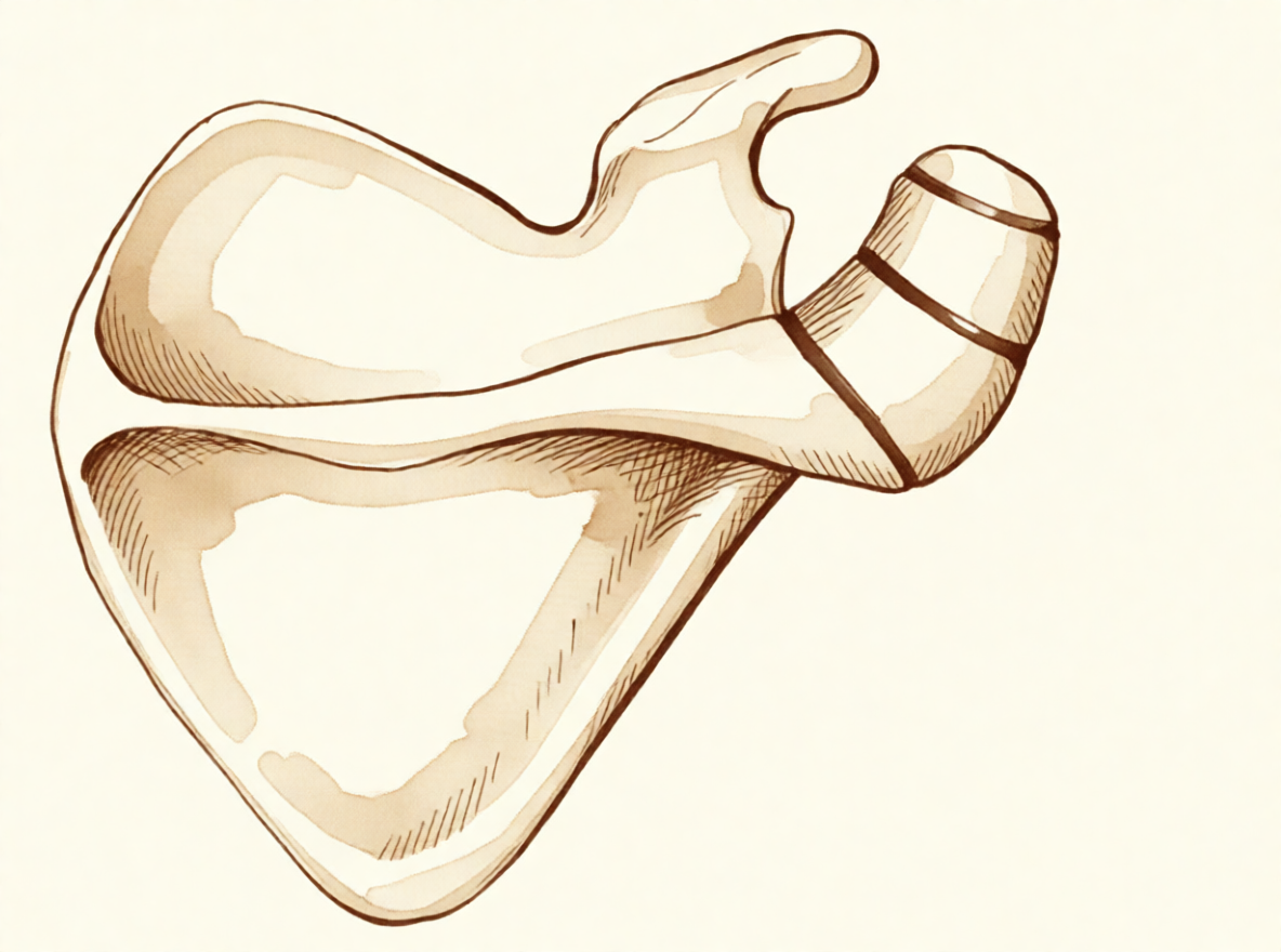
Qué está sintiendo
En el caso de un os acromiale, el dolor suele notarse en la parte frontal y lateral del hombro, sobre el techo óseo de la articulación. Las actividades por encima de la cabeza lo empeoran. Estirarse para alcanzar un estante alto, colgar la ropa o levantar algo por encima de la altura del hombro pueden provocarlo. Muchas personas también notan dolor por la noche, y puede despertarle cuando se acuesta sobre ese lado.
El dolor proviene de un pequeño fragmento óseo en la parte superior del hombro que nunca llegó a unirse al resto durante el crecimiento. Al levantar el brazo, un músculo tira de este fragmento suelto y este se mueve ligeramente, presionando el tendón que está debajo. El punto de unión entre los dos fragmentos óseos también puede doler al tocarlo, y presionarlo directamente puede provocar el dolor.
En el día a día, es posible que note debilidad al levantar el brazo hacia un lado o al girarlo hacia fuera. Es frecuente un arco doloroso: el hombro duele a mitad del recorrido al levantar el brazo y luego se alivia al seguir subiéndolo. Como los síntomas se parecen tanto al desgaste del tendón que está bajo el techo del hombro, esta afección es fácil de confundir con un problema del manguito rotador, y puede seguir causando dolor incluso después de tratar ese problema.
La mayoría de las personas con esta forma del hueso nunca notan nada. A menudo se descubre por casualidad en un estudio de imagen realizado por otro motivo. Cuando sí causa dolor, suele aparecer en adultos jóvenes, a menudo sin ninguna lesión previa, y conviene sospecharlo si el dolor de hombro no se ha aliviado con fisioterapia, con reposo del deporte o con una inyección.
Si sus síntomas no mejoran, empeoran a lo largo de las semanas, le despiertan por la noche o le impiden trabajar o usar el brazo, consulte a su médico de cabecera o solicite una evaluación especializada.
¿Qué está ocurriendo realmente?
El techo óseo del hombro no se forma como una sola pieza de hueso. Comienza como tres centros separados que se van uniendo lentamente; este proceso empieza hacia los 15 años y suele completarse hacia los 25 años. En aproximadamente el 8% de las personas, una de estas uniones nunca se cierra. El espacio se rellena con un tejido resistente, parecido al cartílago, en lugar de hueso, de modo que un pequeño fragmento del techo queda sujeto por una unión flexible en lugar de por hueso sólido.
Puede imaginarlo como una tabla fijada a una pared con un tornillo flojo. Se sostiene, pero se mueve un poco cuando se apoya en ella. El músculo que levanta el brazo hacia un lado se inserta en este techo, de modo que cada vez que levanta el brazo, ese músculo tira del fragmento suelto y este se flexiona ligeramente. Ese movimiento presiona hacia abajo el tendón que pasa por debajo del techo, y el roce lo irrita. Por eso duelen las actividades por encima de la cabeza y por eso el dolor se nota en la parte frontal y lateral del hombro.
La propia unión flexible también puede ser el problema. Puede volverse dolorosa por sí misma, de forma muy parecida a una articulación desgastada, y presionarla provoca el dolor. La mayoría de las personas con esta forma del hueso nunca notan nada, y no parece que por sí sola cause dolor de hombro ni que aumente la probabilidad de una rotura de tendón. Pero cuando el fragmento suelto empieza a moverse más de lo habitual, ya sea por la forma en que se desarrolló o después de un golpe en la zona, puede presionar el tendón cada vez que levanta el brazo. Eso es lo que produce el arco doloroso y el dolor nocturno descritos más arriba.
Como el fragmento suelto se encuentra justo donde está el tendón, ambos problemas suelen aparecer juntos. Si ya le han tratado el hombro por un problema de tendón y el dolor no se ha aliviado, conviene comprobar si existe este fragmento no unido, ya que operar solo el tendón no calmará un fragmento óseo que se mueve por encima de él.
Qué podemos hacer al respecto
El Dr. Kieran Hirpara, cirujano de extremidades superiores en el Mater Private Hospital Rockhampton, comienza con las opciones menos invasivas que se adapten a su condición. Por lo general, los pacientes son derivados a nuestra clínica por su médico de cabecera; si un fisioterapeuta le ha sugerido que nos consulte, igualmente necesitará una derivación de su médico de cabecera para poder acceder al reembolso de Medicare. En su primera visita, tomamos su historia clínica, examinamos su hombro y solicitamos estudios de imagen cuando resultan necesarios.
El primer paso suele ser cambiar la forma en que utiliza el hombro y darle una oportunidad real a la fisioterapia. La fisioterapia busca calmar la irritación bajo el techo óseo y fortalecer los músculos que rodean el hombro para que toleren mejor el trabajo por encima de la cabeza. Pruebe este tratamiento durante un tiempo razonable antes de pensar en cualquier otra opción.
Los analgésicos pueden ayudarle a seguir moviéndose mientras tanto. Los antiinflamatorios sencillos, tomados según le indique su médico de cabecera, alivian las molestias para que pueda hacer sus ejercicios con comodidad. Si el dolor no mejora, empeora a lo largo de las semanas, le despierta por la noche o le impide trabajar o usar el brazo, consulte a su médico de cabecera o solicite una evaluación especializada.
Cuando el tratamiento no quirúrgico no ha producido una mejoría suficiente, consideramos la cirugía. Antes, confirmamos el diagnóstico con radiografías simples, que casi siempre bastan para mostrar el fragmento no unido. Una proyección tomada desde debajo de la axila muestra con claridad la forma de la articulación y la parte frontal del techo, y es importante porque una lesión puede dañar la unión entre los dos fragmentos óseos. La ecografía es una forma sencilla y precisa de revisar el tendón que pasa bajo el techo, y una resonancia magnética ofrece una imagen más completa de ese tendón y de los tejidos blandos que lo rodean.
La operación que ofrecemos con más frecuencia es una evaluación de la articulación mediante cirugía mínimamente invasiva (artroscópica) con una bursectomía, que consiste en retirar el tejido inflamado del espacio que hay bajo el techo, seguida de una pequeña incisión abierta para colocar un injerto óseo y una fijación, es decir, mantener inmóvil el fragmento suelto para que pueda unirse al resto del hueso. Mantenerlo inmóvil permite que los dos fragmentos se suelden. Si el fragmento suelto es pequeño, podemos extirparlo en lugar de fijarlo. Fijar un fragmento más grande requiere cuidado, porque el músculo que levanta el brazo tira de él constantemente y el fragmento es delgado. A veces el fragmento no se une después de la fijación, lo que se denomina falta de consolidación. Hablaremos de qué opción es la más adecuada para su hombro y lo decidiremos juntos.
Qué esperar
La mayoría de las personas con esta forma del hueso nunca tienen ningún problema, y esta variante no necesita ningún tratamiento. Cuando sí causa dolor, el pronóstico depende de si el fragmento suelto es estable y de si es realmente el origen de su dolor.
En el caso de un fragmento doloroso, el tratamiento suele empezar sin cirugía. Cambiar la forma en que utiliza el hombro, la fisioterapia y los analgésicos sencillos alivian el problema a muchas personas. Si esto no ha funcionado después de probarlo durante un tiempo razonable, la cirugía puede reducir de forma real el dolor y mejorar la función del hombro. Cuando el fragmento suelto se mantiene inmóvil y se une al resto del hueso, las personas obtienen mejores resultados en las mediciones de la función del hombro que cuando los dos fragmentos siguen separados. Sin embargo, la cirugía no siempre es la solución. En algunas personas que tienen esta afección y además una rotura de tendón, los resultados tras la cirugía han sido variables, y el 53% obtuvo en conjunto un resultado satisfactorio.
Si no se trata y sigue causándole molestias, el dolor no suele desaparecer por sí solo. Además, puede pasar desapercibido: conviene sospechar esta afección cuando el dolor de hombro no se ha aliviado tras el tratamiento, incluso después de una cirugía por un problema de tendón, ya que operar solo el tendón no calmará un fragmento óseo que se mueve por encima de él.
La recuperación después de la cirugía es gradual. Trabajará con su fisioterapeuta, recuperando primero el movimiento y después la fuerza, y la capacidad de levantar peso por encima de la cabeza volverá poco a poco a lo largo de semanas o meses. El objetivo es un hombro que tolere la vida diaria sin el dolor agudo al levantar el brazo que tenía cuando acudió a consulta.
Si nota que un dedo, la mano o el brazo se pone caliente, rojo, hinchado y doloroso, sobre todo si tiene fiebre, acuda al servicio de urgencias ese mismo día. No necesita una derivación de su médico de cabecera.
¿Cuándo consultar a un especialista?
La mayoría de las personas con esta forma del hueso nunca necesitan atención médica, pero consulte a su médico de cabecera o solicite una evaluación especializada si el dolor de hombro no mejora, empeora a lo largo de las semanas, le despierta por la noche o le impide trabajar o usar el brazo. Solicite una evaluación antes si el dolor se nota en la parte frontal y lateral del hombro, empeora con las actividades por encima de la cabeza o le sigue despertando cuando se acuesta sobre ese lado. También conviene una evaluación si el punto doloroso en la parte superior del hombro duele al presionarlo, si nota debilidad al levantar el brazo hacia un lado o al girarlo hacia fuera, o si el hombro duele a mitad del recorrido al levantar el brazo. Los adultos jóvenes con estos síntomas y sin ninguna lesión previa deben ser evaluados para descartar esta afección, sobre todo si la fisioterapia, el reposo del deporte o una inyección no les han ayudado. Si un dedo, la mano o el brazo se pone caliente, rojo, hinchado y doloroso, sobre todo si tiene fiebre, acuda al servicio de urgencias ese mismo día; no necesita una derivación de su médico de cabecera.
En mayor profundidad
Advanced reading: the deeper science (optional)
Esta sección profundiza más de lo necesario para que usted tome sus propias decisiones de tratamiento. El acromion accesorio merece ser leído con atención, ya que constituye una variante anatómica normal en una proporción significativa de personas; por lo general no provoca ningún problema y, con frecuencia, se le atribuye el dolor en el hombro cuando en realidad no es la causa.
¿Qué es y qué frecuencia tiene?
El acromion, la estructura ósea que forma el techo de la articulación del hombro, se forma a partir de varios centros óseos que normalmente se fusionan a finales de la adolescencia o al inicio de la edad adulta. En algunas personas, uno de esos centros no se fusiona, dejando una unión fibrosa permanente. Eso es lo que se conoce como “os acromiale”, y está presente desde la adolescencia, no se adquiere posteriormente.
La prevalencia varía según la población. Un estudio multicéntrico realizado en 6,842 pacientes determinó que el os acromiale está asociado a lesiones del manguito rotador; además, su prevalencia en la población japonesa estudiada fue menor que en poblaciones no asiáticas, y el tamaño del fragmento tendía a ser más pequeño [1].
Esta asociación con las lesiones del manguito rotador merece ser analizada con detenimiento. Demuestra que ambos fenómenos ocurren juntos con mayor frecuencia de lo que cabría esperar por azar, pero no que el os acromiale cause la rotura del manguito. El mecanismo probable funciona en ambos sentidos: el fragmento no fusionado se mueve ligeramente bajo la acción del músculo deltoides, lo cual podría irritar el tendón subyacente; o bien, la misma mecánica articular que predispone a las lesiones del manguito rotador hace que el fragmento inestable genere síntomas.
La gran incógnita es si realmente es la causa del dolor
Dado que es visible, permanece en el lugar y presenta un aspecto anormal en las imágenes, el hueso acromial suele ser señalado como la causa del dolor en el hombro. Sin embargo, la mayoría de ellos no causan síntomas; de hecho, la mayor parte de las personas que lo tienen ni siquiera lo saben.
Lo que diferencia a los casos sintomáticos es la sensibilidad localizada directamente sobre el fragmento óseo, el dolor que se reproduce al presionarlo y, cuando se realiza una inyección diagnóstica, el alivio que se obtiene al inyectar precisamente en esa zona de unión y no en el espacio subacromial situado debajo. Si no se presentan estos signos, lo más adecuado es considerar este hallazgo como algo incidental.
Los subtipos se clasifican habitualmente según el sitio de fusión afectado [2]; esto es relevante desde el punto de vista quirúrgico, pues el tamaño del fragmento móvil determina si puede extirparse o si es necesario fijarlo.
Por qué complica otras cirugías de hombro
La importancia práctica del os acromiale radica menos en sus propios síntomas que en el efecto que tiene sobre las intervenciones quirúrgicas programadas para otras patologías.
El deltoides se inserta en el acromion; por ello, un fragmento no fusionado queda sometido a esa tracción en una zona anatómica móvil. Al extraer hueso de su superficie inferior, como se hace en la acromioplastia, se reduce aún más el grosor de un fragmento ya de por sí inestable, lo que puede transformar una variante asintomática en una condición dolorosa o provocar una no unión. Este es el motivo principal por el cual debe identificarse mediante estudios de imagen preoperatorios: no para tratarlo, sino para evitar desestabilizarlo.
Cuando sí es necesario tratarlo, se debe optar entre extirpar un fragmento pequeño o fijar uno más grande. La fijación en esta zona resulta difícil: el fragmento es delgado, el deltoides ejerce una tracción constante sobre él y las tasas de no unión son considerables. Esta dificultad constituye, en sí misma, una razón para confirmar que el os acromiale es realmente la causa del dolor antes de proceder a cualquier intervención quirúrgica.
Referencias
[1] Kozono N, Nishii A, Ishitani E, Mizuki Y, Kimura T, Yamamoto S, et al. Prevalencia y factores asociados al os acromiale: un estudio multicéntrico. JSES Int. 2025;9(5):1541-5. https://doi.org/10.1016/j.jseint.2025.05.015
[2] Boehm TD, Matzer M, Brazda D, Gohlke FE. Tratamiento quirúrgico del os acromiale asociado a desgarro del manguito rotador. Revisión de 33 pacientes. J Bone Joint Surg Br. 2003;85(4):545-9. https://doi.org/10.1302/0301-620X.85B4.13634
Evidence & references
This is the clinical evidence summary written for health professionals. It is technical, and it lists the research this page was built from. You do not need to read it to understand your treatment or to make a decision about it.
Overview
- A symptomatic, unstable os acromiale is relatively rare despite the radiographic appearance of an os acromiale being not uncommon in patients presenting with shoulder pain [1].
- An os acromiale should always be suspected in cases of failed rotator cuff surgery or ongoing shoulder pain after failed treatment [2].
- The prevalence of os acromiale was 2.13% in Thai patients with shoulder problems who required MRI evaluation [3].
- Os acromiale is associated with rotator cuff injuries [7].
- The greater the distance of the acromioclavicular joint from the anterior edge of the acromion, the higher the likelihood of an os acromiale [15].
- The authors recommend that the acromion be carefully examined roentgenographically prior to operation in any patient with a rotator cuff tear [10].
- Surgical treatment is usually not indicated for os acromiale in the professional tennis player [9].
- Operative management of a symptomatic os acromiale that has failed initial nonoperative treatment leads to decreased symptoms and improvement in clinical outcomes [4].
- Surgical options for symptomatic os acromiale include arthroscopic sub-total excision, arthroscopic subacromial decompression of stable fragments, and open reduction and internal fixation of unstable fragments [13].
- Patients with a united os acromiale had a significantly better functional outcome as measured by the Constant score (P = .0169) [12].
- The advantages of arthroscopic internal fixation include improved visualization of the undersurface of the acromion, allowing excellent reduction, the possibility of treating concomitant shoulder pathologies, and to avoid increased morbidity of the open approach [14].
- Postoperative local tenderness at the os acromiale can be expected in 1 out of 4 patients but resolves spontaneously over time in the majority of patients [5].
- The presence of os acromiale does not appear to have a negative impact on the clinical outcomes after surgery and rTSA remains a safe and effective treatment option [6].
- The outcome of RTSA does not seem to be negatively affected by the presence of an os acromiale [11].
- Ipsilateral os acromiale may be a relative contraindication to the clavicle hook plate [16].
Anatomy & Pathophysiology
Bony Anatomy
- The acromion has three ossification centers: the metacromion (base), the mesoacromion (middle), and the preacromion (tip) [38].
- Failure of fusion of the acromial ossification centers results in os acromiale [38].
- Os acromiale is defined as incomplete fusion of secondary ossification centers, most commonly between the mesoacromion and meta-acromion [49].
- The unfused segment in os acromiale is most commonly the meso-acromion, a configuration often termed a meso-type os acromiale [22].
- The acromion, coracoacromial ligament, and coracoid process form the coracoacromial arch, a rigid bony-ligamentous structure that imparts stability to the shoulder girdle [35].
- The rotator cuff, subacromial bursa, and subdeltoid bursa pass underneath the coracoacromial arch [35].
- The scapula is attached to the axial skeleton by the acromioclavicular and sternoclavicular joints [37].
- The acromion is a flattened bony process that curves forwards from the scapular spine [37].
Prevalence and Associations
- In Thai patients with shoulder problems who required MRI evaluation, the prevalence of os acromiale was 2.13% [3].
- The radiographic appearance of an os acromiale is not uncommon in patients presenting with shoulder pain [1].
- A symptomatic, unstable os acromiale is relatively rare [1].
Pathophysiology
- Most diagnoses of os acromiale are made incidentally on axillary view radiographs of the shoulder [22].
- Primary shoulder pain is usually unrelated to the unfused os acromiale [22].
- When an os acromiale drives symptoms, the two principal causes are motion at the non-union site or an impingement-type syndrome resulting from flexion of the os fragment during deltoid contraction and arm elevation [22].
- Pain from an os acromiale due to these mechanisms has been reported in a variety of sports, particularly among throwing athletes and swimmers [22].
- Previously stable non-unions can become unstable following an episode of blunt trauma to the region [22].
- The synchondrosis of an os acromiale can be injured following trauma [24].
- Extrinsic impingement occurs when the space available for the rotator cuff is diminished, with examples including acromial fracture or pathologic os acromiale [62].
Epidemiology and Prevalence
- Liberson reviewed 1800 shoulder girdles and identified an incidence of os acromiale of 1.4% [21].
- The lesion was bilateral in 62% of patients in Liberson's review of 1800 shoulder girdles [21].
Associated Pathology and Risk Factors
- The greater the distance of the AC joint from the anterior edge of the acromion, the higher the likelihood of an os acromiale [15].
Diagnostic Considerations
- Awareness of the os acromiale in the young athlete, appropriate clinical examination, and image studies are crucial to confirm diagnosis [19].
- Appropriate radiographic investigation including axillary views is emphasized for the diagnosis of os acromiale injuries [24].
Clinical Presentation
- An os acromiale should always be suspected in cases of failed rotator cuff surgery [2].
- An os acromiale should always be suspected in cases of ongoing shoulder pain after failed treatment [2].
- Postoperative local tenderness at the os acromiale can be expected in 1 out of 4 patients [5].
- Postoperative local tenderness at the os acromiale resolves spontaneously over time in the majority of patients [5].
- The lesion was bilateral in 62% of patients in Liberson's review [21].
- The unfused segment is most commonly the meso-acromion [22].
- Any primary shoulder pain is usually unrelated to the unfused os [22].
- If the os acromiale is driving symptoms, one principle cause is motion at the non-union site [22].
- If the os acromiale is driving symptoms, another principle cause is an impingement-type syndrome resulting from flexion of the os fragment during deltoid contraction and arm elevation [22].
- Pain from an os acromiale by the mechanism of impingement has been reported in a variety of sports [22].
- Pain from an os acromiale by the mechanism of impingement has been reported in particular among throwing athletes and swimmers [22].
- Meta–os acromiale is the rarest subtype of os acromiale [23].
Investigations
Prevalence and Demographics
- Liberson reviewed 1800 shoulder girdles and identified an incidence of os acromiale of 1.4%, with the lesion being bilateral in 62% of patients [21].
- A multicenter study aimed to determine the prevalence of and factors associated with os acromiale in the Japanese population [8].
- The radiographic appearance of an os acromiale is not uncommon in patients presenting with shoulder pain, but a symptomatic, unstable os acromiale is relatively rare [1].
Radiographic Evaluation
- The acromion should be carefully examined roentgenographically prior to operation in any patient with a rotator cuff tear [10].
- Appropriate radiographic investigation for os acromiale includes axillary views [24].
- Awareness of the os acromiale in the young athlete requires appropriate clinical examination and image studies to confirm diagnosis [19].
- The standard shoulder series should include orthogonal views of the shoulder, including a true AP view in the scapular plane, an AP view, an axillary view, and a scapular Y view [60].
- The scapular Y view provides visualization of the coracoacromial arch and can reveal coracoacromial spurs, which have been closely associated with the presence of rotator cuff pathology [60].
- The scapular Y view can also show acromial shape [60].
- At least two X-ray views should be obtained for shoulder imaging: an anteroposterior in the plane of the glenoid and an axillary projection with the arm in abduction to show the relationship of the humeral head to the glenoid [52].
- The purpose of imaging of the shoulder is to help establish the diagnosis, determine the severity of the pathoanatomy, assist in surgical planning, and enable the surgeon to illustrate the condition of the shoulder to the patient [30].
- Standardized plain films are almost always sufficient to garner the information needed for shoulder evaluation [30].
- The first key view for shoulder imaging is the anteroposterior (AP) in the plane of the scapula taken so that the x-ray beam passes through the glenohumeral joint [30].
- The second key view for shoulder imaging is the axillary view taken with the arm in the functional position of elevation in the plane of the scapula and oriented so that both the spinoglenoid notch and the scapular neck are visible [30].
- The axillary view is referred to as the "truth view" because it demonstrates the glenohumeral relationships in the functional position of elevation [30].
- CT scans have the disadvantage of being taken with the arm in the adducted position, unlike the axillary truth view [30].
- The temptation to "overimage" should be resisted, obtaining only the scans or reconstructions that are necessary for the care of the patient [30].
Magnetic Resonance Imaging
- In Thai patients with shoulder problems, os acromiale was identified via Magnetic Resonance Imaging evaluation [3].
- MRI is useful to identify labral tears and rotator cuff tears, although the accuracy for these is enhanced by combining the scan with arthrography [52].
- MRI is the modality of choice for evaluating the rotator cuff, biceps, and subacromial/subdeltoid bursa [59].
- T2-weighted MRI provides better visualization of full thickness rotator cuff tears [59].
- MR arthrography is considered the benchmark for evaluation for labral tears and rarely is indicated for evaluation of rotator cuff pathology [59].
- When MRI or MR arthrography is contraindicated, CT arthrography is indicated [59].
Ultrasonography
- Ultrasonography is a simple and accurate test for identifying rotator cuff tears and calcific tendinitis [52].
- Ultrasonography is a low-cost alternative to MRI and arthrography for evaluating both skeletal and soft-tissue structures of the shoulder [59].
- Ultrasonography can provide immediate, real-time visualization of the rotator cuff, biceps tendon, and calcific deposits [59].
- Ultrasonography can be used to measure the subacromial space and detect atrophy of rotator cuff muscles [59].
- Ultrasonography is highly operator dependent and is not as useful for evaluating labral tears or rotator cuff tears that are very small or larger than 3 cm [59].
- The most commonly performed joint examination using ultrasonography is the shoulder examination, and accuracy depends on the skill of the scanner operator and an awareness of pitfalls that are encountered [50].
Clinical Associations and Diagnostic Implications
- The synchondrosis of an os acromiale can be injured following trauma, though rarely [24].
Treatment
Non-Operative
- A symptomatic os acromiale in a competitive female fastball pitcher was treated successfully with open reduction and internal fixation [27].
Operative
- Symptomatic unstable os acromiale can be effectively treated with autogenous bone-grafting and internal fixation using a rigid tension-band construct and cannulated screws, which results in a high rate of union [65].
- This new arthroscopic technique of fixation of os acromiale with absorbable screws provides promising clinical, cosmetic, and radiologic results with high patient satisfaction [28].
- After 3 to 6 years of follow-up, all 6 shoulders were rated as excellent according to criteria similar to that of the American Shoulder and Elbow Surgeons [18].
- Modified arthroscopic subacromial decompression can provide results similar to standard arthroscopic subacromial decompression for routine impingement [25].
- Meta–os acromiale is the rarest subtype of os acromiale, and special consideration must be given to the type of tension-band construct used to achieve adequate compression and fixation [23].
Complications
Post-operative and Surgical Complications
- Postoperative local tenderness at the os acromiale can be expected in 1 out of 4 patients following reverse total shoulder arthroplasty [5].
Diagnostic and Clinical Implications
Pathophysiology and Mechanisms of Symptom
- Pain from an os acromiale by the mechanism of impingement has been reported in a variety of sports, in particular among throwing athletes and swimmers [22].
Recovery
- After 3 to 6 years of follow-up, all 6 shoulders treated with osteosynthesis of an unstable mesoacromion were rated as excellent according to criteria similar to that of the American Shoulder and Elbow Surgeons [18].
- At a two to five year follow-up, all patients treated for fusion of the unstable mesoacromion had fused [69].
Key Evidence
- [L4] While the radiographic appearance of an os acromiale is not uncommon in patients presenting with shoulder pain, a symptomatic, unstable os acromiale is relatively rare. [1] (10.1097/00132589-200412000-00006)
- [L4] An os acromiale should always be suspected in cases of failed rotator cuff surgery or ongoing shoulder pain after failed treatment. [2] (10.1097/00132589-200609000-00006)
- [L3] In Thai patients with shoulder problems who required MRI evaluation, the prevalence of os acromiale was 2.13%. [3] (10.1177/23259671221078806)
- [L4] Operative management of a symptomatic os acromiale that has failed initial nonoperative treatment leads to decreased symptoms and improvement in clinical outcomes. [4] (10.1016/j.jse.2019.05.047)
- [L3] Postoperative local tenderness at the os acromiale can be expected in 1 out of 4 patients but resolves spontaneously over time in the majority of patients. [5] (10.1177/2325967120965131)
- [L4] The presence of os acromiale does not appear to have a negative impact on the clinical outcomes after surgery and rTSA remains a safe and effective treatment option. [6] (10.1016/j.xrrt.2025.01.002)
- [L3] The study supports previous findings that os acromiale is associated with rotator cuff injuries. [7] (10.1016/j.jseint.2025.05.015)
- [L3] This multicenter study aimed to determine the prevalence of and factors associated with os acromiale in the Japanese population. [8] (10.1016/j.jse.2025.01.008)
- [L4] Surgical treatment is usually not indicated for os acromiale in the professional tennis player. [9] (10.1177/2325967118773723)
- [L4] The authors recommend that the acromion be carefully examined roentgenographically prior to operation in any patient with a rotator cuff tear. [10] (10.2106/00004623-198466080-00029)
- [L4] The outcome of RTSA does not seem to be negatively affected by the presence of an os acromiale. [11] (10.1016/j.jse.2017.02.012)
- [L4] Patients with a united os acromiale had a significantly better functional outcome as measured by the Constant score (P = .0169). [12] (10.1016/s1058-2746(98)90008-8)
- [L5] Surgical options for symptomatic os acromiale include arthroscopic sub-total excision, arthroscopic subacromial decompression of stable fragments, and open reduction and internal fixation of unstable fragments. [13] (10.5435/jaaos-d-17-00011)
- [L5] The advantages of this technique are improved visualization of the undersurface of the acromion, allowing excellent reduction, the possibility of treating concomitant shoulder pathologies, and to avoid increased morbidity of the open approach. [14] (10.1097/bte.0000000000000191)
- [L4] Our data suggest that the greater the distance of the AC joint from the anterior edge of the acromion, the higher the likelihood of an os acromiale. [15] (10.1067/mse.2003.128136)
- [L4] Ipsilateral os acromiale may be a relative contraindication to the clavicle hook plate. [16] (10.1186/s12891-021-04841-1)
- [L4] After 3 to 6 years of follow-up, all 6 shoulders were rated as excellent according to criteria similar to that of the American Shoulder and Elbow Surgeons. [18] (10.1016/s1058-2746(99)90004-6)
- [L4] Awareness of the os acromiale in the young athlete, appropriate clinical examination, and image studies are crucial to confirm diagnosis. [19] (10.1016/j.jseint.2020.02.008)
- [L4] [22] (10.1302/2058-5241.4.180100)
- [L4] Meta–os acromiale is the rarest subtype of os acromiale, and special consideration must be given to the type of tension-band construct used to achieve adequate compression and fixation. [23] (10.1177/03635465211028238)
- [L4] This case highlights that the synchondrosis of an os acromiale can be injured following trauma, though rarely, and emphasizes the need for appropriate radiographic investigation including axillary views and a flexible surgical approach. [24] (10.1016/j.jse.2008.02.012)
- [L5] Increased awareness of the diagnosis and improved techniques have shown that modified arthroscopic subacromial decompression can provide results similar to standard arthroscopic subacromial decompression for routine impingement. [25] (10.1097/00132589-200206000-00003)
- [L4] A symptomatic os acromiale in a competitive female fastball pitcher was treated successfully with open reduction and internal fixation. [27] (10.1177/0363546506288305)
- [L4] This new arthroscopic technique of fixation of os acromiale with absorbable screws provides promising clinical, cosmetic, and radiologic results with high patient satisfaction. [28] (10.1016/j.jse.2011.12.011)
- [L4] Symptomatic unstable os acromiale can be effectively treated with autogenous bone-grafting and internal fixation using a rigid tension-band construct and cannulated screws, which results in a high rate of union. [65] (10.2106/00004623-199809000-00011)
- [L4] At a two to five year followup all had fused. [69] (10.1016/s1058-2746(95)80192-8)
References
[1] Treatment of the Symptomatic Os Acromiale. Techniques in Shoulder and Elbow Surgery. 2004. DOI: 10.1097/00132589-200412000-00006
[2] Management of Os Acromiale. Techniques in Shoulder and Elbow Surgery. 2006. DOI: 10.1097/00132589-200609000-00006
[3] Prevalence of Os Acromiale in Thai Patients With Shoulder Problems: A Magnetic Resonance Imaging Study. Orthopaedic Journal of Sports Medicine. 2022. DOI: 10.1177/23259671221078806
[4] Os acromiale: systematic review of surgical outcomes. Journal of Shoulder and Elbow Surgery. 2020. DOI: 10.1016/j.jse.2019.05.047
[5] Os Acromiale in Reverse Total Shoulder Arthroplasty: A Cohort Study. Orthopaedic Journal of Sports Medicine. 2020. DOI: 10.1177/2325967120965131
[6] Clinical implications of reverse total shoulder arthroplasty with an os acromiale: a systematic review. JSES Reviews, Reports, and Techniques. 2025. DOI: 10.1016/j.xrrt.2025.01.002
[7] Prevalence and factors associated with os acromiale: a multicenter study. JSES International. 2025. DOI: 10.1016/j.jseint.2025.05.015
[8] The prevalence and associated factors of os acromiale: a multicenter study. Journal of Shoulder and Elbow Surgery. 2025. DOI: 10.1016/j.jse.2025.01.008
[9] Os Acromiale in Professional Tennis Players. Orthopaedic Journal of Sports Medicine. 2018. DOI: 10.1177/2325967118773723
[10] Rotator cuff tears associated with os acromiale.. The Journal of Bone & Joint Surgery. 1984. DOI: 10.2106/00004623-198466080-00029
[11] Reverse shoulder arthroplasty in patients with os acromiale. Journal of Shoulder and Elbow Surgery. 2017. DOI: 10.1016/j.jse.2017.02.012
[12] Transacromial approach to obtain fusion of unstable os acromiale. Journal of Shoulder and Elbow Surgery. 1998. DOI: 10.1016/s1058-2746(98)90008-8
[13] Symptomatic, Unstable Os Acromiale. Journal of the American Academy of Orthopaedic Surgeons. 2018. DOI: 10.5435/jaaos-d-17-00011
[14] Arthroscopic Internal Fixation of Symptomatic Os Acromiale. Techniques in Shoulder & Elbow Surgery. 2020. DOI: 10.1097/bte.0000000000000191
[15] Relationship between os acromiale and acromioclavicular joint anatomic position. Journal of Shoulder and Elbow Surgery. 2003. DOI: 10.1067/mse.2003.128136
[16] Os acromiale may be a contraindication of the clavicle hook plate: case reports and literature review. BMC Musculoskeletal Disorders. 2021. DOI: 10.1186/s12891-021-04841-1
[18] Successful osteosynthesis of an unstable mesoacromion in 6 shoulders: A new technique. Journal of Shoulder and Elbow Surgery. 1999. DOI: 10.1016/s1058-2746(99)90004-6
[19] The unstable os acromiale: a cause of pain in the young athlete. JSES International. 2020. DOI: 10.1016/j.jseint.2020.02.008
[21] Types of os acromiale according to Liberson. 2006.
[22] Os acromiale: a review of its incidence, pathophysiology, and clinical management. EFORT Open Reviews. 2019. DOI: 10.1302/2058-5241.4.180100
[23] Rare Symptomatic Meta–Os Acromiale in an Athlete. The American Journal of Sports Medicine. 2021. DOI: 10.1177/03635465211028238
[24] Fracture of an os acromiale with associated rupture of the coracoclavicular ligaments. Journal of Shoulder and Elbow Surgery. 2008. DOI: 10.1016/j.jse.2008.02.012
[25] Arthroscopic Management of the Mesoacromion. Techniques in Shoulder and Elbow Surgery. 2002. DOI: 10.1097/00132589-200206000-00003
[27] Surgical Stabilization of Os Acromiale in a Fast-Pitch Softball Pitcher. The American Journal of Sports Medicine. 2006. DOI: 10.1177/0363546506288305
[28] Arthroscopically assisted internal fixation of the symptomatic unstable os acromiale with absorbable screws. Journal of Shoulder and Elbow Surgery. 2012. DOI: 10.1016/j.jse.2011.12.011
[30] Rockwood And Matsen S The Shoulder. Arthroscopic Management of Prearthritic and Arthritic Conditions of the Shoulder and the Postarthroplasty Shoulder > Radiographic Evaluation.
[35] Rockwood And Matsen S The Shoulder. Shoulder and Elbow Specialty Clinic Workers’ Survey > ANATOMY.
[37] Rockwood And Green S Fractures In Adults. 29: Principles of Nonunion and Bone Defect Treatment > Applied Anatomy Related to Scapular Fractures.
[38] Aaos Comprehensive Orthopaedic Review 3. Anatomy of the Shoulder, Arm, and Elbow > I. Shoulder.
[49] Miller S Review Of Orthopaedics. Genetics of musculoskeletal conditions and abnormalities are summarized in Table 1.27 > UPPER EXTREMITY > SHOULDER.
[50] Orthopaedic Knowledge Update Sports Medicine 6. Diagnostic Ultrasonography and Ultrasonography-Guided Procedures > Annotated References.
[52] Apley And Solomon S Concise System Of Orthopaedics And Trauma. INVESTIGATION.
[59] Orthopaedic Knowledge Update 13 Ebook Without Multimedia. Shoulder Anatomy and Biomechanics, Clinical Evaluation, Imaging > Clinical Evaluation > Imaging.
[60] Aaos Comprehensive Orthopaedic Review 3. Imaging of the Shoulder and Elbow > I. Shoulder.
[62] Campbell S Operative Orthopaedics 4 Volume Set. ANTERIOR CRUCIATE LIGAMENT RECONSTRUCTION WITH BONE-PATELLAR TENDON-BONE GRAFT > Developmental Stages of Impingement Syndrome.
[65] The Treatment of Symptomatic Os Acromiale. The Journal of Bone & Joint Surgery*. 1998. DOI: 10.2106/00004623-199809000-00011
[69] Successful fusion of the unstable mesoacromion: Technique and report of five cases. Journal of Shoulder and Elbow Surgery. 1995. DOI: 10.1016/s1058-2746(95)80192-8