Patients › Elbow
Reconstrucción (estabilización) de los ligamentos del codo
¿Por qué se ha sugerido esta operación?
El Dr. Kieran Hirpara, cirujano de extremidades superiores en el Mater Private Hospital Rockhampton, adapta el tratamiento a su lesión específica. Por lo general, los pacientes son derivados a nuestra clínica por su médico de cabecera; si un fisioterapeuta le ha recomendado que nos consulte, igualmente necesitará una derivación de su médico de cabecera para poder acceder al reembolso de Medicare. En su consulta, tomamos su historia clínica, examinamos su codo y, si es necesario, solicitamos estudios de imagen para determinar cuál es el problema.
La reconstrucción de los ligamentos del codo es una intervención mediante la cual se repara un ligamento desgastado o roto utilizando un fragmento de tendón de donante, con el fin de mantener la articulación estable. Normalmente la recomendamos cuando, tras otros tratamientos, el codo sigue mostrando tendencia a “ceder” o no permanece en su posición correcta. También se indica cuando el codo solo puede mantenerse en su sitio si se mantiene casi completamente estirado, o cuando una fractura cerca de la articulación ha provocado inestabilidad. El objetivo es lograr un codo estable que pueda moverse, usarse y confiarse sin dolor. La reconstrucción con injerto alogénico restaura la estabilidad del codo en aproximadamente el 85 % de los casos de inestabilidad rotatoria posterolateral. Discutiremos todo esto con usted y decidiremos conjuntamente si este procedimiento se adapta a su codo y a sus objetivos.
Antes de la operación
En las semanas previas a la cirugía, finalizamos la planificación mediante estudios como radiografías, resonancias magnéticas (un estudio que muestra los tejidos blandos, como los ligamentos) o ecografías. El día de la operación, no debe comer ni beber durante siete horas antes. Pedimos que se respeten esas siete horas para poder adelantar su intervención si la lista de quirófanos lo permite; su cirujano le confirmará el tiempo exacto de ayuno. Es posible que deba suspender algunos medicamentos antes de la cirugía; le daremos instrucciones claras sobre cuáles y por cuánto tiempo. Lleve una lista por escrito de todos los fármacos que toma, incluyendo pastillas, gotas y cremas. Organice que alguien lo lleve a casa después de la operación. Use ropa holgada y cómoda, cuyas mangas puedan deslizarse por encima del codo. Si padece otras enfermedades, es posible que también necesite análisis de sangre o una consulta con el anestesista (el médico encargado de administrar la anestesia).
El día de la intervención
Llega usted a la unidad de admisiones quirúrgicas del hospital, donde se le registrará y preparará para el quirófano. Allí conocerá al anestesista. Esta operación se realiza bajo anestesia general. En ocasiones se añade un bloqueo nervioso regional para aliviar el dolor postoperatorio; el anestesista hablará con usted al respecto ese mismo día. A continuación, se le lleva al quirófano, donde se lleva a cabo la intervención. Una vez finalizada, despierta usted en la sala de recuperación, donde las enfermeras le vigilan mientras la anestesia va desapareciendo. Cuando su estado sea estable, será trasladado a la planta de hospitalización o podrá volver a casa, según el tipo de procedimiento y su recuperación.
El objetivo de este día es sencillo: devolver suficiente estabilidad al codo para que pueda empezar a moverse poco después de la cirugía. Mantener el codo inmóvil durante mucho tiempo después de la operación suele provocar rigidez, por lo que el movimiento temprano forma parte del plan de tratamiento.
Qué implica la operación
Los pasos exactos dependen de qué estructuras alrededor del codo están dañadas; planificamos todo esto a partir de sus estudios de imagen antes del día de la intervención. Si hay una fractura, el cirujano repara primero los fragmentos óseos mediante tornillos o una placa pequeña. En caso de que un fragmento óseo en la parte superior del antebrazo no pueda repararse, podría sustituirse por un implante metálico. Una vez que el hueso queda estabilizado, se reparan o reconstruyen los ligamentos desgarrados.
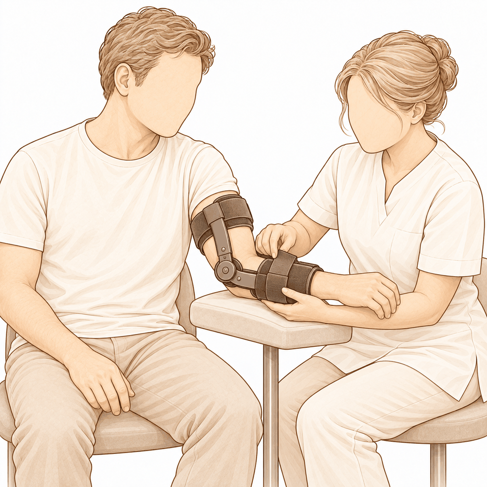
La reconstrucción de un ligamento consiste en crear uno nuevo a partir de un fragmento de tendón; este puede provenir de tejido de donante o de su propio cuerpo. Pequeñas anclas fijan el nuevo ligamento a los huesos en los lugares adecuados, y el cirujano verifica que el codo mantenga su articulación en toda su amplitud de movimiento antes de finalizar. En ocasiones, se añade una cinta de sutura resistente junto a la reparación, que actúa como un refuerzo interno y sostiene el ligamento mientras cicatriza. Si el codo sigue sin mantenerse estable, se puede colocar una bisagra temporal para mantenerlo en posición mientras todo se asienta.
Con frecuencia, la operación se realiza mediante incisiones pequeñas en lugar de una sola incisión larga. Para ciertas reconstrucciones ligamentosas, una incisión de unos 2 a 3 cm es suficiente. Realizar la intervención a través de incisiones pequeñas protege los tendones y músculos sanos alrededor de la articulación, evitando alterar el propio revestimiento articular.
Una vez finalizada la reparación, las incisiones se cierran con puntos de sutura y se cubren con un vendaje. El objetivo de toda la operación es obtener un codo estable que pueda comenzar a moverse poco después, ya que mantenerlo inmóvil durante demasiado tiempo suele provocar rigidez.
Después de la operación
Despertará en la sala de recuperación, donde las enfermeras lo vigilarán de cerca mientras el efecto de la anestesia desaparece. Su brazo estará en un cabestrillo o apoyado en almohadas, con un vendaje sobre las heridas. Le administraremos analgésicos y comprobaremos que surtan efecto antes de permitirle moverse. Alguien debe acompañarlo durante las primeras 24 horas después de volver a casa. Su equipo le indicará si podrá irse a casa el mismo día o si deberá permanecer una noche en el hospital. Dejamos el vendaje puesto durante unos 10 días; por favor, no lo retire antes de ese plazo a menos que se lo indiquemos. Lo cambiamos o lo retiramos cuando venga a la consulta. La mayoría de las personas pueden caminar y realizar tareas ligeras de inmediato, pero debe mantener el codo apoyado en el cabestrillo mientras se mueva.
Recuperación
Durante los primeros días, el codo le dolerá y se hinchará. Esto mejora gradualmente. El descanso, mantener el brazo elevado y los analgésicos que le proporcionamos aliviarán la molestia. Es normal sentir cierto dolor al empezar a mover la articulación; este dolor disminuye con el paso de las semanas.
Se irá a casa con el brazo en un cabestrillo. Podrá caminar y realizar tareas ligeras de inmediato, pero deberá mantener el codo apoyado en el cabestrillo mientras esté de pie. La mano y la muñeca pueden moverse desde el principio; los ejercicios suaves para ellas suelen comenzar a los pocos días. La terapia de la mano tras la cirugía la llevará a cabo Ruby Doolan en Extend Rehabilitation. Ruby es terapeuta especializada en mano: ella le guiará en los ejercicios y le confeccionará cualquier férula que necesite. El objetivo de la terapia es lograr un movimiento temprano y constante, pues mantener el codo inmóvil durante mucho tiempo suele provocar rigidez.
A medida que la hinchazón disminuya, podrá utilizar más el brazo: para comer, escribir y realizar tareas domésticas ligeras. Una vez que el cirujano considere que la reparación es segura, se retirará el cabestrillo y comenzará a doblar y estirar el codo por sí mismo. Al principio, el movimiento suele mejorar rápidamente; después, el progreso se vuelve más lento. Cuando pueda agarrar y sostener objetos sin dolor, las actividades cotidianas le resultarán más fáciles. No es seguro conducir mientras el brazo permanezca en cabestrillo; además, deberá ser capaz de sujetar el volante con ambas manos y reaccionar en caso de una frenada de emergencia, sin depender de fuertes analgésicos. Nuestra guía sobre conducción tras una cirugía de miembro superior explica cuándo puede volver a conducir.
Cada persona sana a su propio ritmo. Su cronograma personal puede variar; su cirujano y terapeuta le guiarán durante todo el proceso.
Qué puede salir mal
La mayoría de los pacientes evolucionan bien, pero en ocasiones pueden surgir problemas. Su cirujano y el equipo lo vigilarán de cerca para detectar cualquier incidencia a tiempo.
El nervio que discurre por la parte interna del codo puede irritarse después de la cirugía. Es posible que note hormigueo, sensación de pinchazos o entumecimiento en el dedo anular y el meñique. Con frecuencia esto se resuelve por sí solo; sin embargo, avísenos si no mejora o si los dedos parecen débiles.
En algunos casos, el codo permanece flojo o da la impresión de volver a “ceder”. Si siente inestabilidad en el codo o si éste se disloca, comuníquese con la clínica de inmediato.
El codo también puede volverse rígido. Es posible que le resulte difícil estirar o doblar el brazo por completo; el movimiento podría sentirse bloqueado en lugar de doloroso. Mencione esto en su revisión, pues la terapia adicional o un tratamiento posterior pueden ser de ayuda.
Las infecciones son poco frecuentes pero graves. Esté atento a un dolor profundo y pulsátil que no ceda con analgésicos comunes, a enrojecimiento que se extienda desde la herida o a secreción de líquido. Si observa alguno de estos síntomas, llame a la clínica ese mismo día; si presenta fiebre o malestar general, acuda a urgencias.
Los pequeños anclajes o tornillos metálicos utilizados para fijar la reparación pueden irritar los tejidos, o una articulación temporaria podría aflojarse. Es posible que note un nuevo chasquido, sensación de “traba” o un bulto bajo la piel. Coméntelo en su próxima revisión.
Cuando se emplea un fragmento de su propio tendón para reconstruir el ligamento, esa zona puede permanecer sensible o dolorida durante un tiempo. Avísenos si el dolor empeora en lugar de mejorar.
En ocasiones se forma hueso donde no debería, alrededor o dentro de la articulación. Esto puede limitar el movimiento o provocar sensación de “traba” y fricción. Si el codo deja de mejorar, indíquelo en su revisión.
En raras ocasiones, la cicatriz puede abrirse o supurar. Si la herida se abre, se enrojece o comienza a secretar líquido, comuníquese con nosotros sin esperar.
En la tabla de complicaciones de esta página se detallan las tasas típicas, por si desea conocer los datos específicos.
¿Cuándo deben llamarnos?
La mayoría de los problemas aparecen en las primeras etapas; preferimos que nos informen lo antes posible. Llámenos si tiene fiebre, si la herida se vuelve más roja o comienza a supurar líquido, o si el dolor empeora en lugar de mejorar. Acuda a urgencias si presenta hinchazón o dolor en la pantorrilla, o si experimenta dificultad respiratoria repentina. Llámenos de inmediato si sus dedos anular y meñique se entumecen, si siente debilidad en la mano o si no puede mover el brazo en absoluto. Si su codo se disloca o parece volver a “ceder”, comuníquese con la clínica ese mismo día.
Evidence & references
This is the clinical evidence summary written for health professionals. It is technical, and it lists the research this page was built from. You do not need to read it to understand your treatment or to make a decision about it.
Overview
- Surgical ligament reconstruction or repair is the most favorable treatment option for restoration of normal elbow function in posterolateral rotatory instability [4].
- Repair and reconstruction of the lateral ulnar collateral ligament offers practical and often successful solutions for patients with posterolateral rotatory instability of the elbow [10].
- Improving arthroscopic techniques provide a satisfactory alternative in stabilizing the elbow [7].
- The gravity varus test has biomechanical plausibility and confirms that pronation stabilizes the lateral elbow [1].
- An all-arthroscopic reconstruction technique for the lateral ulnar collateral ligament is reproducible and avoids residual instability [2].
- Open posterolateral ligament plication and lateral ulnar collateral ligament repair using an all-suture construct allows for complete posterolateral stabilization of the elbow with a single implant and bone preservation [3].
- Suture-augmented lateral ulnar collateral ligament and radial collateral ligament reconstruction provides a reproducible, anatomically based construct that restores posterolateral elbow stability and addresses the complex spectrum of lateral-sided injuries observed in posterolateral rotatory instability [5].
- Further follow-up and wider experience of the Surgilig/LockDown technique is required to assess its long-term functional outcome in comparison with more established surgical techniques for stabilization of posterolateral elbow instability [6].
- Arthroscopic lateral ulnar collateral ligament plication/reconstruction with augmented lateral collateral ligament imbrication allows effective management of elbow instability while promoting quicker patient recovery and long-term functional restoration [8].
- Acute repair of the lateral ulnar collateral ligament with a cortical button resulted in satisfactory clinical outcomes and successful restoration of elbow stability [9].
- The use of suture button fixation for repair of the lateral ulnar collateral ligament in terrible triad injuries has not been previously described [11].
- Corrective open-wedge osteotomy with structural bone allograft restored radial head alignment, eliminated dynamic subluxation, and achieved stable elbow function without the need for ligament reconstruction in complex posterolateral rotatory instability due to malunited radius fracture [12].
- Reconstruction of the lateral ulnar collateral ligament with a tendon graft offers an alternative that restores stability through a dynamic “sling effect” rather than rigid constraint [13].
Anatomy & Pathophysiology
Bony Anatomy
- The elbow is a trocho-ginglymoid joint consisting of medial and lateral articulations that provide bony stability [22].
- The ulnohumeral joint is formed by the articulation of the trochlea with the ulna within the greater sigmoid notch [22].
- The ulnohumeral joint provides highly congruent anatomy through almost 180° of articular contact, except for a bare area of the greater sigmoid notch devoid of cartilage [22].
- The radiocapitellar joint is formed by the articulation of the capitellum and the radial head [22].
- The radial head is a concave elliptical structure covered with articular cartilage along the radiocapitellar joint and approximately 270° of the articular margin [22].
- The radius is held in close approximation to the ulna at the proximal radioulnar joint by the annular ligament [22].
- The distal humeral articulation is angled 30° from the longitudinal axis of the humerus [22].
- The axis of rotation is angulated 5° to 7° in the coronal plane relative to the epicondylar axis, with the medial side more distal than the lateral side [22].
- The articular surface of the distal humerus is angled 30 degrees anterior to the humeral shaft axis [28].
- In full extension, 60% of axial load is transmitted through the radiocapitellar joint [28].
- The medial column of the distal humerus diverges from the humeral shaft at a 45-degree angle in the coronal plane [30].
- The lateral column of the distal humerus diverges from the humeral shaft at a 20-degree angle in the coronal plane [30].
- The trochlea is covered by articular cartilage anteriorly, inferiorly, and posteriorly, creating an arc of almost 270 degrees [33].
- The olecranon fossa and coronoid fossa are separated by a thin bony septum which is occasionally absent [33].
- The ulna medially bends approximately 8° at 8 cm from the tip of the olecranon [22].
- The articulation to the tip of the coronoid is approximately 30° from the long axis of the ulna in the sagittal plane [22].
Ligamentous Anatomy
- The lateral collateral ligament complex consists of the radial collateral ligament, the lateral ulnar collateral ligament (LUCL), and the annular ligament [33].
- The radial collateral ligament originates from an isometric point on the lateral epicondyle and fans out to attach to the annular ligament [33].
- The LUCL arises from an isometric point on the lateral epicondyle and attaches to the crista supinatoris of the proximal ulna [33].
- The annular ligament attaches to the anterior and posterior margins of the lesser sigmoid notch [33].
- The LUCL origin center is located 10.7 mm from the lateral epicondyle [21].
- The LUCL insertion is located 3.3 mm from the apex of the supinator crest [21].
- The LUCL acts as a posterior buttress for the radial head and the ulna to prevent subluxation [17].
- The LUCL is considered the principal constraint of the elbow joint against posterolateral rotatory instability (PLRI) [17].
- The lateral collateral ligament contributes 14% of varus stability of the elbow with the joint in full extension [37].
- The lateral collateral ligament contributes 9% of varus stability of the elbow with the joint in 90 degrees of flexion [37].
- The medial ulnar collateral ligament (MUCL) is the primary restraint to valgus stress within functional elbow range of motion [28].
- The MUCL originates on the posterior medial epicondyle and inserts on the sublime tubercle of the medial coronoid process [28].
- The anterior bundle of the MUCL is the primary restraint to valgus stress within functional elbow range of motion [28].
- The posterior bundle of the MUCL is the primary restraint to valgus stress with the elbow in maximal flexion [28].
- The common extensor tendon is a secondary stabilizer of the lateral elbow [16].
- The joint capsule is a secondary stabilizer of the elbow [19].
- The radiocapitellar articulation is a secondary stabilizer of the elbow [19].
Biomechanics & Pathophysiology
- Elbow stability is determined by primary and secondary stabilizers, with injury to these structures causing instability [19].
- The three primary stabilizers of the elbow are the ulnohumeral articulation, the MUCL, and the LUCL complex [19].
- Secondary stabilizers of the elbow include the radiocapitellar articulation, common flexor tendon, common extensor tendon, and joint capsule [19].
- Tensile forces are present at the medial elbow while compressive forces are present at the lateral elbow [23].
- The capsule allows maximum distension at approximately 70 to 80 degrees of flexion [23].
- The anterior capsule attaches at a point approximately 6 mm distal to the tip of the coronoid [23].
- Pronation stabilizes the lateral elbow [1].
- The LUCL is taut in both flexion and extension [37].
- Disruption of the LUCL results in posterolateral rotatory instability [37].
- Failure of primary stabilizers results in increased stress on secondary stabilizers, leading to capsular contractures, chondromalacia, osteophytes, and loose bodies [37].
- Ulnar nerve symptoms may develop from traction, scarring, or osteophyte impingement following primary stabilizer failure [37].
- The typical injury pattern for traumatic elbow dislocation involves a valgus internal rotation force of the forearm on a slightly flexed extremity [37].
- In traumatic elbow dislocation, structures are disrupted on the lateral side first, progressing medially as more force is applied [37].
- Posterolateral structures are most commonly affected when recurrence or persistence of instability results from traumatic dislocation [37].
- A coronoid fracture in association with disruption of the posterior band of the ulnar collateral ligament can result in symptomatic posteromedial instability [37].
- The normal elbow has a range of motion from 0° to 140° from extension to flexion [19].
- The normal elbow has a range of motion of 75° and 85° in pronation and supination respectively [19].
- A functional arc for elbow flexion and extension is 100° [19].
- A functional arc for forearm rotation is 100° [19].
- The normal valgus carrying angle of the elbow is 5 to 10 degrees for men and 10 to 15 degrees for women [28].
- The overall valgus angle of the elbow in extension, including the ulna, is 10 to 17 degrees [33].
- The distal humerus articular surface is internally rotated 3 to 8 degrees axially [33].
- As the elbow flexes, it internally rotates resulting in slight varus alignment [33].
Classification
- The lateral ulnar collateral ligament (LUCL) acts as a posterior buttress for the radial head and the ulna to prevent subluxation [17].
- Pronation stabilizes the lateral elbow in a cadaveric model of posterolateral rotatory instability [1].
Clinical Presentation
- The gravity varus test has biomechanical plausibility for evaluating posterolateral rotatory instability of the elbow [1].
- Complex posterolateral rotatory instability can be caused by a malunited radius fracture [12].
- Four-dimensional computed tomography scan can be used to diagnose complex posterolateral rotatory instability due to malunited radius fracture [12].
- Dynamic subluxation is a feature of complex posterolateral rotatory instability associated with malunited radius fracture [12].
Investigations
Physical Examination
- Pronation stabilizes the lateral elbow during the gravity varus test [1].
- Elbow stability is determined by primary stabilizers, which include the ulnohumeral articulation, the medial ulnar collateral ligament (MUCL), and the lateral ulnar collateral ligament (LUCL) complex [19].
- Secondary stabilizers of the elbow include the radiocapitellar articulation, the common flexor tendon, the common extensor tendon, and the joint capsule [19].
- The physical examination for elbow pathology is directed by the history and the location of patient pain in the anterior, posterior, medial, or lateral aspect of the elbow [19].
- The LUCL is considered the principal constraint of the elbow joint against posterolateral rotatory instability [17].
- The lateral ulnar collateral ligament injury is the cause of recurrent instability following elbow dislocation [41].
Imaging
- Plain radiographs remain the hallmark and best screening test for elbow evaluation [19].
- Standard radiographic views for elbow evaluation include AP, lateral, and oblique views [36].
- CT is helpful when assessing for malunion architecture and the location and pattern of osteophytes or loose bodies [36].
- Three-dimensional CT is used to check for heterotopic ossification [36].
- CT is not necessary when elbow stiffness is entirely soft-tissue related [36].
- CT is beneficial if any joint incongruity or abnormal bony anatomy is present [36].
- MRI can be used to evaluate ligaments and tendons, but it is rarely indicated for elbow stiffness [36].
- Magnetic resonance evaluation of the elbow includes imaging of ligament complexes [20].
- MRI findings in acute elbow dislocation provide insight into the mechanism of injury [20].
- MR evaluation of instability patterns emphasizes the soft-tissue lesions that result from dislocation [20].
- Four-dimensional computed tomography scan can be used for diagnosis in complex posterolateral rotatory instability due to malunited radius fracture [12].
Treatment
Operative Techniques and Constructs
- Surgical ligament reconstruction or repair is the most favorable treatment option for restoration of normal elbow function [4].
- Repair and reconstruction of the lateral ulnar collateral ligament (LUCL) offers practical and often successful solutions for patients with posterolateral rotatory instability (PLRI) of the elbow [10].
- An all-arthroscopic reconstruction technique for the LUCL is reproducible and achieves reconstruction while avoiding residual instability [2].
- Open posterolateral ligament plication and LUCL repair using an all-suture construct allows for complete posterolateral stabilization of the elbow with a single implant and bone preservation [3].
- A suture-augmented LUCL and radial collateral ligament reconstruction method provides a reproducible, anatomically based construct that restores posterolateral elbow stability and addresses the complex spectrum of lateral-sided injuries observed in PLRI [5].
- Acute repair of the LUCL with a cortical button resulted in satisfactory clinical outcomes and successful restoration of elbow stability [9].
- Arthroscopic LUCL plication/reconstruction with augmented lateral collateral ligament imbrication is a minimally invasive method that allows effective management of elbow instability while promoting quicker patient recovery and long-term functional restoration [8].
- Arthroscopic-assisted LUCL reconstruction provides less insult and dissection to the soft tissue at the lateral side of the elbow while being an excellent tool to diagnose any concomitant intraarticular pathologies [15].
- A knotless, onlay technique for LUCL reconstruction performs reconstruction with a tendon graft without violation of the extensor origin and soft tissue envelop [16].
- The use of knotless anchors and an onlay technique shortens operative time, reduces the required surgical exposure, and removes the risk of tunnel osteolysis or fracture and resultant graft failure while maintaining a broad bone surface for graft incorporation [16].
- A minimally-invasive dissection prevents iatrogenic injury to the common extensor origin, an important secondary stabilizer of the lateral elbow, and the articular capsule [16].
- Remaining extracapsular with a minimally-invasive technique avoids plication of the capsular structures or risk of formation of intra-articular adhesions, theoretically reducing the risk of any postoperative loss of range of motion [16].
Outcomes and Considerations
- The docking technique originally described by Jones et al. in 2012 is the most common method in use in contemporary practice for LUCL reconstruction [16].
- The docking technique has shown recurrent instability rates as high as 25% [16].
- A minimally-invasive approach allows for earlier rehabilitation and return of range of motion, reduced postoperative pain, and reduced operative time as a repair of the common extensor origin is not required following LUCL reconstruction [16].
- Fluoroscopic guidance can help to reduce injuries to unintended structures that could be foreseen due to a limited exposure [16].
- Postoperative stiffness is a known complication of LUCL reconstruction and occurs not uncommonly [16].
Non-Ligamentous Interventions
- Corrective open-wedge osteotomy with structural bone allograft restored radial head alignment, eliminated dynamic subluxation, and achieved stable elbow function without the need for ligament reconstruction in cases of complex PLRI due to malunited radius fracture [12].
Instability and Recurrence
- The docking technique for LUCL reconstruction has shown recurrent instability rates as high as 25% [16].
- The knotless, onlay technique for LUCL reconstruction removes the risk of tunnel osteolysis or fracture and resultant graft failure [16].
- The arthroscopic technique for LUCL reconstruction achieves reconstruction and avoids residual instability [2].
- The all-suture construct for posterolateral ligament plication and LUCL repair allows for complete posterolateral stabilization of the elbow [3].
- The suture-augmented LUCL and radial collateral ligament reconstruction method restores posterolateral elbow stability [5].
- Acute repair of the LUCL with a cortical button resulted in successful restoration of elbow stability [9].
- Repair and reconstruction of the LUCL offers practical and often successful solutions for patients with PLRI of the elbow [10].
- Reconstruction of the lateral ulnar collateral ligament with a tendon graft restores stability through a dynamic “sling effect” rather than rigid constraint [13].
Stiffness and Range of Motion
- The minimally-invasive, extracapsular technique for LUCL reconstruction theoretically reduces the risk of postoperative loss of range of motion by avoiding plication of capsular structures or risk of formation of intra-articular adhesions [16].
- The minimally-invasive method for managing elbow instability promotes quicker patient recovery and long-term functional restoration [8].
Iatrogenic Injury and Soft Tissue
- The minimally-invasive dissection for LUCL reconstruction prevents iatrogenic injury to the common extensor origin, an important secondary stabilizer of the lateral elbow [16].
- The minimally-invasive dissection for LUCL reconstruction prevents iatrogenic injury to the articular capsule [16].
- The arthroscopic-assisted LUCL reconstruction provides less insult and dissection to the soft tissue at the lateral side of the elbow [15].
Long-Term Outcomes
Recovery
- The minimally invasive arthroscopic method allows effective management of elbow instability while promoting quicker patient recovery and long-term functional restoration [8].
- The all-arthroscopic reconstruction technique is reproducible and achieves the reconstruction of the lateral ulnar collateral ligament as well as avoids residual instability [2].
- The open posterolateral ligament plication and lateral ulnar collateral ligament repair technique allows for complete posterolateral stabilization of the elbow with a single implant and bone preservation [3].
- The suture-augmented lateral ulnar collateral ligament and radial collateral ligament reconstruction method provides a reproducible, anatomically based construct that restores posterolateral elbow stability and addresses the complex spectrum of lateral-sided injuries observed in posterolateral rotatory instability [5].
- Reconstruction of the lateral ulnar collateral ligament with a tendon graft offers an alternative, restoring stability through a dynamic “sling effect” rather than rigid constraint [13].
- Clinical practice supports formal reconstruction of the lateral ulnar collateral ligament for chronic late presenting injury with posterolateral rotatory instability in the patient with open physes, without concern for causing significant growth arrest [14].
Key Evidence
- [L5] These findings support the biomechanical plausibility of the test and confirm that pronation stabilizes the lateral elbow. [1] (10.1016/j.jseint.2026.101792)
- [L5] The presented arthroscopic technique is reproducible and achieves the reconstruction of the LUCL of the elbow as well as avoids residual instability. [2] (10.1016/j.eats.2024.103096)
- [L5] The technique allows for complete posterolateral stabilization of the elbow with a single implant and bone preservation. [3] (10.1016/j.eats.2024.103172)
- [L4] Surgical ligament reconstruction or repair is the most favorable treatment option for restoration of normal elbow function. [4] (10.1016/s1058-2746(03)00091-0)
- [L5] The described method provides a reproducible, anatomically based construct that restores posterolateral elbow stability and addresses the complex spectrum of lateral-sided injuries observed in PLRI. [5] (10.1016/j.eats.2025.103797)
- [L4] Further follow-up and wider experience of the technique is required to assess its long-term functional outcome in comparison with more established surgical techniques for stabilization of posterolateral elbow instability. [6] (10.1097/bte.0000000000000025)
- [L5] Improving arthroscopic techniques provide a satisfactory alternative in stabilizing the elbow. [7] (10.1016/s0278-5919(05)70246-5)
- [L5] This minimally invasive method allows effective management of elbow instability while promoting quicker patient recovery and long-term functional restoration. [8] (10.1016/j.eats.2025.103529)
- [L4] The study found that acute repair of the LUCL with a cortical button resulted in satisfactory clinical outcomes and successful restoration of elbow stability. [9] (10.1097/bte.0000000000000178)
- [L5] Repair and reconstruction of the LUCL now offers practical and often successful solutions for patients with PLRI of the elbow. [10] (10.1016/j.csm.2004.06.010)
- [L4] The use of suture button fixation for repair of lateral ulnar collateral ligament has not been previously described. [11] (10.1016/j.eats.2023.10.004)
- [L5] Corrective open-wedge osteotomy with structural bone allograft restored radial head alignment, eliminated dynamic subluxation, and achieved stable elbow function without the need for ligament reconstruction. [12] (10.1016/j.xrrt.2026.100840)
- [L5] Reconstruction of the lateral ulnar collateral ligament with a tendon graft offers an alternative, restoring stability through a dynamic “sling effect” rather than rigid constraint. [13] (10.1002/atn2.70037)
- [L5] Clinical practice supports formal reconstruction of the LUCL for chronic late presenting injury with PLRI in the patient with open physes, without concern for causing significant growth arrest. [14] (10.1097/bth.0b013e3181dd88f3)
- [L5] It provides less insult and dissection to the soft tissue at the lateral side of the elbow while being an excellent tool to diagnose any concomitant intraarticular pathologies. [15] (10.1016/j.eats.2024.103101)
- [L5] [16] (10.1002/atn2.70135)
- [L5] On the basis of these morphological features, the LUCL might act a posterior buttress for the radial head and the ulna to prevent its subluxation, and therefore it is considered to be the principal constraint of the elbow joint against PLRI. [17] (10.1177/1558944716660555mm)
References
[1] Biomechanical Evaluation of the Gravity Varus Test in a Cadaveric Model of Posterolateral Rotatory Instability of the Elbow. JSES International. 2026. DOI: 10.1016/j.jseint.2026.101792
[2] Posterolateral Elbow Dislocation: An All‐Arthroscopic Reconstruction of the Lateral Ulnar Collateral Ligament. Arthroscopy Techniques. 2024. DOI: 10.1016/j.eats.2024.103096
[3] Open Posterolateral Ligament Plication and Lateral Ulnar Collateral Ligament Repair in Posterolateral Rotatory Instability of the Elbow Using an All‐Suture Construct. Arthroscopy Techniques. 2024. DOI: 10.1016/j.eats.2024.103172
[4] Surgical reconstruction for posterolateral rotatory instability of the elbow. Journal of Shoulder and Elbow Surgery. 2003. DOI: 10.1016/s1058-2746(03)00091-0
[5] Suture‐Augmented Lateral Ulnar Collateral Ligament and Radial Collateral Ligament Reconstruction for Subacute and Chronic Posterolateral Rotatory Instability. Arthroscopy Techniques. 2025. DOI: 10.1016/j.eats.2025.103797
[6] Surgical Repair of Posterolateral Rotatory Instability of the Elbow Using Surgilig/LockDown. Techniques in Shoulder & Elbow Surgery. 2014. DOI: 10.1097/bte.0000000000000025
[7] Posterolateral Rotatory Instability of the Elbow. Clinics in Sports Medicine. 2001. DOI: 10.1016/s0278-5919(05)70246-5
[8] Arthroscopic Lateral Ulnar Collateral Ligament Plication/Reconstruction With Augmented Lateral Collateral Ligament Imbrication. Arthroscopy Techniques. 2025. DOI: 10.1016/j.eats.2025.103529
[9] Clinical Outcomes of Lateral Ulnar Collateral Ligament Repair of the Elbow Using a Cortical Button. Techniques in Shoulder & Elbow Surgery. 2019. DOI: 10.1097/bte.0000000000000178
[10] PLRI: posterolateral rotatory instability of the elbow. Clinics in Sports Medicine. 2004. DOI: 10.1016/j.csm.2004.06.010
[11] Suture Button Repair for Lateral Ulnar Collateral Ligament in Terrible Triad Injuries: Surgical Technique. Arthroscopy Techniques. 2024. DOI: 10.1016/j.eats.2023.10.004
[12] Complex posterolateral rotatory instability due to malunited radius fracture: diagnosis with four-dimensional computed tomography scan and corrective osteotomy. JSES Reviews, Reports, and Techniques. 2026. DOI: 10.1016/j.xrrt.2026.100840
[13] Arthroscopic Lateral Collateral Ligament Reconstruction With Tendon Graft in High‐Grade Atraumatic Posterolateral Rotatory Instability in Elbows. Arthroscopy Techniques. 2026. DOI: 10.1002/atn2.70037
[14] Surgical Treatment of Posterolateral Rotatory Instability of the Elbow in Children. Techniques in Hand & Upper Extremity Surgery. 2010. DOI: 10.1097/bth.0b013e3181dd88f3
[15] Arthroscopic‐Assisted Lateral Ulnar Collateral Ligament Reconstruction for Posterolateral Rotatory Instability of the Elbow: A Technical Note. Arthroscopy Techniques. 2024. DOI: 10.1016/j.eats.2024.103101
[16] Lateral Ulnar Collateral Ligament Reconstruction Through a Mini‐Invasive Approach. Arthroscopy Techniques. 2026. DOI: 10.1002/atn2.70135
[17] Morphology of the Lateral Ulnar Collateral Ligament of the Elbow. HAND. 2016. DOI: 10.1177/1558944716660555mm
[19] Orthopaedic Knowledge Update 13 Ebook Without Multimedia. Anatomy, Biomechanics, Physical Examination, and Imaging of the Elbow > Summary and Conclusions.
[20] Orthopaedic Knowledge Update Sports Medicine 6. Magnetic Resonance Imaging of the Elbow > Annotated References.
[21] Orthopaedic Knowledge Update 13 Ebook Without Multimedia. Anatomy, Biomechanics, Physical Examination, and Imaging of the Elbow > Annotated References.
[22] Orthopaedic Knowledge Update 13 Ebook Without Multimedia. Anatomy, Biomechanics, Physical Examination, and Imaging of the Elbow > Anatomy > Bony Anatomy.
[23] Miller S Review Of Orthopaedics. SECTION 16 PATELLAR TRACKING IN TOTAL KNEE ARTHROPLASTY > TABLE 2.3 Shoulder Spaces.
[28] Miller S Review Of Orthopaedics. ELBOW.
[30] A Lange Medical Book Current Diagnosis Treatment In Orthopedics Fifth Edition. 2Musculoskeletal Trauma Surgery > INJURIES AROUND THE ELBOW.
[33] Rockwood And Green S Fractures In Adults. 29: Principles of Nonunion and Bone Defect Treatment > Pathoanatomy and Applied Anatomy Related to Distal Humerus Fractures.
[36] Aaos Comprehensive Orthopaedic Review 3. Elbow Stiffness* > IV. Evaluation.
[37] Campbell S Operative Orthopaedics 4 Volume Set. POSTERIOR SURGICAL APPROACH FOR QUADRILATERAL SPACE SYNDROME > MCLAUGHLIN PROCEDURE > ARTHROSCOPIC SURGERY.
[41] A Lange Medical Book Current Diagnosis Treatment In Orthopedics Fifth Edition. 2Musculoskeletal Trauma Surgery > ELBOW DISLOCATION.