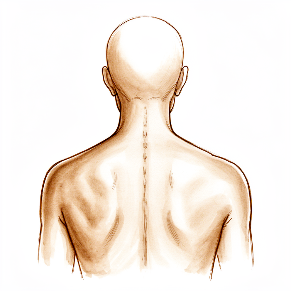
Qué está sintiendo
La ciática es un dolor que se extiende desde la parte baja de la espalda hasta una de las piernas. Por lo general afecta a una sola pierna, aunque a veces ambas duelen, siendo una más dolorosa que la otra. En raras ocasiones, el dolor puede cambiar de una pierna a la otra. La mayoría de las personas también experimentan un dolor sordo y molesto en la propia zona lumbar. El dolor de espalda junto con el dolor en la pierna es el patrón más habitual; el dolor en las piernas sin ningún dolor de espalda es poco común.
El dolor de espalda suele aparecer tras una flexión o esfuerzo de levantamiento. Tiende a empeorar cuando se utiliza la espalda, por ejemplo al levantar compras, al levantarse de una silla baja o al agacharse junto a la bañera. El descanso lo alivia, al menos en parte. Durante el primer episodio de dolor, este suele aparecer y desaparecer en lugar de ser constante.
El dolor en la pierna puede ser agudo o como una sensación de ardor, y a veces se extiende por debajo de la rodilla. Es posible que note hormigueo o entumecimiento en el pie. La intensidad del dolor en la pierna no indica cuánto se está comprimiendo el nervio. Algunas personas notan que el dolor empeora por la noche o al levantarse por primera vez. Las tareas cotidianas que requieren flexión, estar sentado durante largos periodos o cargar pesos pueden volverse difíciles de realizar.
Si algo de esto se asemeja a lo que usted siente, le conviene anotar cuándo comenzó el dolor, qué lo mejora o empeora y qué partes de la pierna se ven afectadas. Lleve esas notas a su consulta; le ayudarán a su cirujano a determinar si el nervio de la pierna es la causa del dolor, ya que varias otras afecciones pueden provocar síntomas similares.
¿Qué está ocurriendo realmente?
Su columna vertebral está formada por una serie de huesos, con un cojín entre cada par de ellos. Ese cojín se llama disco intervertebral. Imagíneselo como una rosquilla rellena de mermelada: un centro blando y gelatinoso envuelto en un anillo exterior resistente. El disco actúa como un amortiguador, absorbiendo las cargas de la vida diaria y permitiendo que la espalda se doble.
La ciática se produce cuando el centro blando de un disco se desplaza a través del anillo exterior y comprime un nervio. Los nervios que bajan por las piernas salen de la columna en la zona lumbar; por eso, un abultamiento allí puede irritar a uno de ellos. El nervio también se inflama en el punto donde es comprimido. Esta inflamación está estrechamente relacionada con la intensidad del dolor ciático.
Esto explica los síntomas que acaba de leer. El dolor en la espalda proviene del propio disco sobrecargado. El dolor en la pierna, los hormigueos o la entumecimiento se deben al nervio irritado; el nervio afectado determina qué partes de la pierna se ven afectadas. El descanso ayuda porque reduce la inflamación alrededor del nervio. El tamaño del abultamiento no guarda relación directa con la intensidad del dolor en la pierna; por eso, un problema aparentemente leve puede causar un dolor intenso.
En la mayoría de las personas, el problema mejora con el tiempo y mediante cuidados sencillos. Si el dolor persiste entre 4 y 12 meses, una cirugía para extraer la parte del disco que comprime el nervio puede aliviar el dolor más que el tratamiento no quirúrgico por sí solo. Una inyección cerca de la raíz nerviosa, en la que se administra medicación cerca del nervio irritado, también puede ser útil cuando la ciática se repite o cuando resulta difícil identificar la causa exacta.
Qué podemos hacer al respecto
La resonancia magnética suele ser el método preferido para el dolor ciático, ya que muestra con claridad los discos, los nervios y los orificios por donde pasan.
En la mayoría de los casos, el dolor ciático mejora sin necesidad de cirugía. Su médico de cabecera puede explicarle qué está ocurriendo, revisar sus medicamentos para el dolor y animarlo a mantenerse activo y seguir trabajando. La fisioterapia constituye un pilar fundamental de este tratamiento: combina educación con ejercicios de estiramiento, fortalecimiento y acondicionamiento, con el objetivo de permitirle seguir moviéndose mientras el nervio se recupera. Un programa típico incluye hasta 9 sesiones en los primeros 3 meses, más tres sesiones de refuerzo en el cuarto, quinto y sexto meses. Algunas personas también prueban la descompresión espinal no quirúrgica junto con la fisioterapia; este método estira suavemente la columna y se ha asociado con una reducción del dolor y un aumento en la altura del disco tras 4 semanas de tratamiento.
Los medicamentos pueden ayudarle a mantenerse activo mientras su cuerpo se recupera. Su médico de cabecera podría revisar y ajustar sus fármacos analgésicos, incluidos los antiinflamatorios, para que pueda moverse sin molestias. En ocasiones se emplean inyecciones para el dolor ciático: la inyección cerca de la raíz nerviosa coloca el fármaco cerca del nervio irritado, resultando útil cuando el dolor reaparece con frecuencia o cuando la causa no es fácil de identificar. La inyección epidural de plasma rico en plaquetas, que utiliza un preparado elaborado a partir de su propia sangre, constituye una alternativa para problemas discales en un solo nivel. Otra opción son las inyecciones de cortisona en el espacio que rodea los nervios de la zona lumbar, indicadas para el dolor ciático agudo durante las primeras semanas.
La cirugía se contempla cuando el tratamiento no quirúrgico no brinda suficiente alivio. Evaluamos casos en los que el dolor proviene de la compresión nerviosa, es persistente o se repite con frecuencia, limita su vida cotidiana y va acompañado de signos de afectación nerviosa como entumecimiento o debilidad. En caso de que un disco comprima un nervio, la operación consiste en extirpar la parte del disco que ejerce presión. Para la estenosis espinal o una vértebra desplazada, la cirugía puede incluir la liberación del nervio y, en algunos casos, la fusión de dos huesos para estabilizar la columna. Analizaremos conjuntamente si la cirugía es adecuada para usted y tomaremos la decisión juntos.
Qué esperar
En la mayoría de las personas, el dolor ciático mejora con el tiempo y con cuidados sencillos. En las primeras semanas, el dolor suele aparecer y desaparecer en lugar de mantenerse constante. La duración exacta varía mucho de una persona a otra; incluso los especialistas tienen dificultades para predecir un plazo concreto para cada paciente.
Si el dolor persiste entre 4 y 12 meses, la cirugía para extraer la parte del disco que comprime el nervio puede reducir el dolor más que el tratamiento no quirúrgico por sí solo. La cirugía también resulta útil cuando el dolor ciático se vuelve crónico y otros tratamientos no han surtido efecto. Tras una intervención por un problema de disco, la mayoría de los pacientes refieren una mejora real. En un grupo de pacientes, el 82 % dejó de sentir dolor en la pierna después de la operación, mientras que el 13 % solo experimentaba dolor ocasional. No obstante, no todos logran un alivio total; un pequeño porcentaje necesita una segunda intervención en el mismo disco dentro de los cinco años posteriores a la primera cirugía.
Sin tratamiento, el dolor ciático que ya dura varios meses tiende a persistir en lugar de desaparecer por sí solo. El pronóstico suele ser mejor en personas más jóvenes. En adolescentes y adultos jóvenes, casi todos los problemas de disco se resuelven con tratamiento no quirúrgico; además, la cirugía en este grupo de edad también conlleva mejoras tanto a corto como a largo plazo.
La recuperación suele ser gradual, no repentina. Se puede esperar que el dolor en la pierna disminuya primero, aunque es posible que persistan ciertas molestias o pinchazos ocasionales. Mantenerse activo y seguir con la rutina habitual en la medida de lo posible ayuda a que el nervio se recupere. Si el dolor empeora bruscamente o se vuelve intenso sin mejorar, es conveniente acudir a un médico de inmediato, ya que podría indicar otro problema subyacente.
Algunos aspectos son difíciles de predecir únicamente a partir de los estudios de imagen. Lo que se observa en una resonancia magnética no nos permite determinar con certeza cuánto durará el dolor de espalda ni si aparecerá en absoluto. La salud general del paciente también es relevante; por eso, su cirujano evaluará el cuadro completo, no solo la columna vertebral, para decidir qué tratamiento le resultará más beneficioso.
¿Cuándo consultar a un especialista?
La mayoría de los casos de ciática mejoran con el tiempo y con cuidados básicos; por eso, la primera consulta debe ser con el médico de cabecera para síntomas típicos. Acuda a su médico si el dolor persiste más de unas pocas semanas, si no disminuye con el reposo o si le impide dormir o trabajar. Solicite una evaluación especializada si el dolor en la pierna reaparece con frecuencia, si nota entumecimiento o hormigueo nuevo en el pie, o si una pierna se debilita en comparación con la otra.
Acuda de inmediato a urgencias si pierde el control de la vejiga o los intestinos, si la zona de la ingle o el ano se entumece, o si ambas piernas se vuelven débiles o pesadas. Estos pueden ser signos del síndrome de cola de caballo, en el cual el haz de nervios situado en la base de la columna vertebral queda comprimido. Requiere evaluación el mismo día, pues la presión continua sobre esos nervios puede provocar problemas duraderos, incluidas alteraciones en la función sexual que algunas personas padecen posteriormente.
Informe a su médico si tiene más de 50 años y desarrolla ciática. En esa misma consulta, vale la pena preguntar por la vacuna contra el herpes zóster, ya que el riesgo de padecerlo es mayor en personas de su edad con esta afección.
Evidence & references
This is the clinical evidence summary written for health professionals. It is technical, and it lists the research this page was built from. You do not need to read it to understand your treatment or to make a decision about it.
Anatomy & Pathophysiology
Osseous Anatomy
- The bony anatomy of the spine consists of 7 cervical vertebrae, 12 thoracic vertebrae, 5 lumbar vertebrae, 5 fused sacral vertebrae, and 4 or 5 fused coccygeal vertebrae [3].
- The vertebral body consists of a fairly cylindrical mass of bone connected by pedicles to the posterior arch, which consists of the lamina and spinous process [3].
- The spinal canal is formed by the vertebral body anteriorly, the lamina posteriorly, and the pedicles laterally [3].
- The vertebral bodies function primarily to bear weight and transfer forces to the pelvis and hips [3].
- The posterior elements provide protection to the neural structures and function as a tension band [3].
- The thoracic spine represents two transitional zones: from the highly mobile cervical spine into the more rigid thoracic region, and then back to the more mobile lumbar spine [5].
- The thoracic spine, in conjunction with the ribs and sternum, forms a bony "cube" that is an inherently stable structure providing protection to the heart and lungs [5].
- The vertebral bodies of the thoracic spine are larger than those of the cervical spine but smaller than the lumbar vertebrae [5].
- The pedicles of the thoracic spine arise more superiorly from the posterior vertebral body than in the cervical or lumbar spine and project obliquely from superodorsal to inferoventral [5].
- The spinal canal is narrowest in the thoracic region of the spine [5].
- The spinous processes of the midthoracic spine project sharply obliquely, overlapping the lamina and spinous processes inferiorly [5].
- The rib heads articulate with the lateral aspect of the vertebral bodies, with a shared articulation at the level of the disk space referred to as a demifacet [5].
- The transverse processes of the thoracic spine project obliquely superolaterally, with the costotransverse joint located along their ventral aspect [5].
- There is no costotransverse articulation at T11 or T12 [5].
- The vertebral column comprises 33 vertebrae divided into five sections: 7 cervical, 12 thoracic, 5 lumbar, 5 sacral, and 4 coccygeal [11].
- The sacral and coccygeal vertebrae are fused, which typically allows for 24 mobile segments [11].
- Each mobile vertebral body increases in size when moving from cranial to caudal [11].
- A typical vertebra comprises an anterior body and a posterior arch that enclose the vertebral canal [11].
- The neural arch is composed of two pedicles laterally and two laminae posteriorly that are united to form the spinous process [11].
- The articular processes articulate with adjacent vertebrae to form synovial joints [11].
- The relative orientation of the articular processes accounts for the degree of flexion, extension, or rotation possible in each segment of the vertebral column [11].
- The spinous and transverse processes serve as levers for the numerous muscles attached to them [11].
- The length of the vertebral column averages 72 cm in men and 7 to 10 cm less in women [11].
- The vertebral canal extends throughout the length of the column and provides protection for the spinal cord, conus medullaris, and cauda equina [11].
- The vertebral body is composed of an inner region of cancellous bone surrounded by a thin shell of cortical bone [8].
- The cervical spine is composed of seven vertebrae and assumes a lordotic curvature [8].
- The thoracic spine is composed of 12 vertebrae and assumes a kyphotic curvature [8].
- The lumbar spine is composed of five vertebrae and assumes a lordotic curvature [8].
- The five fused sacral vertebrae form a portion of the pelvis [8].
- Four small, fused vertebrae form the coccyx at the most caudal extent of the spinal column [8].
Intervertebral Disc Anatomy
- The intervertebral disc (IVD) separates each successive vertebral body except between the atlas (C1) and the axis (C2) [8].
- The IVD provides a unique combination of compressive stiffness and flexibility to support normal spine biomechanics [8].
- The IVD is composed of an inner nucleus pulposus (NP) and an outer ring termed the anulus fibrosus (AF) [8].
- The nucleus pulposus serves as an osmotic pump to attract water and generate hydraulic pressure when subjected to significant loads during activities of daily living [8].
- The anulus fibrosus encapsulates the gelatinous nucleus pulposus and provides mechanical support to contain NP pressure and constrain intervertebral rotations [8].
- The outer anulus fibrosus is integrated with the vertebral rim via a fibrocartilage enthesis that consists of a thin layer of calcified cartilage, or "tidemark" [8].
- The end plate is a bilayer of cartilage and bone that separates the disk from adjacent vertebrae [8].
- The cartilage end plate integrates with the inner anulus fibrosus to fully encapsulate the nucleus pulposus [8].
- The end plate must be strong and thick to resist significant loads but must also be permeable to favor chemical transport and disk cellular vitality [8].
- The anulus fibrosus consists of 10-25 layers [8].
Ligaments and Soft Tissue
- Additional soft-tissue structures providing passive support include the anterior longitudinal ligament, the posterior longitudinal ligament, the ligamentum flavum, the facet joint capsule, the interspinous ligament, and the supraspinous ligaments [8].
- The erector spinae muscle runs longitudinally on the dorsal surface of the spinal column and functions to extend the spine [8].
- The psoas muscle runs longitudinally on the ventrolateral surface of the spinal column and serves to flex the hip (bilateral contraction) or laterally bend the trunk (unilateral contraction) [8].
- The multifidus muscle connects intersegmentally to stabilize the spine by acting like a bowstring to maintain lordosis [8].
Biomechanics and Alignment
- Normal cervical alignment is approximately 15° of lordosis [7].
- The thoracic spine generally ranges from 20° to 40° of kyphosis [7].
- The lumbar spine has approximately 40° to 50° of lordosis [7].
- The sacrum is kyphotic [7].
- Kyphotic segments (thoracic, sacral) are considered "primary" curvatures as they are already present in utero and at birth [7].
- The lordotic curvatures of the cervical and lumbar spine develop secondarily later in life to allow the growing child to develop an upright posture [7].
- The center of gravity of the spinal column runs from the odontoid process proximally through the sacral promontory caudally [7].
- Changes in sagittal balance that shift the center of gravity too far ventrally can result in significant pain and disability [7].
- The basic motion segment of the spine, the "functional spinal unit," consists of two vertebrae, the disk between them, and the facet joints (and their capsules) [7].
- The functional spinal unit serves to limit motion of the spine within the confines of protecting the neural structures contained therein [7].
- Vertebral bodies are loaded in series, with more caudal levels supporting more weight than more cranial segments [7].
- The vertebral bodies bear 70% to 90% of the static axial load of the spine [7].
- The facet joints support 10% to 20% of axial load in a standing, neutral alignment [7].
- In extension, the facet joints may bear up to 30% of the axial load [7].
- In flexion, the facet joints may be burdened with up to 50% of the anterior shear load [7].
- As compressive forces are applied to the disk, the nucleus pulposus deforms, redistributing axial forces radially [7].
- The radial pressure generated by the nucleus pulposus is resisted by the tensile properties of the alternating bands of fibers within the anulus fibrosus [7].
- The spinous processes and transverse processes act as lever arms, providing mechanical advantage for the muscles that insert along their surfaces [7].
Vascular Anatomy
- The thoracic and lumbar levels are supplied by paired segmental arteries which originate directly from the aorta along its posterior surface [12].
- Branches of the segmental arteries supply the vertebral body, the paraspinal musculature, and the spinal cord [12].
- The cervical spine derives its circulation primarily from the vertebral arteries [12].
- The vertebral arteries arise from the subclavian arteries on either side and typically enter the transverse foramen at the C6 level [12].
- The vertebral arteries run proximally through the transverse foramina to C1, then course posteriorly over the superior aspect of the C1 ring before entering the foramen magnum [12].
- In the foramen magnum, the vertebral arteries merge to form the basilar artery [12].
- Segmental branches to each cervical vertebra arise from the vertebral artery and the deep cervical branch of the costocervical trunk [12].
- Typically one side of the vertebral artery is more dominant, having a larger diameter than the other [12].
- Occasionally, the vertebral artery enters through the transverse foramen of C7 rather than C6 [12].
- The vascular supply of the spinal cord is primarily from the medullary branches of the segmental spinal arteries [12].
- Medullary branches merge to feed the anterior spinal artery, which is responsible for supplying approximately 80% of the vascular supply to the spinal cord [12].
- Typically, three anterior medullary arteries supply the cervical region, one or two supply the thoracic region, and one supplies the lumbosacral spinal cord [12].
- The arteria medullaris magna (AMM), also known as the arteria radicularis magna or artery of Adamkiewicz, is the largest anterior segmental artery [12].
- The AMM typically arises on the left side anywhere between the T8 and L1 level, although right-sided origins are not uncommon [12].
Neural Anatomy
- A typical mixed spinal nerve has three distinct components: motor, sensory, and sympathetic [13].
- Motor rootlets leave the anterolateral sulcus of the spinal cord and unite to form each motor root [13].
- Motor fibers traversing these roots arise from the anterior horn cells and innervate the skeletal muscles [13].
- Sensory fibers arise from pain, thermal, tactile, and stretch receptors [13].
- Cell bodies for sensory fibers are located within the dorsal root ganglia with axons entering the posterolateral sulcus of the cord via several rootlets [13].
- Fibers conveying joint or position sensibility and some tactile fibers turn cephalad in the dorsal columns and do not synapse before reaching the gracile and cuneate nuclei at the cervicomedullary junction [13].
- Pain and temperature fibers synapse in the substantia gelatinosa and cross to ascend in the dorsal spinothalamic tract [13].
- Tactile fibers enter, synapse, and cross to ascend in the ventral spinothalamic tract [13].
- The sympathetic component of all 31 mixed spinal nerves leaves the spinal cord along only 14 motor roots [13].
- The cells of origin for sympathetic fibers are in the intermediolateral cell column that extends throughout the thoracic and upper lumbar cord segments [13].
- Sympathetic fibers exit from the cord with the 12 thoracic and first two lumbar motor roots, enter the respective mixed spinal nerve, and promptly emerge from it as white rami [13].
- White rami pass anteriorly to the corresponding sympathetic ganglion [13].
- Postganglionic fibers pass back to the mixed spinal nerve as a gray ramus [13].
- Mixed spinal nerves, having left the intervertebral foramina, receive their sympathetic component and promptly branch into anterior and posterior primary rami [13].
- The posterior primary rami are directed posteriorly and supply the paraspinal musculature and the skin along the posterior aspect of the trunk, the neck, and the head [13].
- The upper three cervical posterior rami are larger than their corresponding anterior rami, supplying relatively large areas of the scalp posteriorly and the musculature around the craniocervical junction [13].
- With exceptions for the upper three cervical nerves, posterior primary rami are small, and the major part of each spinal nerve continues laterally in an anterior primary ramus to enter a plexus or to become an intercostal nerve [13].
- Anterior primary rami of all the cervical, the first thoracic, and all the lumbosacral nerves join in the formation of plexuses [13].
- The upper four cervical anterior rami form the cervical plexus [13].
- The lower four cervical and first thoracic anterior rami form the brachial plexus [13].
- The first three and a part of the fourth lumbar anterior rami form the lumbar plexus [13].
- The sacral anterior rami along with the fifth lumbar and a part of the fourth join to form the lumbosacral plexus [13].
- The area of skin supplied by the fibers of a single spinal root is called a dermatome [13].
- Segmental dermatomal patterns are well preserved in the thoracic region but not in the limbs [13].
- C1 emerges between the skull and C1 vertebra [9].
- C2–7 emerge superior to pedicles [9].
- C8 emerges inferior to the pedicle of C7 vertebra [9].
- T1–Co emerge inferior to pedicles of their respective vertebrae [9].
- The L4 nerve root is associated with the tibialis anterior muscle and the patellar reflex [9].
- The L5 nerve root is associated with toe extensors [9].
- The S1 nerve root is associated with the peroneal muscle and the Achilles reflex [9].
Pathophysiology of Stenosis
- Spinal stenosis can be categorized according to the anatomic area of the spine affected, the region of each vertebral segment affected, and the specific pathologic entity involved [17].
- Spinal stenosis is most common in the lumbar region, but cervical stenosis also occurs frequently [17].
- Spinal stenosis has been rarely reported in the thoracic spine [17].
- Degeneration of the disc occurs with disc narrowing and subsequent ligamentous redundancy, which compromises the spinal canal area [17].
- Instability may ensue from disc degeneration and ligamentous redundancy [17].
- Relative hypermobility precipitates the formation of facet overgrowth and ligamentous hypertrophy [17].
- The ligamentum flavum may be markedly thickened into the lateral recess where it attaches to the facet capsule, causing nerve root compression [17].
- Central spinal stenosis denotes involvement of the area between the facet joints, which is occupied by the dura and its contents [17].
- Stenosis in the central region is usually caused by protrusion of a disc, bulging anulus, osteophyte formation, or buckled or thickened ligamentum flavum [17].
- Symptomatic central spinal stenosis results in neurogenic claudication with generalized leg pain [17].
- The lateral canal contains the nerve roots, and compression in this region results in radiculopathy [17].
- The lateral recess, also known as "Lee's entrance zone," begins at the medial border of the superior articular process and extends to the medial border of the pedicle [17].
- The borders of the lateral recess are the pedicle laterally, the superior articular facet dorsally, the posterior ligamentous complex to disc and floor of the canal, and the central canal medially [17].
- Facet arthritis most frequently causes stenosis in the lateral recess zone, along with vertebral body spurring and disc or anulus pathology [17].
- "Lee's midzone" describes the foraminal region, which lies ventral to the pars [17].
- The borders of the foraminal region are the lateral recess medially, the posterior vertebral body and disc ventrally, the pars and intertransverse ligament dorsally, and the lateral border of the pedicle laterally [17].
- The foramen is essentially the area between the cephalad and caudal pedicles [17].
- The dorsal root ganglion and ventral motor root occupy 30% of the foraminal space [17].
- The foramen is the point where the dura becomes confluent with the nerve root as epineurium [17].
- Causes of stenosis in the foraminal area are pars fracture with proliferative fibrocartilage or a lateral disc herniation [17].
- Thickening of the ligamentum flavum sometimes extends into the foramen and can be associated with a spur from the undersurface of the pars, especially if foraminal height is less than 15 mm and posterior intervertebral disc height is less than 4 mm [17].
- The exit zone is identified as the area lateral to the facet joint [17].
- The nerve root in the exit zone can be compressed by a "far lateral" disc, spondylolisthesis and associated subluxation, or facet arthritis [17].
- The most common type of spinal stenosis is caused
Investigations
Magnetic Resonance Imaging (MRI)
- MRI is the procedure of choice for screening patients with low back or sciatic pain after routine radiography [22].
- In the lumbar and thoracic spine, MRI has supplanted CT myelography because it is noninvasive and less expensive [22].
- MRI provides ideal evaluation of intervertebral discs, nerve roots, posterior longitudinal ligament, and intervertebral foramen due to high soft-tissue contrast and resolution [22].
- MRI provides excellent assessment of the spinal cord [22].
- CT myelography is reserved for patients who have contraindications to MRI or who have equivocal MRI examinations [22].
- Sagittal images provide an initial evaluation of the intervertebral discs and posterior longitudinal ligament [22].
- A normal intervertebral disc exhibits signal hyperintensity on T2-weighted images due to its high water content [22].
- The aging process results in gradual desiccation of disc material and loss of T2-weighted signal hyperintensity [22].
- Disc herniations or extrusions appear as convex or polypoid masses extending posteriorly into the ventral epidural space [22].
- Disc herniations or extrusions frequently maintain a signal intensity similar to that of the disc of origin [22].
- Sagittal T2-weighted or gradient-echo images create a “myelographic” effect useful for evaluating compromise of the subarachnoid space [22].
- Sagittal T1-weighted images should be examined to identify narrowing of the neuroforamina [22].
- Normal T1-weighted hyperintense perineural fat in the foramina provides excellent contrast to darker displaced disc material [22].
- Far lateral disc herniations are best seen on selected axial images localized through disc levels [22].
- Free disc fragments appear discontinuous with the intervertebral disc and usually have intermediate T1-weighted signal in contrast to hypointense cerebrospinal fluid [22].
- MRI can detect significant spinal cord compromise, with edema within the cord demonstrated as hyperintensity on T2 weighting [22].
- MRI is superior to CT for identification of infections, tumors, and degenerative changes within the discs [23].
- MRI is superior for imaging the disc and directly imaging neural structures [23].
- MRI typically shows the entire region of the spine being evaluated (cervical, thoracic, or lumbar) [23].
- MRI allows imaging of the nerve root in the foramen, which is difficult with postmyelography CT because contrast does not extend fully through the foramen [23].
- MRI and CT, with or without myelography, can be used in a complementary fashion in certain circumstances [23].
- MRI evidence of disc degeneration has been reported in the cervical spine in 25% of patients younger than 40 years [23].
- MRI evidence of disc degeneration has been reported in the cervical spine in 60% of patients 60 years and older [23].
- Lumbar disc degeneration was found in 35% of patients aged 20 to 39 years [23].
- Lumbar disc degeneration was found in 100% of patients older than 50 years [23].
- MRI findings must be carefully correlated with the clinical impression because anatomy may be abnormal but asymptomatic [23].
- Meaningful clinical information from MRI is obtained by posing a specific question derived from history and physical examination before the study [23].
- The specific question for MRI interpretation should be posed using the parameters of neural compression, instability, and deformity [23].
- The specific location of the abnormality should be suspected before MRI and confirmed with the study [23].
- Only abnormalities in categories of neural compression, instability, or deformity are important for operative treatment [23].
- Failure to interpret MRI in the context of neural compression, instability, or deformity leads to poor clinical choices and outcomes [23].
- MRI is superior to CT in most circumstances for advanced imaging of the spine [26].
- Diffusion tensor imaging has been reported to demonstrate spinal cord impairment in patients with early stage cervical spondylosis before it is visible on plain MRI scans [26].
Computed Tomography (CT)
- CT myelography is invasive and more costly than MRI [22].
- CT has largely supplanted plain radiographs as the initial screening study of choice for spine injuries due to high sensitivity and specificity [24].
- CT of the spine should be obtained in the setting of a high-risk mechanism, acute thoracic or lumbar pain after trauma, fractures identified on plain radiographs, or suspected spine injury with neurologic deficit [24].
- A dedicated spine CT consists of 2- to 3-mm wide axial slices of the thoracic and lumbar spine, typically reformatted into sagittal and coronal images [24].
- CT allows for identification of subtler fractures that might have remained undiagnosed on plain radiographs [24].
- CT provides additional three-dimensional detail, including the degree of canal compromise and the amount of fracture comminution [24].
- CT is particularly useful in differentiating compression fractures from burst fractures [24].
- CT identifies subtle features of injury such as the presence of facet widening [24].
- CT evaluation is essential in determining the stability of thoracic and lumbar spine fractures [24].
- The primary disadvantage of CT imaging compared to MRI is that it does not provide as good a visualization of soft tissues [24].
Terminology
- A bulge is defined as a circumferential, symmetric extension of the disc beyond the interspace around the endplates [22].
- A protrusion is defined as a focal or asymmetric extension of the disc beyond the interspace, with the base against the disc of origin broader than any other dimension of the protrusion [22].
- An extrusion is defined as a more extreme extension of the disc beyond the interspace, with the base against the disc of origin narrower than the diameter of the extruding material itself or with no connection between the material and the disc of origin [22].
- A sequestration specifically refers to a disc fragment that has completely separated from the disc of origin [22].
References
[3] Orthopaedic Knowledge Update 13 Ebook Without Multimedia. Spinal Anatomy > Osseous Anatomy.
[5] Orthopaedic Knowledge Update 13 Ebook Without Multimedia. Spinal Anatomy > Osseous Anatomy > Thoracic Vertebrae.
[7] Orthopaedic Knowledge Update 13 Ebook Without Multimedia. Spinal Anatomy > Biomechanics.
[8] Orthopaedic Basic Science Fifth Edition Print Ebook. Biology and Mechanics of the Skeletal Extracellular Matrix > Anatomy.
[9] Miller S Review Of Orthopaedics. Genetics of musculoskeletal conditions and abnormalities are summarized in Table 1.27 > SURGICAL APPROACHES TO THE SPINE (Table 2.46).
[11] Campbell S Operative Orthopaedics 4 Volume Set. RECONSTRUCTION OF THE PATELLOFEMORAL AND PATELLOTIBIAL LIGAMENTS WITH A SEMITENDINOSUS TENDON GRAFT > ANATOMY OF VERTEBRAL COLUMN.
[12] Orthopaedic Knowledge Update 13 Ebook Without Multimedia. Spinal Anatomy > Vascular Anatomy.
[13] Campbell S Operative Orthopaedics 4 Volume Set. PERIPHERAL NERVE INJURIES OF THE UPPER AND LOWER EXTREMITIES > ANATOMY OF THE SPINAL NERVES > COMPONENTS OF MIXED SPINAL NERVES.
[17] Campbell S Operative Orthopaedics 4 Volume Set. OVERVIEW OF LUMBAR AND THORACIC DISC DEGENERATION AND HERNIATION > STENOSIS OF THE THORACIC AND LUMBAR SPINE > ANATOMY.
[22] Campbell S Operative Orthopaedics 4 Volume Set. INTERVERTEBRAL DISC DISEASE.
[23] Campbell S Operative Orthopaedics 4 Volume Set. OVERVIEW OF LUMBAR AND THORACIC DISC DEGENERATION AND HERNIATION > MAGNETIC RESONANCE IMAGING.
[24] Rockwood And Green S Fractures In Adults. Imaging of Cervical Spine Fractures and Dislocations > Computed Tomography.
[26] Campbell S Operative Orthopaedics 4 Volume Set. POSTERIOR APPROACH TO THE LUMBAR SPINE, L1 TO L5 > MAGNETIC RESONANCE IMAGING.