Qué está sintiendo
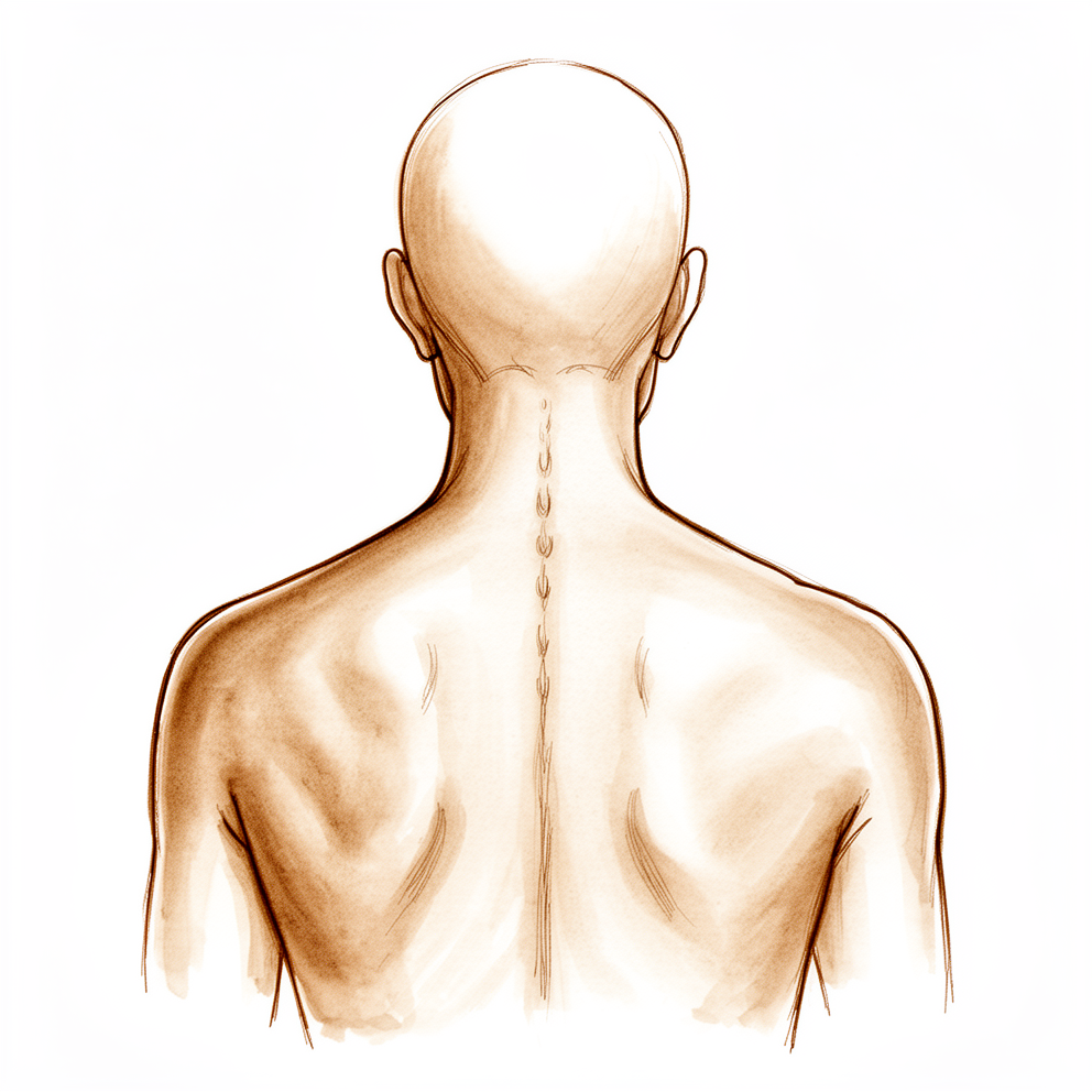
Una fractura osteoporótica ocurre cuando un hueso de la columna vertebral, llamado vértebra, se vuelve tan delgado y frágil que se rompe bajo el esfuerzo cotidiano. El dolor suele localizarse en la zona media de la espalda o en el punto donde esta se une a la zona lumbar. Con frecuencia aparece de forma repentina, a veces tras actividades tan simples como agacharse, levantar algo o tropezar levemente.
Al principio el dolor es agudo y luego se transforma en una molestia sorda. Tiende a intensificarse al permanecer de pie o caminar durante un rato, al girar el torso o al inclinarse hacia adelante. Incorporarse desde la posición acostada puede resultar difícil, y el dolor suele empeorar al despertar o después de estar de pie durante mucho tiempo. Acostarse puede aliviarlo. Las tareas cotidianas que implican carga para la columna se vuelven complicadas: llevar las compras, levantarse de una silla baja, hacer la cama o subir escaleras.
Si la fractura afecta la parte inferior de una vértebra, a veces puede irritar un nervio que desciende por la pierna. Eso provoca dolor, hormigueo o entumecimiento en dicha pierna. Asimismo, algunas personas tensan la fuerte capa de tejido que rodea los músculos de la zona lumbar, lo cual aumenta la molestia.
Estas fracturas pueden afectar a más de una vértebra, y es fácil que pasen desapercibidas en las radiografías simples. Si usted tiene más de 60 años y experimenta dolor de espalda repentino, es conveniente realizarse una evaluación adecuada. Una fractura indica que sus huesos son más débiles de lo normal, por lo que existe una probabilidad real de sufrir otra fractura —incluida una fractura de cadera— en los próximos años. Este riesgo aumenta aún más si ya ha tenido más de una fractura vertebral.
La mayoría de estas fracturas mejoran con el tiempo y no requieren cirugía. No obstante, si el dolor sigue siendo intenso a pesar de los analgésicos, existen procedimientos que pueden ayudar; los describiremos en las siguientes secciones.
¿Qué está ocurriendo realmente?
Su columna vertebral está formada por una serie de huesos llamados vértebras; cada una cuenta con un bloque sólido en la parte anterior que soporta su peso. El hueso sano funciona como un andamiaje vivo que se reconstruye constantemente: el hueso viejo se elimina y se deposita hueso nuevo. En la osteoporosis, este equilibrio se rompe: el hueso se elimina más rápido de lo que se reemplaza, por lo que el andamiaje se vuelve delgado y poroso, similar a un panal de abejas con huecos cada vez más grandes.
El bloque anterior de cada vértebra está diseñado para soportar la carga de compresión del cuerpo, un poco como un ladrillo que sostiene el peso de la pared situada encima. Cuando ese ladrillo se vuelve poroso, cargas cotidianas como agacharse o levantar objetos pueden aplastarlo. El hueso se comprime y se deforma, por lo que la vértebra pierde altura en su parte anterior y la columna se curva ligeramente hacia adelante en ese punto. Ese aplastamiento es la fractura que usted siente; el dolor proviene tanto del propio hueso fracturado como de la tensión que genera en los tejidos circundantes.
Estas fracturas ocurren con mayor frecuencia en la zona donde la espalda media se une a la lumbar, pues esa unión soporta una mayor parte de las fuerzas de flexión de la columna. Si se aplastan varias vértebras, la curvatura hacia adelante se acentúa, lo que altera la forma en que toda la columna distribuye la carga y puede intensificar el dolor y la rigidez. Por lo general, el hueso se recupera por sí solo en un plazo de semanas a meses; sin embargo, dado que es delgado, la vértebra reparada puede seguir comprimiéndose lentamente, y esa misma fragilidad hace posible otra fractura en otro lugar.
Solo alrededor de una de cada tres personas con estas fracturas vertebrales siente dolor, razón por la cual suelen pasar desapercibidas. La mayoría se resuelven solo con el tiempo y los analgésicos. Si, a pesar de ello, el dolor sigue siendo intenso, existen procedimientos que pueden estabilizar el hueso fracturado y aliviar el dolor; los describiremos a continuación.
Qué podemos hacer al respecto
La mayoría de estas fracturas sanan sin cirugía. El primer paso es mantenerse activo: el ejercicio suave y el movimiento favorecen la cicatrización ósea, evitan mayor pérdida de masa ósea y le permiten seguir haciendo las actividades que disfruta. La fisioterapia guía este proceso; su objetivo es recuperar la fuerza y la confianza mientras la fractura se consolida. Debe darle varias semanas o meses, pues normalmente el hueso sana por sí solo en ese plazo. Se puede utilizar un corsé para sostener la espalda durante la curación. Los analgésicos le ayudarán a seguir moviéndose durante este período.
Paralelamente, cuidamos del hueso en sí. Los medicamentos para la osteoporosis pueden fortalecerlo y reducir el riesgo de nuevas fracturas. La deficiencia de vitamina D es frecuente en estos casos; por ello la evaluamos y la corregimos antes de cualquier intervención quirúrgica. Este cuidado de la salud ósea es fundamental: mejora la cicatrización, disminuye las complicaciones y reduce el riesgo de nuevas fracturas. A menudo se pasa por alto, pero constituye una parte esencial de la recuperación tras cualquier fractura provocada por la osteoporosis.
Si el dolor persiste de forma intensa tras haber probado tratamientos no quirúrgicos, podríamos plantear un procedimiento llamado aumento vertebral. Se trata de una operación mínimamente invasiva que estabiliza la vértebra fracturada mediante cemento óseo. Existen dos variantes: en la vertebroplastia se inyecta el cemento directamente; en la cifoplastia, primero se usa un pequeño balón para elevar el hueso colapsado y luego se rellena el espacio con cemento, lo cual también puede corregir la curvatura hacia adelante causada por la fractura. La cifoplastia es la opción que preferimos cuando los tratamientos no quirúrgicos no han sido suficientes. Estos procedimientos se realizan a través de pequeñas incisiones, no de una incisión grande, lo que minimiza los riesgos en pacientes de edad avanzada. En ocasiones el cemento puede filtrarse fuera del hueso durante la intervención; esto ocurre con mayor frecuencia en la vertebroplastia, por lo que empleamos técnicas adaptadas a su fractura para prevenirlo. Existe una página específica sobre esta operación, donde se detalla lo que sucederá el día de la intervención y cómo será la recuperación.
Qué esperar
En la mayoría de las personas, el dolor provocado por este tipo de fracturas disminuye en cuestión de semanas o meses a medida que el hueso se va curando por sí solo. El dolor agudo suele desaparecer primero, mientras que el dolor sordo tarda más en ceder. Mantenerse activo y conservar la fuerza durante este período resulta beneficioso; en cambio, periodos prolongados de reposo pueden ser perjudiciales, ya que debilitan aún más los huesos y aumentan el riesgo de caídas y nuevas lesiones.
Si se controla bien el dolor, se puede volver a las actividades cotidianas a medida que la fractura se estabiliza. Si el dolor es intenso y no mejora con los analgésicos, un procedimiento para estabilizar el hueso puede aliviar ese dolor; los beneficios de dicho procedimiento, en comparación con el uso exclusivo de medicamentos, pueden mantenerse durante el primer año. Estos procedimientos también buscan sostener la vértebra aplastada para evitar que la columna siga curvándose hacia adelante en esa zona.
Es importante conocer ciertas limitaciones. Debido a que sus huesos son frágiles, existe la posibilidad de sufrir una nueva fractura en otra vértebra; este riesgo es mayor si usted tiene más edad, si ya ha tenido fracturas previas o si su densidad ósea es baja. El riesgo disminuye cuando la osteoporosis se trata con medicamentos además de la atención específica para la fractura. Asimismo, en algunas personas la vértebra colapsada puede ir perdiendo altura gradualmente con el tiempo; esto forma parte del comportamiento natural de los huesos frágiles y no indica que el tratamiento haya fracasado.
Si se deja sin tratar una fractura y la curvatura hacia adelante de la columna se acentúa, especialmente cuando hay varias fracturas, este cambio de forma puede afectar la forma en que la columna soporta la carga y seguir generando problemas. Cuidar la salud ósea, mantenerse activo y seguir el tratamiento para la osteoporosis son las mejores medidas para lograr una buena recuperación y seguir manteniéndose en pie.
¿Cuándo consultar a un médico?
Si tiene más de 60 años y experimenta un dolor de espalda repentino, incluso después de un pequeño tropiezo o una simple flexión, consulte sin demora a su médico de cabecera. Estas fracturas suelen pasar desapercibidas en las radiografías convencionales; por eso, pregunte si es necesario realizar estudios de imagen adicionales. Acuda a urgencias si aparece nueva debilidad, entumecimiento u hormigueo en las piernas, o si tiene dificultades para controlar la vejiga o el intestino. Solicite una evaluación especializada si el dolor persiste de forma intensa a pesar de tomar analgésicos durante varias semanas; si ya ha sufrido una de estas fracturas y el dolor vuelve o se desplaza a otro nivel; o si se sabe que tiene huesos frágiles y presenta cualquier tipo de dolor de espalda nuevo.
Evidence & references
This is the clinical evidence summary written for health professionals. It is technical, and it lists the research this page was built from. You do not need to read it to understand your treatment or to make a decision about it.
Anatomy & Pathophysiology
Vertebral Column Anatomy
- The vertebral column comprises 33 vertebrae divided into five sections: 7 cervical, 12 thoracic, 5 lumbar, 5 sacral, and 4 coccygeal [16].
- The sacral and coccygeal vertebrae are fused, typically allowing for 24 mobile segments [16].
- A typical vertebra comprises an anterior body and a posterior arch that enclose the vertebral canal [16].
- The neural arch is composed of two pedicles laterally and two laminae posteriorly that are united to form the spinous process [16].
- To either side of the arch of the vertebral body is a transverse process and superior and inferior articular processes [16].
- The articular processes articulate with adjacent vertebrae to form synovial joints [16].
- The relative orientation of the articular processes accounts for the degree of flexion, extension, or rotation possible in each segment of the vertebral column [16].
- The spinous and transverse processes serve as levers for the numerous muscles attached to them [16].
- The length of the vertebral column averages 72 cm in men and 7 to 10 cm less in women [16].
- The vertebral canal extends throughout the length of the column and provides protection for the spinal cord, conus medullaris, and cauda equina [16].
Intervertebral Disk Anatomy
- The intervertebral disk connects adjacent vertebral bodies [6].
- The adjacent vertebral bodies, the disk, and the facet joints constitute the functional spinal unit that provides mechanical stability and allows physiologic motion [6].
- The intervertebral disks run between vertebral bodies from C2 to S1 and function to resist loads on the spine and provide stability [18].
- The disks contribute up to one-third of the height of the spinal column [18].
- The disk consists of the cartilaginous end plates of the vertebral bodies, the outer anulus fibrosus, and the inner nucleus pulposus [18].
- The end plate serves as a point of attachment of the disk to the superior and inferior surfaces of the vertebral bodies [18].
- The end plate is a thin layer of hyaline cartilage [18].
- The nucleus pulposus is centrally located and confined by the end plates and the anulus fibrosus [6].
- The nucleus pulposus resists compressive loads, dampens mechanical loads, and evenly distributes forces onto the end plates [6].
- The nucleus pulposus is the remnant of the embryonic notochord and comprises the gelatinous center of the intervertebral disk [18].
- The extracellular matrix of the nucleus pulposus is composed primarily of type II collagen and aggrecan, which makes it relatively hydrophilic [18].
- The nucleus pulposus serves to resist axial loads as well as provide height to the intervertebral disk [18].
- The anulus fibrosus is peripheral to the nucleus pulposus and confines the nucleus pulposus [6].
- The anulus fibrosus is designed to resist tensile loads, allow spinal motion, provide mechanical connection between the vertebrae, and confine the nucleus pulposus [6].
- The anulus fibrosus consists of concentrically layered fibrous cartilage lamellae primarily composed of type I collagen [18].
- The fibers of the anulus fibrosus run in alternating oblique trajectories [18].
- The anulus fibrosus resists tensile forces within the spine, including those due to the compression of the nucleus pulposus [18].
- The end plates form the interface between the vertebrae and the disk and define the upper and lower boundaries of the disk [21].
- The central portion of the end plate provides a major pathway for nutrients from the vertebral bodies to diffuse into the disk [21].
Disk Physiology and Pathophysiology
- In a normal healthy lumbar disk, large aggregating proteoglycans (aggrecan and versican) constitute a high percentage of the dry weight in the nucleus [21].
- Glycosaminoglycan molecules (keratan sulfate and chondroitin sulfate) decorate the aggrecan and versican core protein and are highly negatively charged [21].
- The highly negatively charged glycosaminoglycans create a highly hydrophilic matrix that attracts H2O molecules [21].
- The attraction of H2O molecules provides swelling pressure that counteracts the axial loads encountered by the disk [21].
- The matrix is viscoelastic and therefore dissipates mechanical energy and is subject to creep [21].
- Disk height is less at the end of each day due to creep [21].
- The adult intervertebral disk is avascular and derives its nutrition through diffusion from terminal capillaries in the vertebral bodies just below the end plates [18].
- In the adult, the blood supply ends at the bony end plate of the vertebral body and the outer anulus fibrosus [21].
- Most of the disk is considered immunologically isolated due to its avascularity [21].
- Nutrients are supplied to the disk cells primarily through diffusion [21].
- As the disk gets larger during development, the distances that nutrition must diffuse across become larger, further impeding nutritional supply to the disk cells [21].
- The decrease in nutritional transport is thought to contribute to disk degeneration [21].
- Innervation of the intervertebral disk is confined to the peripheral anulus fibrosus [21].
- The sinuvertebral nerve, which arises from the dorsal root ganglion, innervates the outer anulus fibrosus [21].
- In some degenerated disks with fissures, nerve fibers may be found deeper in the anulus fibrosus [21].
- The normal nucleus pulposus is not innervated [21].
- Pain sensation from the disk arises only from the anulus fibrosus [21].
- The nucleus pulposus can generate molecules such as cytokines and proteinases that can lead to pain [21].
- With normal ageing, glycosaminoglycan production diminishes, leading to gradual desiccation of the disc [19].
- The annulus fibrosus develops fissures and disc nuclear material may prolapse through during degeneration [19].
- The discs lose height and bulge beyond the margins of the vertebral bodies during degeneration [19].
- Disc protrusion against the ligaments causes formation of marginal osteophytes [19].
- Adjacent vertebral end plates ossify and become sclerotic while fatty change occurs in the subchondral bone marrow during spondylosis [19].
- Disc degeneration results in altered biomechanics and increased loading forces on the facet joints [19].
- Facet joint osteoarthritis may lead to osteophyte encroachment into the canal causing spinal lateral recess stenosis [19].
- The ligamentum flavum thickens, which contributes to stenosis, while the disc bulges from anteriorly into the spinal canal [19].
Osteoporosis Pathophysiology
- Osteoporosis is the most common metabolic bone disease resulting from imbalance between bone formation and bone resorption [2].
- Spinal fractures are the most common manifestation of osteoporosis [2].
- Only one in three patients with spinal fractures are symptomatic [2].
- In elderly patients, osteoporosis-related spinal fractures have similar morbidity and mortality as hip fractures [2].
- Osteoporosis and low bone mass are present in about 50% of elective spine patients older than 50 years [2].
- Osteoporosis and low bone mass negatively affect clinical outcomes and are associated with increased complications and revision surgery [2].
- Vitamin D deficiency is present in the majority of spine patients [2].
Clinical Presentation
- A history of fragility fracture significantly increases the risk of another fracture [15].
- The relative risk of a subsequent fracture is two to three times higher in women with a prior fragility fracture [15].
- The relative risk of a subsequent fracture is up to six times higher in men with a prior fragility fracture [15].
- A history of greater than a single vertebral fracture significantly increases the risk of further vertebral fractures [15].
- In a meta-analysis of nontreated patients enrolled in vertebroplasty studies, 18% had a secondary fracture within 12 months [15].
Investigations
Imaging Modalities
- Dual energy x-ray absorptiometry (DXA) is the benchmark to assess bone mineral density (BMD) [25].
- DXA measures areal BMD in g/cm² of the proximal femur, lumbar spine, and distal radius [25].
- DXA is accurate and precise when calibrated and performed properly [25].
- DXA involves a very low radiation dose [25].
- BMD is reported as a standard deviation difference to a reference standard [25].
- T-score is the reference standard for young healthy females [25].
- Z-score is the reference standard for age- and gender-matched subjects [25].
- Vertebral fracture assessment (VFA) can be performed at the time of DXA [25].
- VFA identifies occult vertebral fracture in 20% to 30% of cases [25].
- VFA is useful to change a diagnosis from low bone mass (osteopenia) to osteoporosis [25].
- Opportunistic CT uses CT data to estimate bone status [25].
- CT is based on the attenuation coefficient called the Hounsfield unit (HU) [25].
- Hounsfield units are related to BMD and can be used to estimate BMD and the presence of osteoporosis [25].
- PACS tools can calculate mean HU for any elliptical region of interest [25].
- Most osteoporotic vertebral fractures can be diagnosed by plain radiographs or CT [26].
- MRI can show edema in the vertebral body to indicate a recent fracture when age determination is required [26].
- An acute measured height change of greater than 1 inch is suggestive of an acute vertebral fracture [26].
Laboratory and Preoperative Assessment
- Greater than 75% of patients older than 50 years having elective spine surgery have vitamin D insufficiency or deficiency [14].
- Vitamin D deficiency is linked to increased pain severity in spinal stenosis [14].
- Ravindra demonstrated slower fusion and lower rates of success in vitamin D–deficient patients [14].
- Kim found that baseline low vitamin D was associated with worse Oswestry Disability scores at follow-up [14].
- Vitamin D deficiency is treatable but takes 6 weeks [14].
- Osteoporosis is linked to subsidence of cages, pedicle screw loosening, proximal junction failure, poorer fusion rates, increased spondylolisthesis after laminectomy, and higher rates of revision surgery [14].
- Bjerke reviewed 140 lumbar fusion patients and found that 10 were osteoporotic and 59% had low bone mass [14].
- Nonunion was 50% in osteoporotic patients compared with 18% in those with low bone mass or normal BMD [14].
- Osteoporosis-related complications occurred in 23% of patients with normal BMD, 28% of patients with low bone mass, and 46% of osteoporotic patients [14].
- Complication rates worsened relative to BMD when more than single-level fusion was performed [14].
- The AOA suggests using the same principles as outlined for secondary fracture prevention for preoperative bone health optimization [14].
- Inclusion criteria for preoperative bone health optimization assessment include all patients having thoracolumbar surgery who are older than 50 years [14].
- Preoperative patients are recommended to consume vitamin D3 2,000 U daily and 1,200 mg calcium in the diet [14].
- Preoperative patients should be assessed for fall risk and considered for physical therapy if warranted [14].
- Patients who do not need DXA or who screen negative for DXA should undergo surgery [14].
- Patients with low bone mass or osteoporosis should be considered for preoperative bone health optimization [14].
- If medically indicated, patients are treated with diphosphonate or anabolic medication before surgery [14].
- The duration of preoperative treatment is unknown, but effects of bone strength and physiology are seen within 2 months [14].
- A minimum of 3 months of preoperative treatment is recommended if clinically possible before surgery [14].
- Longer treatment up to 9 months should be considered for multilevel or high-risk cases such as osteotomy [14].
- Antiosteoporosis medications should be continued postoperatively [14].
- Multiple RCTs comparing biphosphonates and placebo in patients undergoing lumbar spine fusion show improved clinical outcomes, radiologic outcomes, and lower risk of complications [14].
- Biphosphonates do not appear to be associated with any impairment of bone health [14].
- One RCT and multiple cohort studies show that teriparatide can lead to improved clinical outcomes, radiologic outcomes, and lower risk of complications in spinal fusion patients [14].
Treatment
Non-Operative Management
- The initial treatment of osteoporotic vertebral fractures is nonsurgical [2].
- Secondary fracture prevention is performed in less than 20% of cases following osteoporosis-related fractures [2].
- Secondary fracture prevention includes education, assessment of bone health, nutritional supplements, weight-bearing exercise, fall prevention, and pharmaceutical medications if indicated [2].
- A history of fragility fracture increases the relative risk of another fracture by two to three times in women and up to six times in men [15].
- Secondary treatment was given in 10% of patients in a Medicare database analysis but was associated with a reduced risk of fracture by 40% [15].
- The American Orthopaedic Association (AOA) Own the Bone program is located in all states and 150 sites in the United States [15].
- The AOA Own the Bone program significantly improves compliance with treatment recommendations after fragility fracture [15].
- A prospective randomized controlled non-inferiority trial found that rigid brace, soft brace, and no brace treatments for osteoporotic compression fractures without neurologic injury had comparable outcomes [1].
- Vitamin D deficiency is present in the majority of spine patients and should be corrected after fracture and before elective surgery [2].
- Routine use of vitamin D (1,000 to 2,000 IU/day) and calcium supplementation for all spine fusion patients aged 65 years or older may help avoid nonunion and infection [28].
- Target serum 25-hydroxyvitamin D levels are 30 ng/mL [28].
Operative Management: Cement Augmentation
- Vertebroplasty or kyphoplasty is considered for osteoporotic vertebral fracture in patients who are hospitalized for pain, those who do not improve with nonoperative care, or those who have progressive collapse [2].
- A 2009 multicenter trial by Kallmes et al. found that vertebroplasty and a simulated procedure without cement resulted in similar improvements in pain [29].
- A 2009 multicenter, randomized, double-blinded placebo-controlled trial by Buchbinder et al. identified no clinical benefit to vertebroplasty compared to a simulated procedure at follow-up visits ranging from 1 week to 6 months [29].
- A multicenter European randomized control trial by Klazen et al. concluded that vertebroplasty was safe and provided immediate and sustained pain relief and improvement in quality of life to a significantly greater degree than nonsurgical treatment [29].
- A randomized control study by Boonen et al. showed that kyphoplasty and vertebroplasty significantly reduced pain, improved early mobility, and enhanced short-term quality of life compared to noninvasive management options [29].
- A systematic review by Taylor et al. showed that kyphoplasty and vertebroplasty significantly reduced pain, improved early mobility, and enhanced short-term quality of life compared to noninvasive management options [29].
- Balloon kyphoplasty may correct the loss of vertebral body height, a predictor of progressive kyphosis, but this has only been confirmed on a limited basis for kyphoplasty and not for vertebroplasty [29].
- Vertebroplasty and kyphoplasty are largely equivalent in their ability to provide pain relief and functional improvement, with little advantage gained by performing kyphoplasty given its significantly greater cost [29].
- A Medicare database analysis by Edidin et al. identified lower mortality (39% vs. 50%) in patients treated with vertebroplasty compared to patients treated noninvasively [29].
- A 2018 Cochrane Review by Buchbinder et al. analyzing 21 randomized and quasi-randomized controlled trials concluded that current high- to moderate-quality evidence does not support a role for vertebroplasty in the routine treatment of acute or subacute osteoporotic vertebral fractures [29].
- The 2018 Cochrane Review by Buchbinder et al. could not identify sufficient evidence to support any clinically relevant benefit to vertebroplasty compared to placebo [29].
- Subgroup analyses in the 2018 Cochrane Review showed that results were not affected by whether the duration of pain was less than or greater than 6 weeks [29].
- Adverse events identified in the 2018 Cochrane Review included thromboembolic events, cement leakage, spinal cord compression, neurologic injury, respiratory failure, and osteomyelitis [29].
- A randomized study by Firanescu et al. comparing vertebroplasty with a placebo (simulated procedure) control group found a similar, statistically significant reduction in pain as measured by visual analog scale (VAS) scores for both groups during 12-month follow-up [29].
Preoperative Bone Health Optimization
- Greater than 50% of patients older than 50 years having elective spine surgery have low bone mass or osteoporosis [14].
- Vitamin D deficiency is treatable but will take 6 weeks [14].
- In a review of 140 lumbar fusion patients by Bjerke, nonunion was 50% in osteoporotic patients compared with 18% in those with low bone mass or normal BMD [14].
- In a review of 140 lumbar fusion patients by Bjerke, osteoporosis-related complications occurred in 23% of patients with normal BMD, 28% in patients with low bone mass, and 46% in osteoporotic patients [14].
- Inclusion criteria for preoperative bone health optimization include all patients having thoracolumbar surgery who are older than 50 years of age [14].
- Preoperative patients are recommended to consume vitamin D3 2,000 U daily and 1,200 mg calcium in diet [14].
- Preoperative patients should be assessed for fall risk and consider physical therapy if warranted [14].
- Patients who have low bone mass or osteoporosis should be considered for preoperative bone health optimization [14].
- The author recommends a minimum of 3 months of preoperative treatment if clinically possible, as effects of bone strength and physiology are seen within 2 months [14].
- For multilevel or high-risk cases such as osteotomy, longer treatment up to 9 months should be considered [14].
Complications
- Osteoporosis and vitamin D deficiency are associated with poorer outcomes, recurrent fractures, and higher complication risks [2].
- Osteoporosis and low bone mass negatively affect clinical outcomes and are associated with increased complications and revision surgery in elective spine patients older than 50 years [2].
- Preoperative and postoperative treatment of osteoporosis in spine surgery patients has been shown to improve outcomes and reduce complications [2].
References
[1] Aaos Comprehensive Orthopaedic Review 3. Assessment and Treatment of Osteoporosis and Vertebral Fractures* > XV. Conclusion > Bibliography.
[2] Aaos Comprehensive Orthopaedic Review 3. Assessment and Treatment of Osteoporosis and Vertebral Fractures* > XV. Conclusion.
[6] Aaos Comprehensive Orthopaedic Review 3. Intervertebral Disk > I. Function.
[14] Aaos Comprehensive Orthopaedic Review 3. Assessment and Treatment of Osteoporosis and Vertebral Fractures* > XIV. Preoperative Bone Health Optimization.
[15] Aaos Comprehensive Orthopaedic Review 3. Assessment and Treatment of Osteoporosis and Vertebral Fractures* > XII. Secondary Fracture Prevention.
[16] Campbell S Operative Orthopaedics 4 Volume Set. RECONSTRUCTION OF THE PATELLOFEMORAL AND PATELLOTIBIAL LIGAMENTS WITH A SEMITENDINOSUS TENDON GRAFT > ANATOMY OF VERTEBRAL COLUMN.
[18] Orthopaedic Knowledge Update 13 Ebook Without Multimedia. Spinal Anatomy > Intervertebral Disk.
[19] Apley And Solomon S Concise System Of Orthopaedics And Trauma. INTERVERTEBRAL DISC LESIONS.
[21] Aaos Comprehensive Orthopaedic Review 3. Intervertebral Disk > II. Anatomy.
[25] Aaos Comprehensive Orthopaedic Review 3. Assessment and Treatment of Osteoporosis and Vertebral Fractures* > V. Diagnosis of Osteoporosis.
[26] Aaos Comprehensive Orthopaedic Review 3. Assessment and Treatment of Osteoporosis and Vertebral Fractures* > XIII. Osteoporotic Vertebral Fractures.
[28] Rockwood And Green S Fractures In Adults. Imaging of Cervical Spine Fractures and Dislocations > Special Considerations in Thoracolumbar Spine Fractures and Dislocations > Treatment Options for Osteoporotic Vertebral Fractures.
[29] Rockwood And Green S Fractures In Adults. Imaging of Cervical Spine Fractures and Dislocations > Operative Treatment of Osteoporotic Vertebral Fractures > Cement Augmentation.