Patients › Rehabilitation
प्रॉक्सिमल ह्यूमरस फ़िक्सेशन (ORIF)
Rehabilitation after locking-plate fixation of a proximal humerus fracture, gated on radiographic healing at review.
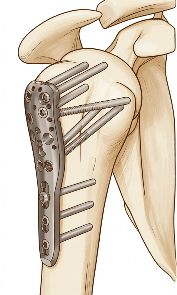
यह प्रोटोकॉल माटर प्राइवेट हॉस्पिटल, रॉकहैम्पटन में डॉ. कीरन हिरपरा द्वारा प्रॉक्सिमल ह्यूमरस फ्रैक्चर (बांह की ऊपरी हड्डी का कंधे के पास टूटना, जिसे लॉकिंग प्लेट और स्क्रू (पेंच) से जोड़ा जाता है; इसे ओपन रिडक्शन एंड इंटरनल फ़िक्सेशन, यानी ORIF कहते हैं) के ऑपरेशन के बाद के रिहैबिलिटेशन (ताकत और हरकत वापस लाने की प्रक्रिया) के बारे में है। अपनी पहली फिजियोथेरेपी मुलाक़ात पर यह पेज या इसका पीडीएफ साथ लाएं, ताकि आपका रिहैबिलिटेशन तालमेल के साथ चले। आपका फ्रैक्चर कैसे जुड़ता है, इसके अनुसार आपका फिजियोथेरेपिस्ट नीचे दिए गए चरणों में आपका रिहैबिलिटेशन व्यक्तिगत रूप से आगे बढ़ाता है।
सर्जरी के बाद अगर आपको अपने घाव को लेकर कोई भी चिंता हो, तो क्लिनिक से संपर्क करें। अक्सर घाव की फ़ोटो लेकर उसे जांच के लिए ईमेल करना मददगार होता है।
क्या उम्मीद करें
फ्रैक्चर को जोड़ने के ऑपरेशन के बाद रिहैबिलिटेशन, कंधे के ज़्यादातर पहले से तय ऑपरेशनों के बाद होने वाले रिहैबिलिटेशन से अलग तरह से चलता है। प्लेट और स्क्रू टूटी हुई हड्डी को उसकी जगह पर थामे रखते हैं, लेकिन हड्डी को ख़ुद तो जुड़ना ही होता है, और हड्डी जुड़ने की रफ़्तार हर व्यक्ति में काफ़ी अलग होती है। इसी कारण नीचे दी गई समय-सारणी एक तय कार्यक्रम नहीं, बल्कि आम तौर पर लगने वाले समय की गाइड है: आपके प्रोग्राम में हर अगला कदम दोनों बातों पर निर्भर करता है, सर्जरी के बाद कितने सप्ताह बीते हैं और आपके एक्स-रे में फ्रैक्चर कैसे जुड़ रहा है, जिसकी पुष्टि डॉ. हिरपरा के साथ आपकी फॉलो-अप जांचों में होती है। केवल कैलेंडर देखकर ख़ुद अगले चरण पर न जाएं; अपनी फॉलो-अप जांच का इंतज़ार करें।
एक और बात इस ऑपरेशन को अलग बनाती है। ह्यूमरस (बांह की ऊपरी हड्डी) के ऊपरी सिरे पर मौजूद हड्डीदार उभार (ट्यूबरोसिटी) वह जगह हैं जहां रोटेटर कफ़ (कंधे को घुमाने वाली मांसपेशियों और टेंडनों का समूह) के टेंडन (कंडरा) जुड़ते हैं, और कई प्रॉक्सिमल ह्यूमरस फ्रैक्चर में ये टुकड़े भी टूटे होते हैं और इन्हें प्लेट से या टांकों से वापस जोड़ा जाता है। रोटेटर कफ़ की मांसपेशियों से बहुत ज़्यादा और बहुत जल्दी काम लेने पर, इन टुकड़ों के जुड़ने से पहले ही उन पर खींच पड़ सकती है। आधुनिक लॉकिंग प्लेट से पहले दिन से ही जोड़ मज़बूत होता है, इसलिए शुरुआत में हल्की हरकत को बढ़ावा दिया जाता है: आप शुरुआत से ही आराम की सीमा में कंधे को एक्टिव-असिस्टेड (दूसरी बांह की मदद से) और धीरे से एक्टिव (ख़ुद) हिला सकते हैं, और बांह को बिना दर्द वाली रेंज में ऊपर उठा सकते हैं। सावधानी वाली बात रोटेटर कफ़ पर भार डालना है: जहां ट्यूबरोसिटी के टुकड़े फ्रैक्चर का हिस्सा नहीं हैं, या मज़बूती से जुड़े हुए हैं, वहां आपका सर्जन आपको एक्टिव रूप से घुमाने और रोटेटर कफ़ का काम जल्दी शुरू करने की अनुमति दे सकता है; जहां ट्यूबरोसिटी प्रभावित हैं और जुड़ रही हैं, वहां रोटेटर कफ़ का एक्टिव काम और ज़बरदस्ती बाहर की ओर घुमाना थोड़ा और लंबे समय तक रोक कर रखा जाता है। डॉ. हिरपरा आपको बताएंगे कि आप पर इनमें से कौन-सी बात लागू होती है।
आपके एक्सरसाइज़ प्रोग्राम में तीन तरह की हरकतें होती हैं, और आपकी टीम बताएगी कि इनमें से कौन-सी आप पर लागू होती हैं:
- एक्टिव रेंज ऑफ मोशन (जोड़ कितना हिल-डुल सकता है): बिना किसी सहारे या मदद के हरकत करने की अनुमति है।
- एक्टिव-असिस्टेड रेंज ऑफ मोशन: बांह को हिलाने में मदद के लिए दूसरी बांह या किसी चीज़ का इस्तेमाल करना।
- पैसिव रेंज ऑफ मोशन: बांह पूरी तरह ढीली रहती है, और दूसरी बांह या बाहरी ज़ोर से 100% काम किया जाता है।
सर्जरी के बाद जब आप जागेंगे, तो आपकी बांह स्लिंग (बांह को सहारा देने वाली पट्टी) में होगी। पहले लगभग तीन सप्ताह तक इसे पूरे समय पहनें, फिर उसके बाद के सप्ताहों में आराम के अनुसार धीरे-धीरे इसे छोड़ें; प्रकाशित प्रोटोकॉल में बांह को स्थिर रखने की औसत अवधि लगभग तीन सप्ताह है, और प्रमाण स्लिंग में ज़्यादा समय रहने के बजाय जल्दी हरकत शुरू करने के पक्ष में हैं। आपको इसे पहनकर सोने की ज़रूरत नहीं है। अपनी एक्सरसाइज़ के लिए और नहाने-धोने और कपड़े पहनने के लिए इसे दिन में कई बार उतारें, और घर पर शांति से बैठे होने पर आप बांह को स्लिंग से बाहर रखकर आराम दे सकते हैं। कंधे के किसी भी ऑपरेशन के बाद कम से कम छह सप्ताह तक गाड़ी न चलाएं; आपका सर्जन आपको गाड़ी चलाने की अनुमति देगा, आम तौर पर छह सप्ताह की फॉलो-अप जांच में।
एक नज़र में पूरा सफ़र:
- चरण I — सुरक्षा और शुरुआती हरकत: सप्ताह 0–6
- चरण II — एक्टिव हरकत वापस पाना: सप्ताह 6–12
- चरण III — ताकत बढ़ाना: लगभग 3 से 4½ महीने
- चरण IV — पूरी गतिविधि पर लौटना: लगभग 4½–6 महीने से
चरण I — सुरक्षा और शुरुआती हरकत (सप्ताह 0–6)
Kieran Hirpara 4.0
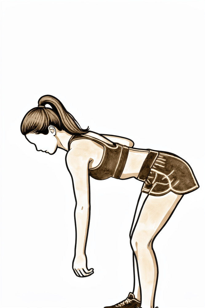
पेंडुलम एक्सरसाइज़
आगे की ओर झुकें, अपने दूसरे हाथ से मेज़ का सहारा लें, और ऑपरेशन वाली बांह को ढीला लटकने दें। अपने शरीर को धीरे-धीरे हिलाकर छोटे-छोटे गोले बनाएं, पहले घड़ी की सुई की दिशा में, फिर उलटी दिशा में। बांह अपने-आप (पैसिव तरीके से) झूलती है; हरकत आपके शरीर से आती है, कंधे की मांसपेशियों से नहीं।
दिन में कई बार
Kieran Hirpara 4.0
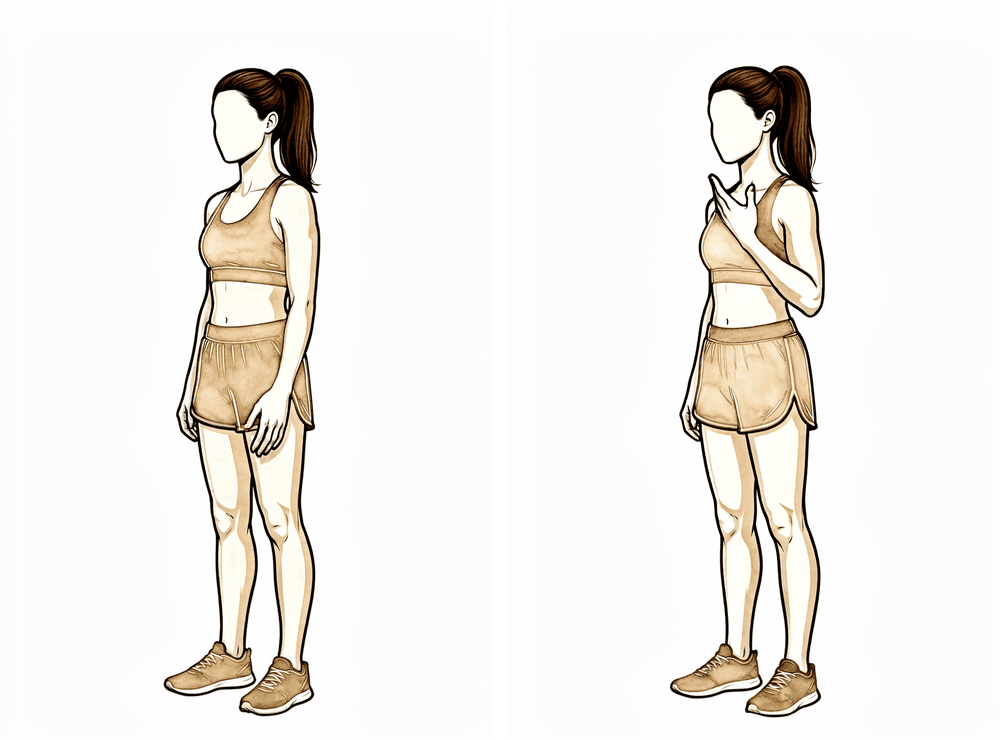
कोहनी को ख़ुद मोड़ना (एक्टिव)
बांह को स्लिंग से बाहर निकालकर और बांह के ऊपरी हिस्से को शरीर के बगल में टिकाकर, कोहनी को जितना आराम से मुड़ सके उतना मोड़ें, फिर उसे पूरी तरह सीधा करें। केवल कोहनी हिलती है: कंधे को स्थिर रखें और बांह को शरीर के बगल में रखें।
दिन में कई बार
Kieran Hirpara 4.0
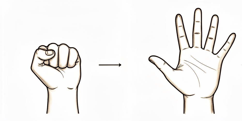
बॉल दबाना
बांह को सहारा देकर, एक नरम गेंद दबाएं या कसकर मुट्ठी बनाएं, फिर हाथ को पूरी तरह खोलें। हर बार जब बांह स्लिंग से बाहर हो, उंगलियों, कलाई और हाथ को हिलाते रहें।
दिन में कई बार
Kieran Hirpara 4.0
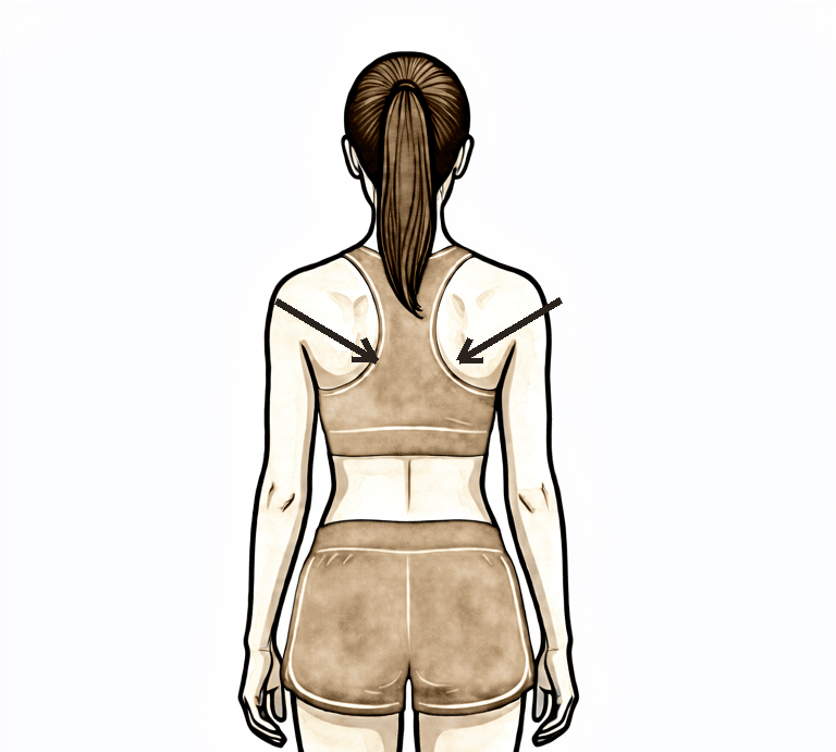
स्कैपुलर सेटिंग
सीधे तनकर बैठें या खड़े हों, बांहों को हिलाए बिना अपनी कंधे की चपटी हड्डियों को धीरे से नीचे और पीछे की ओर आपस में खींचें, कुछ सेकंड रोककर रखें, फिर ढीला छोड़ दें। अपने कान, कंधे और कूल्हे एक सीध में रखें।
जैसा आपका फिजियोथेरेपिस्ट बताए
Kieran Hirpara 4.0
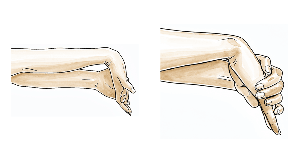
कलाई की हरकतें
बांह को स्लिंग से बाहर निकालकर, कलाई को आगे, पीछे और धीरे से अगल-बगल मोड़कर हिलाते रहें। इससे कंधे के सुरक्षित रहने के दौरान पूरी बांह काम करती रहती है।
दिन में कई बार
Kieran Hirpara 4.0
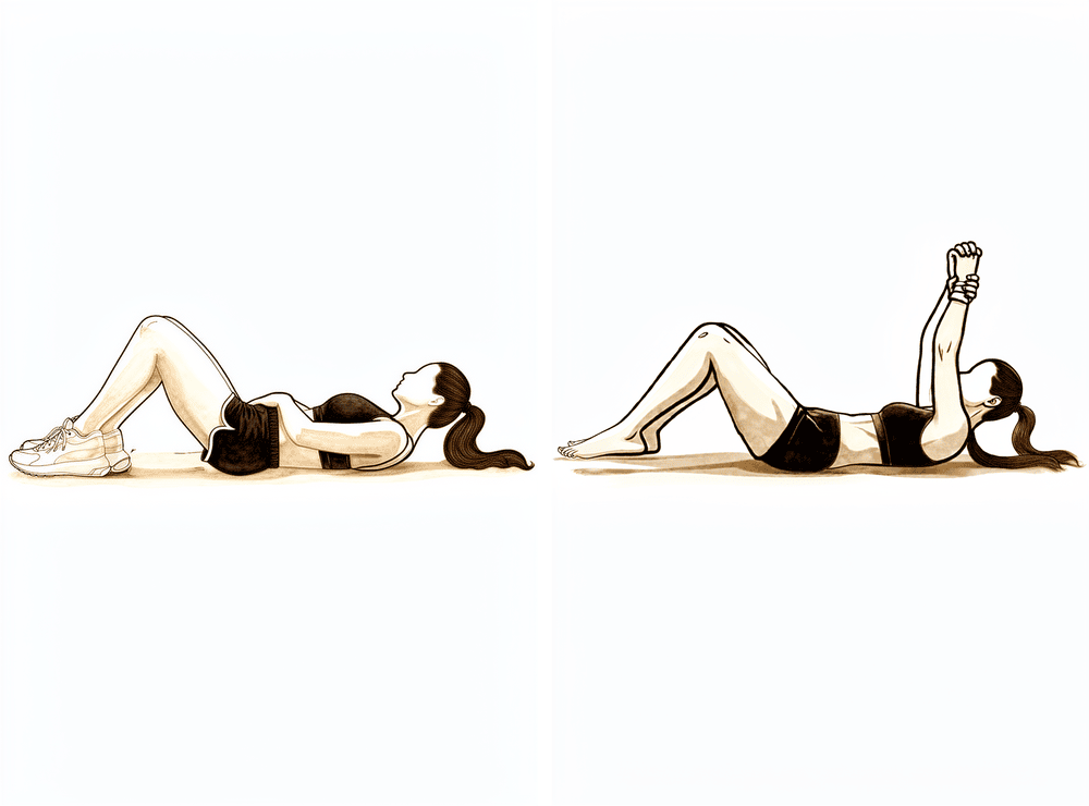
मदद से बांह आगे उठाना (लेटकर)
पीठ के बल लेटें और अपनी ठीक वाली बांह से ऑपरेशन वाली बांह को अपने सामने ऊपर उठाने में मदद करें, फिर ठीक वाली बांह की मदद जारी रखते हुए उसे धीरे से नीचे लाएं। पीठ के बल शुरू करने से गुरुत्वाकर्षण मदद करता है और शुरुआत में बांह उठाना आसान होता है। आरामदायक, बिना दर्द वाली रेंज में रहें और जहां खिंचाव महसूस होने लगे वहीं रुक जाएं: ज़बरदस्ती न करें।
दिन में कई बार, बिना दर्द वाली रेंज में
Kieran Hirpara 4.0
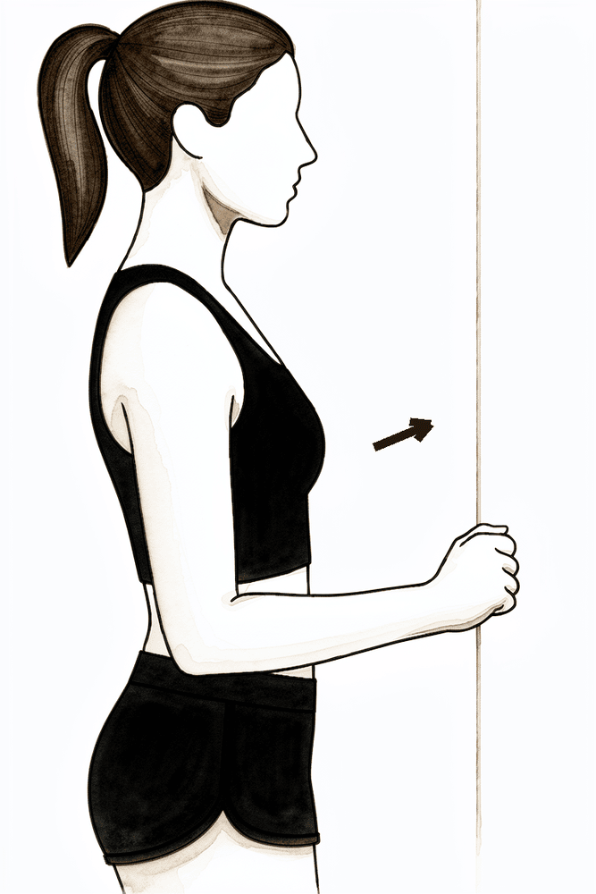
हल्का फ़ॉरवर्ड-एलिवेशन आइसोमेट्रिक (आगे की ओर दबाना)
दीवार की ओर मुंह करके खड़े हों, ऑपरेशन वाली बांह शरीर के बगल में हो और हल्की बंद मुट्ठी सामने दीवार से सटी हो। बांह को हिलने दिए बिना हाथ को धीरे से आगे दीवार में दबाएं, कुछ सेकंड रोककर रखें, फिर ढीला छोड़ दें। इससे जुड़ते हुए फ्रैक्चर पर ज़ोर डाले बिना कंधे को ऊपर उठाने वाली मांसपेशियां सक्रिय होती हैं। इसे केवल तभी करें जब आपके फिजियोथेरेपिस्ट ने आपको इसे शुरू कराया हो।
जैसा आपका फिजियोथेरेपिस्ट बताए
पहले छह सप्ताह का मकसद है जोड़े गए फ्रैक्चर की सुरक्षा करना, और साथ ही बाक़ी बांह (और ख़ुद कंधे के जोड़) को जकड़ने से बचाना। क्योंकि लॉकिंग प्लेट शुरुआत से ही हड्डी को मज़बूती से थामे रखती है, इसलिए छह सप्ताह इंतज़ार करने के बजाय शुरुआत में ही हल्की हरकत को बढ़ावा दिया जाता है। घर जाने से पहले एक फिजियोथेरेपिस्ट आपको हल्की एक्सरसाइज़ शुरू कराएगा: पेंडुलम एक्सरसाइज़ (शरीर को धीरे-धीरे हिलाकर बांह को लटकने और धीरे से झूलने देना), और दिन में कई बार स्लिंग से बाहर निकालकर कोहनी, कलाई और हाथ को हिलाते रहना। शुरुआत से ही, आरामदायक और बिना दर्द वाली रेंज के भीतर, आप अपने दूसरे हाथ की मदद से (एक्टिव-असिस्टेड) और धीरे से बांह की अपनी ताकत से (एक्टिव) बांह को हिलाना शुरू कर सकते हैं, और आराम के अनुसार उसे अपने सामने ऊपर उठा सकते हैं। सावधानी वाला हिस्सा रोटेटर कफ़ पर भार डालना है: बांह को ज़बरदस्ती बाहर की ओर घुमाने (एक्सटर्नल रोटेशन) से बचें, और प्रतिरोध के साथ रोटेटर कफ़ का काम करने से तब तक बचें जब तक आपकी ट्यूबरोसिटी जुड़ न जाएं, जिसकी पुष्टि डॉ. हिरपरा करेंगे। दर्द से राहत के लिए बर्फ़ लगाएं, और अपनी एक्सरसाइज़ और फिजियोथेरेपी अपॉइंटमेंट से पहले अपनी दर्द निवारक दवाएं ले लें।
आपके फिजियोथेरेपिस्ट के लिए:
लक्ष्य
- फ़िक्सेशन की सुरक्षा करना और हड्डी के जुड़ने को बेहतर बनाना
- दर्द और सूजन को शांत करना
- बिना दर्द वाली रेंज में, शुरुआती एक्टिव-असिस्टेड एलिवेशन (ऊपर उठाना) स्थापित करना और उससे एक्टिव एलिवेशन की ओर बढ़ना
- गर्दन, कोहनी, कलाई और हाथ की पूरी हरकत बनाए रखना
प्रबंधन
- ~3 सप्ताह तक पूरे समय स्लिंग, फिर आराम के अनुसार धीरे-धीरे छुड़ाना (साहित्य में प्रचलित तरीका); एक्सरसाइज़ और साफ़-सफ़ाई के लिए दिन में कई बार उतारें; बिस्तर में ज़रूरी नहीं
- पेंडुलम / कॉडमैन एक्सरसाइज़ दिन में कई बार
- स्थिर फ़िक्सेशन होने पर शुरुआत से ही, आरामदायक और बिना दर्द वाली रेंज के भीतर, शुरुआती एक्टिव-असिस्टेड और हल्का एक्टिव फ़ॉरवर्ड एलिवेशन; पीठ के बल लेटकर शुरू करके, नियंत्रण के अनुसार सीधे बैठकर/खड़े होकर एलिवेशन की ओर बढ़ें; आराम के अनुसार रेंज बढ़ाएं
- जहां अभी एक्टिव हरकत सहन न हो, वहां ज़रूरत के अनुसार पैसिव रेंज ऑफ मोशन: स्कैपुलर प्लेन में फ़ॉरवर्ड एलिवेशन लगभग 90° तक, इंटरनल रोटेशन (अंदर की ओर घुमाना) पेट तक (पीठ के पीछे नहीं)
- एक्सटर्नल रोटेशन को बांह शरीर के बगल में रखते हुए लगभग 30–40° की हल्की शुरुआती डिफ़ॉल्ट सीमा तक रखें; जहां ट्यूबरोसिटी प्रभावित न हों या मज़बूती से जुड़ी हों, वहां सर्जन इसे आगे बढ़ा सकते हैं
- स्लिंग से बाहर गर्दन, कोहनी, कलाई और हाथ की एक्टिव रेंज ऑफ मोशन; पकड़ के लिए बॉल दबाना
- स्कैपुलर सेटिंग और स्कैपुला (कंधे की चपटी हड्डी) की मोबिलिटी का काम (एलिवेशन, डिप्रेशन, रिट्रैक्शन, प्रोट्रैक्शन)
- आराम के अनुसार डेल्टॉइड (कंधे को ढकने वाली बड़ी मांसपेशी) और पेरीस्कैपुलर (कंधे की चपटी हड्डी के आसपास की) मांसपेशियों के हल्के आइसोमेट्रिक्स शुरू किए जा सकते हैं
- सेशन से पहले क्रायोथेरेपी (बर्फ़ से सिकाई) और दर्द निवारक दवा; घाव भर जाने के बाद निशान की मालिश (स्कार मोबिलाइज़ेशन) और डीसेंसिटाइज़ेशन (छूने पर होने वाली अति-संवेदनशीलता कम करना)
सावधानियां
- जब तक ट्यूबरोसिटी के जुड़ने की पुष्टि न हो जाए (सर्जन के मार्गदर्शन में), ज़बरदस्ती या प्रतिरोध के साथ एक्सटर्नल रोटेशन और रोटेटर कफ़ पर भारी भार से बचें; बिना दर्द वाली रेंज में एक्टिव एलिवेशन की अनुमति है
- पीठ के पीछे इंटरनल रोटेशन नहीं; कोरोनल प्लेन में एब्डक्शन (बांह को सीधे बगल से ऊपर उठाना) से बचें
- शुरुआती हरकत को बिना दर्द के रखें: हल्की और आराम की सीमा में, ज़बरदस्ती नहीं
- ऑपरेशन वाली बांह से लगभग 0.5–1 किलो से ज़्यादा वज़न नहीं उठाना
- ऑपरेशन वाली बांह के ज़रिए कोई वज़न नहीं डालना (कुर्सी या बिस्तर से हाथों के बल ज़ोर लगाकर नहीं उठना)
- छह सप्ताह तक गाड़ी नहीं चलाना (यह कंधे के किसी भी ऑपरेशन पर लागू होता है)
- रेंज के आख़िरी छोर पर कोई ज़बरदस्ती वाली या दर्द वाली हरकत नहीं
अगले चरण में जाने के मानदंड
- एक्स-रे पर हड्डी के जुड़ने की प्रगति का प्रमाण, जिसकी पुष्टि लगभग 6 सप्ताह पर डॉ. हिरपरा के साथ फॉलो-अप जांच में हो
- दर्द अच्छी तरह काबू में हो
- कोहनी, कलाई और हाथ की पूरी हरकत बनी रहे
- बिना दर्द वाली रेंज के भीतर आरामदायक एक्टिव-असिस्टेड से लेकर एक्टिव एलिवेशन
चरण II — एक्टिव हरकत वापस पाना (सप्ताह 6–12)
Kieran Hirpara 4.0
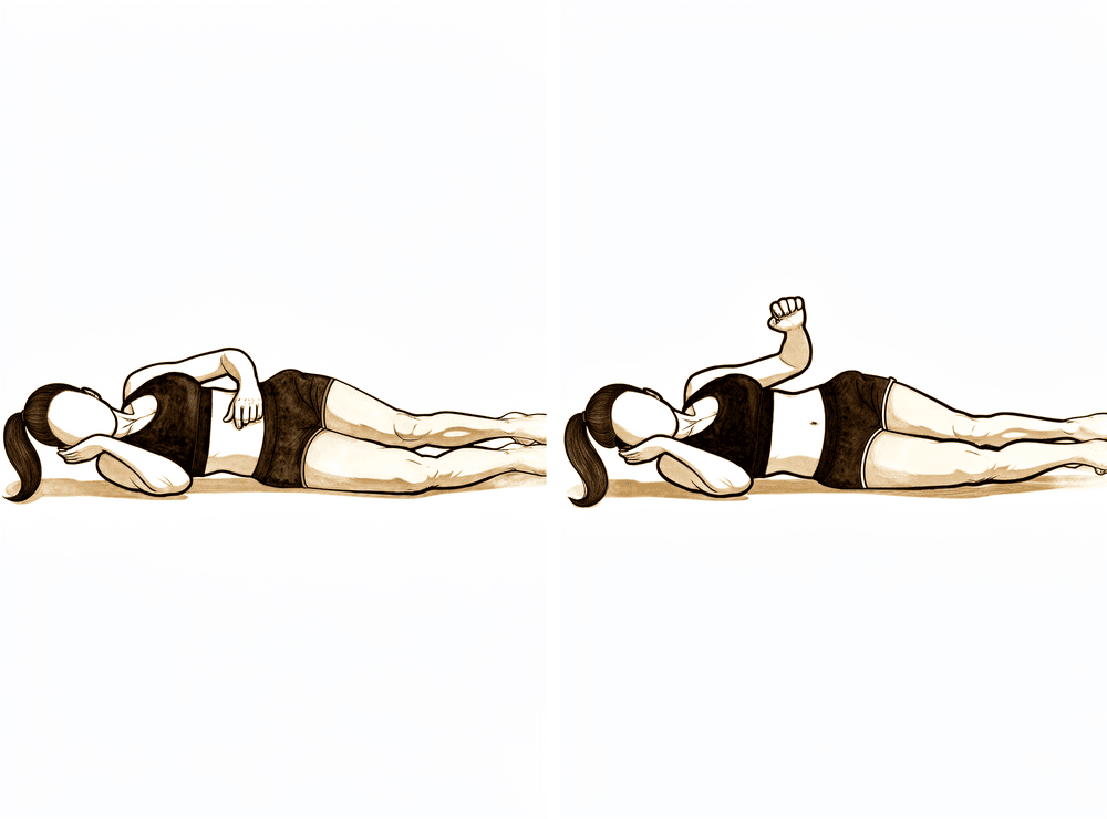
करवट लेटकर एक्सटर्नल रोटेशन
बिना ऑपरेशन वाली करवट लेटें, ऑपरेशन वाली बांह ऊपर हो, कोहनी 90 डिग्री पर मुड़ी हुई और शरीर से सटी हो। कोहनी को सटाए रखते हुए, अग्रबाहु (कोहनी से कलाई तक का हिस्सा) को पेट से दूर ऊपर की ओर जितना आराम से हो सके घुमाएं, फिर नियंत्रण के साथ नीचे लाएं। कोई अतिरिक्त वज़न नहीं: बांह अपनी ताकत से हिलती है।
जैसा आपका फिजियोथेरेपिस्ट बताए
Kieran Hirpara 4.0
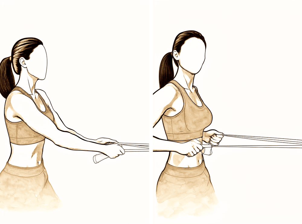
इलास्टिक बैंड के साथ लो रो
इलास्टिक बैंड को अपने सामने कमर की ऊंचाई पर बांधें। सीधे तनकर खड़े होकर बैंड को अपनी ओर खींचें, कंधे की चपटी हड्डी को धीरे से नीचे और पीछे ले जाते हुए, फिर नियंत्रण के साथ छोड़ें। प्रतिरोध हल्का रखें।
जैसा आपका फिजियोथेरेपिस्ट बताए
Kieran Hirpara 4.0
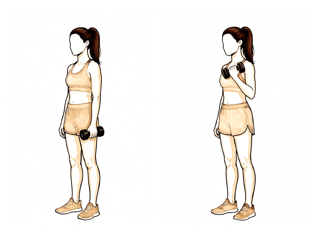
बाइसेप्स कर्ल
हथेली ऊपर की ओर रखकर हल्का वज़न पकड़ें, कोहनी मोड़कर वज़न को अपने कंधे की ओर लाएं, फिर धीरे-धीरे नीचे करें। बांह के ऊपरी हिस्से को शरीर के बगल में स्थिर रखें, और वज़न हल्का रखें: इस चरण में लगभग 1–2 किलो से ज़्यादा नहीं।
जैसा आपका फिजियोथेरेपिस्ट बताए
Kieran Hirpara 4.0
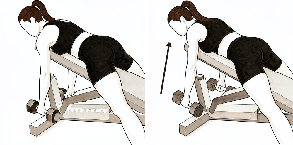
सहारे के साथ रो
अपनी ठीक वाली बांह को मेज़ पर टिकाकर आगे झुकें, और ऑपरेशन वाली बांह को हल्का वज़न पकड़े लटकने दें। कंधे की चपटी हड्डी को पीछे खींचें और वज़न को कूल्हे की ओर ऊपर खींचें (रो करें), फिर नियंत्रण के साथ नीचे लाएं। धड़ को स्थिर रखें: हरकत कंधे की चपटी हड्डी और बांह से आती है, पीठ से नहीं।
जैसा आपका फिजियोथेरेपिस्ट बताए
Kieran Hirpara 4.0
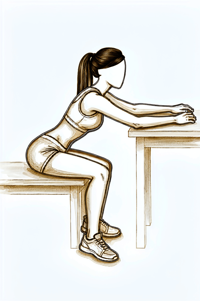
टेबल स्लाइड (मेज़ पर हाथ सरकाना)
मेज़ के पास बैठें, हाथ किसी तौलिये या कपड़े पर टिका हो। आगे झुकें और हाथ को मेज़ पर अपने से दूर सरकाएं, जिससे बांह सामने और आगे तक पहुंचे, फिर उसे वापस सरकाएं। बांह का वज़न मेज़ संभालती है, इसलिए आगे की हरकत वापस पाने का यह एक हल्का तरीका है।
जैसा आपका फिजियोथेरेपिस्ट बताए
Kieran Hirpara 4.0
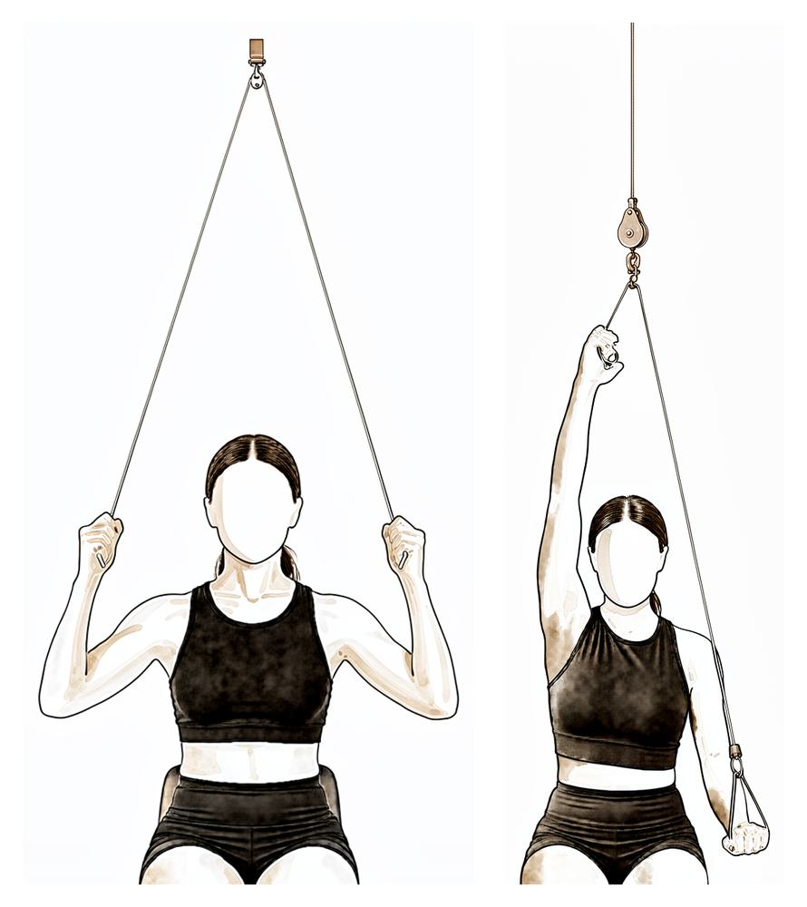
दरवाज़े के ऊपर लगी पुली
दरवाज़े के ऊपर लगी पुली (रस्सी और घिरनी) के नीचे बैठें, दोनों हाथों में एक-एक हैंडल हो। ठीक वाली बांह से नीचे खींचकर ऑपरेशन वाली बांह को जितना आराम से जा सके उतना ऊपर उठाएं, फिर उसे धीरे-धीरे नीचे लाएं। काम ठीक वाली बांह को करने दें और आरामदायक रेंज में रहें।
जैसा आपका फिजियोथेरेपिस्ट बताए
Kieran Hirpara 4.0
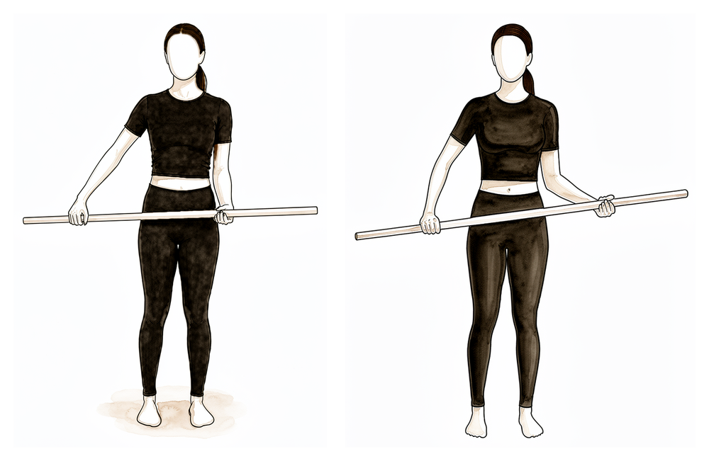
छड़ी की मदद से एक्सटर्नल रोटेशन
पीठ के बल लेटें, कोहनी शरीर के बगल से सटी और समकोण पर मुड़ी हो। दोनों हाथों में एक छड़ी पकड़ें और ठीक वाली बांह से ऑपरेशन वाले हाथ को बाहर की ओर घुमाएं। जुड़ने की पुष्टि हो जाने के बाद, बाहर की ओर घुमाने को धीरे से थोड़ा और आगे ले जाएं, लेकिन उसी रेंज के भीतर रहें जो आपके सर्जन और फिजियोथेरेपिस्ट ने तय की है।
जैसा आपका फिजियोथेरेपिस्ट बताए
Kieran Hirpara 4.0
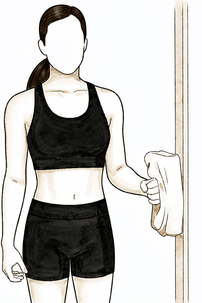
आइसोमेट्रिक एक्सटर्नल रोटेशन (बाहर की ओर दबाना)
कोहनी को शरीर के बगल से सटाकर और समकोण पर मोड़कर खड़े हों, हाथ का पिछला हिस्सा (हथेली की उलटी तरफ़) दीवार या दरवाज़े की चौखट से सटा हो। बांह को हिलने दिए बिना धीरे से बाहर की ओर दबाएं, कुछ सेकंड रोककर रखें, फिर ढीला छोड़ दें। इससे बिना किसी हरकत के रोटेटर कफ़ सक्रिय होना शुरू होता है: इसे केवल तब शुरू करें जब डॉ. हिरपरा आपकी हड्डी जुड़ने की पुष्टि कर दें।
जैसा आपका फिजियोथेरेपिस्ट बताए
Kieran Hirpara 4.0
ट्राइसेप्स एक्सटेंशन
हल्का वज़न पकड़कर, कोहनी को मोड़ें और फिर सीधा करें, ताकि बांह के ऊपरी हिस्से के पीछे की मांसपेशी काम करे। बांह के ऊपरी हिस्से को स्थिर रखें और वज़न हल्का रखें: इस चरण में लगभग 1–2 किलो से ज़्यादा नहीं।
जैसा आपका फिजियोथेरेपिस्ट बताए
Kieran Hirpara 4.0
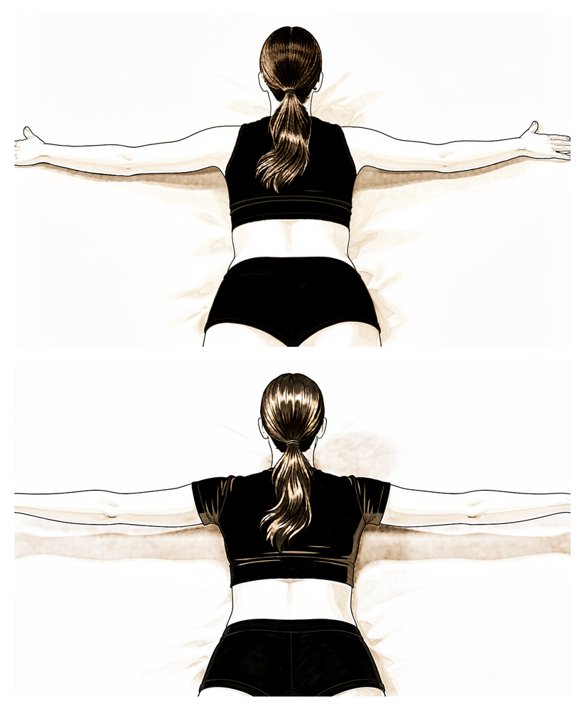
पेट के बल लेटकर स्कैपुलर रो
पेट के बल लेटें, आपकी बांह बिस्तर या बेंच के किनारे से नीचे लटकी हो। कंधे की चपटी हड्डी को पीछे खींचें और बांह को थोड़ा बगल की ओर उठाएं, कंधे की चपटी हड्डियों को आपस में भींचते हुए, फिर नियंत्रण के साथ नीचे लाएं। इससे वे मांसपेशियां मज़बूत होती हैं जो कंधे की चपटी हड्डी को स्थिर रखती हैं।
जैसा आपका फिजियोथेरेपिस्ट बताए
लगभग छह सप्ताह पर आपकी फॉलो-अप जांच में डॉ. हिरपरा आपके एक्स-रे देखते हैं। अगर हड्डी उम्मीद के अनुसार जुड़ रही है, तो स्लिंग का बचा हुआ इस्तेमाल बंद हो जाता है और आप बांह को ख़ुद हिलाना शुरू करते हैं, पहले मदद के साथ (दूसरी बांह, छड़ी या पुली, यानी रस्सी और घिरनी, का इस्तेमाल करके), फिर एक्टिव रूप से। बांह को ख़ुद उठाना आम तौर पर पीठ के बल लेटकर शुरू करना सबसे अच्छा रहता है, जहां गुरुत्वाकर्षण कम बाधा डालता है, और नियंत्रण बेहतर होने पर बैठकर और खड़े होकर करने की ओर बढ़ें। डॉ. हिरपरा द्वारा जुड़ने की पुष्टि हो जाने के बाद, इस चरण में रोटेटर कफ़ के लिए हल्की मसल-सेटिंग (आइसोमेट्रिक) एक्सरसाइज़ शुरू होती हैं; प्रतिरोध के विरुद्ध एक्सरसाइज़ बाद में आती हैं। आप फिर से गाड़ी चलाना तब शुरू कर सकते हैं जब आप स्लिंग से बाहर हों, सुरक्षित रूप से गाड़ी चलाने के लिए बांह में पर्याप्त हरकत और नियंत्रण हो, और आप तेज़ दर्द निवारक दवाएं न ले रहे हों; अगर संदेह हो, तो अपनी फॉलो-अप जांच में इस पर बात करें।
आपके फिजियोथेरेपिस्ट के लिए:
लक्ष्य
- पूरी पैसिव रेंज ऑफ मोशन वापस लाना
- सभी दिशाओं में एक्टिव-असिस्टेड से एक्टिव रेंज ऑफ मोशन की ओर बढ़ना
- सामान्य स्कैपुलोह्यूमरल रिदम (कंधे और कंधे की चपटी हड्डी की तालमेल वाली हरकत) फिर से स्थापित करना और क्षतिपूरक पैटर्न कम से कम करना
- रोज़मर्रा की सामान्य हल्की गतिविधियों पर लौटना
प्रबंधन
- स्लिंग ज़्यादा से ज़्यादा 6 सप्ताह की फॉलो-अप जांच तक पूरी तरह छोड़ दी जाए
- एक्टिव-असिस्टेड रेंज ऑफ मोशन: लॉन-चेयर प्रोग्रेशन, टेबल / वॉल स्लाइड, पुली, छड़ी वाली एक्सरसाइज़, आराम के अनुसार चरण I की सीमाओं से आगे बढ़ते हुए
- लगभग 6–8 सप्ताह से एक्टिव रेंज ऑफ मोशन: पीठ के बल लेटकर फ्लेक्शन से सीधे बैठकर/खड़े होकर एलिवेशन की ओर; करवट लेटकर एक्सटर्नल रोटेशन और फ्लेक्शन; लो रो / लो पंच
- लगभग 6–8 सप्ताह से, जुड़ने की पुष्टि होने के बाद, बांह शरीर के बगल में रखते हुए, रोटेटर कफ़ और डेल्टॉइड के अधिकतम से कम ज़ोर वाले आइसोमेट्रिक्स
- कोहनी के हल्के आइसोटोनिक्स (बाइसेप्स कर्ल, ट्राइसेप्स एक्सटेंशन) और स्कैपुलर ताकत का काम (रिट्रैक्शन, पेट के बल लेटकर रो)
- ज़रूरत के अनुसार ग्लेनोह्यूमरल और स्कैपुलोथोरेसिक मोबिलाइज़ेशन, हड्डी के जुड़ने के साथ ग्रेड बढ़ाते हुए
- पोस्चर सुधारना; पसंद के अनुसार सेशन के आसपास सिकाई / बर्फ़ और दर्द निवारक दवा जारी रखें
सावधानियां
- जुड़ने की पुष्टि होने तक प्रतिरोध वाला (आइसोटोनिक) रोटेटर कफ़ स्ट्रेंथनिंग नहीं, आम तौर पर 8–12 सप्ताह से पहले नहीं
- रेंज के आख़िरी छोर पर कोई ज़ोरदार दबाव (ओवरप्रेशर) या आक्रामक पैसिव स्ट्रेचिंग नहीं
- ऑपरेशन वाली बांह से वज़न उठाना लगभग 1–2 किलो तक सीमित
- एलिवेशन के दौरान कंधा उचकाने और धड़ के झुकने जैसी क्षतिपूर्ति पर नज़र रखें और उसे सुधारें
अगले चरण में जाने के मानदंड
- पूरी, या लगभग पूरी, पैसिव रेंज ऑफ मोशन
- अच्छी तकनीक के साथ एक्टिव एलिवेशन, कम से कम कंधे की ऊंचाई से नीचे
- आइसोमेट्रिक्स बिना दर्द भड़के अच्छी तरह सहन हों
- एक्स-रे पर हड्डी का जुड़ना आगे बढ़ रहा हो, जिसकी पुष्टि डॉ. हिरपरा के साथ आपकी फॉलो-अप जांच में हो
चरण III — ताकत बढ़ाना (लगभग 3 से 4½ महीने)
Kieran Hirpara 4.0
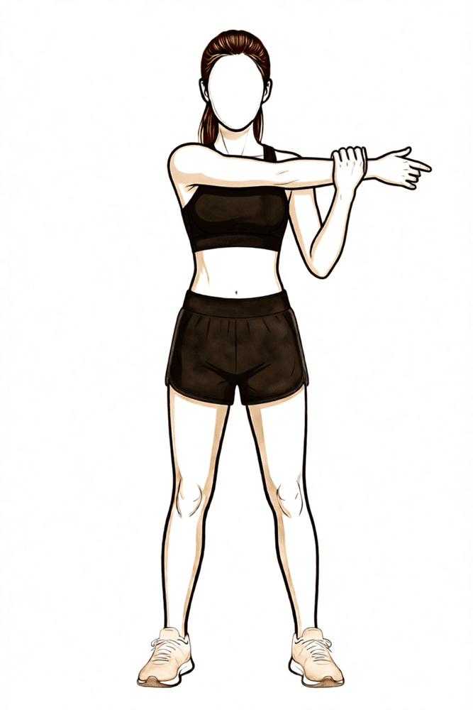
क्रॉस-बॉडी स्ट्रेच
अपने दूसरे हाथ से ऑपरेशन वाली बांह को अपनी छाती के आर-पार तब तक खींचें जब तक कंधे के पिछले हिस्से में खिंचाव महसूस न हो। थोड़ी देर रोकें, फिर छोड़ दें। केवल स्पष्ट असहजता तक स्ट्रेच करें, तेज़ दर्द तक कभी नहीं।
जैसा आपका फिजियोथेरेपिस्ट बताए
Kieran Hirpara 4.0
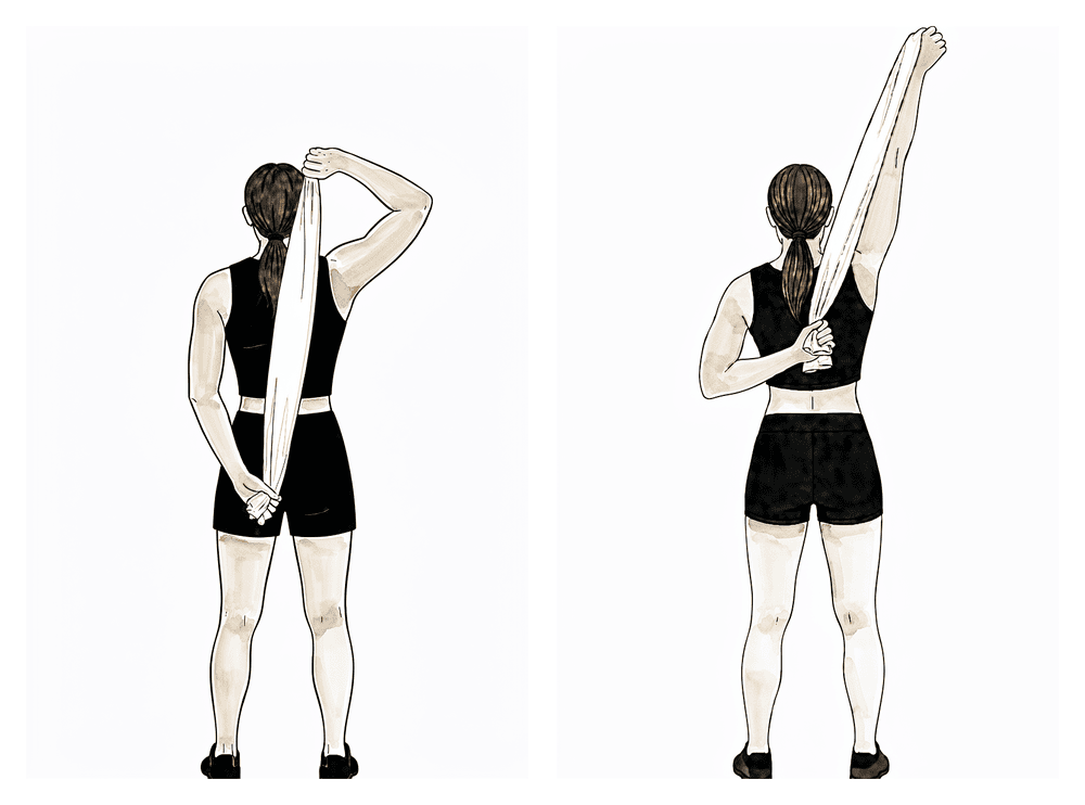
पीठ के पीछे स्ट्रेच
पीठ के पीछे एक तौलिया पकड़ें, ठीक वाली बांह ऊपर और ऑपरेशन वाली बांह नीचे। ऊपर वाले हाथ से नीचे वाले हाथ को धीरे से पीठ पर ऊपर की ओर खींचें, जब तक आरामदायक खिंचाव महसूस न हो, रोकें, फिर छोड़ दें। रेंज धीरे-धीरे बढ़ाएं: स्ट्रेच के लिए कभी ज़बरदस्ती न करें और तेज़ दर्द तक न ले जाएं।
जैसा आपका फिजियोथेरेपिस्ट बताए
Kieran Hirpara 4.0
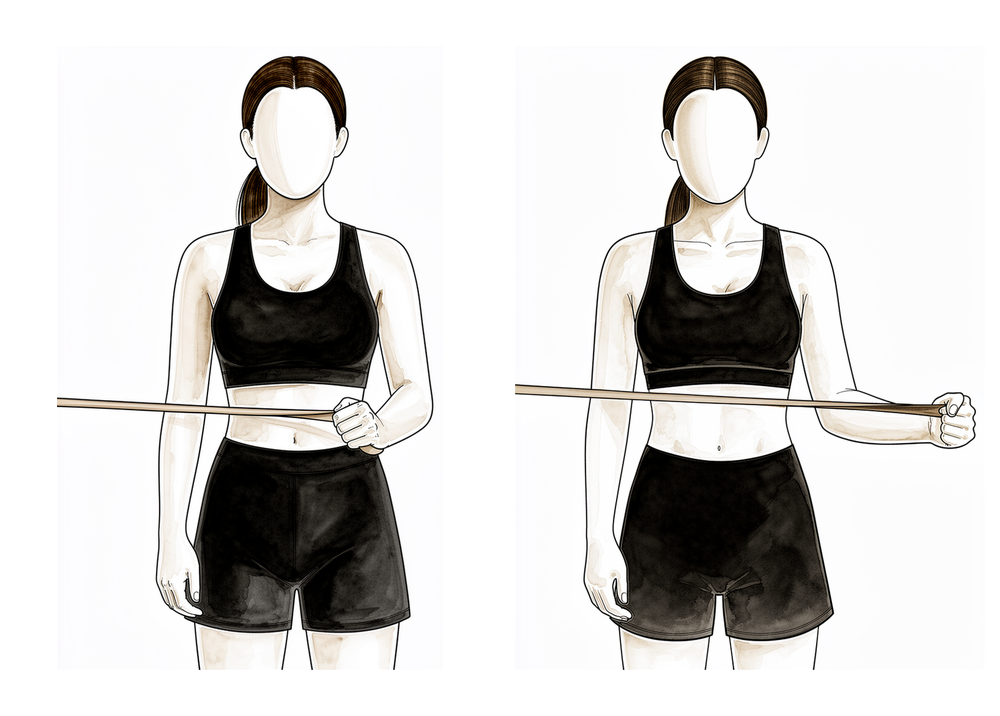
बैंड के साथ एक्सटर्नल रोटेशन
कोहनी को शरीर के बगल से सटाकर और समकोण पर मोड़कर खड़े हों, कमर की ऊंचाई पर बंधा इलास्टिक बैंड पकड़े हुए। कोहनी को बगल में रखते हुए, अग्रबाहु को बैंड के विरुद्ध बाहर की ओर घुमाएं, फिर धीरे-धीरे वापस लाएं। इससे बांह बगल में रखते हुए रोटेटर कफ़ मज़बूत होता है, जो घुमाने की ताकत बढ़ाने का शुरुआती बिंदु है।
2 से 3 सेट, हर सेट में 8 से 12 बार, कम प्रतिरोध
Kieran Hirpara 4.0
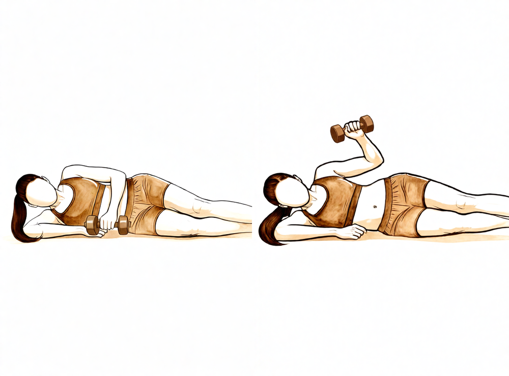
करवट लेटकर वज़न के साथ एक्सटर्नल रोटेशन
बिना ऑपरेशन वाली करवट लेटें, ऊपर वाली कोहनी समकोण पर मुड़ी हुई और शरीर से सटी हो। हल्का वज़न पकड़कर अग्रबाहु को छत की ओर ऊपर घुमाएं, फिर धीरे-धीरे नीचे लाएं। जैसे-जैसे आपके रोटेटर कफ़ की ताकत वापस बनती है, भार हल्का और दोहराव ज़्यादा रखें।
2 से 3 सेट, हर सेट में 8 से 12 बार, कम भार
Kieran Hirpara 4.0
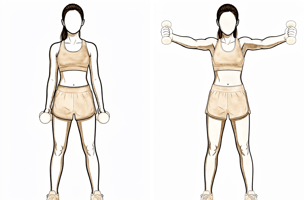
फ़ुल-कैन रेज़ (स्कैप्शन)
अंगूठा ऊपर की ओर रखते हुए हल्का वज़न पकड़ें, और बांह को आगे-और-बाहर की तिरछी दिशा में उठाएं, सीधे बगल की ओर नहीं, लगभग कंधे की ऊंचाई तक, फिर धीरे-धीरे नीचे लाएं। इससे कंधे का अगला हिस्सा और रोटेटर कफ़ साथ-साथ काम करते हैं। वज़न हल्का रखें, और अगर इससे ऐसा दर्द हो जो देर तक बना रहे, तो रुक जाएं।
2 से 3 सेट, हर सेट में 8 से 12 बार, कम भार
Kieran Hirpara 4.0
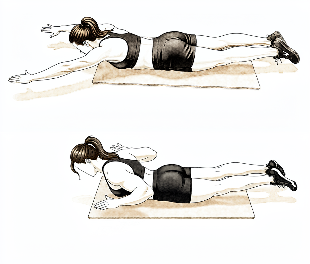
पेट के बल लेटकर रो
पेट के बल लेटें, ऑपरेशन वाली बांह फ़र्श की ओर नीचे लटकी हो। अपनी कोहनी को छत की ओर ऊपर खींचें, कंधे की चपटी हड्डी को रीढ़ की ओर भींचते हुए, फिर धीरे-धीरे नीचे लाएं। बिना वज़न के शुरू करें और हरकत को नियंत्रित रखें।
जैसा आपका फिजियोथेरेपिस्ट बताए
Kieran Hirpara 4.0
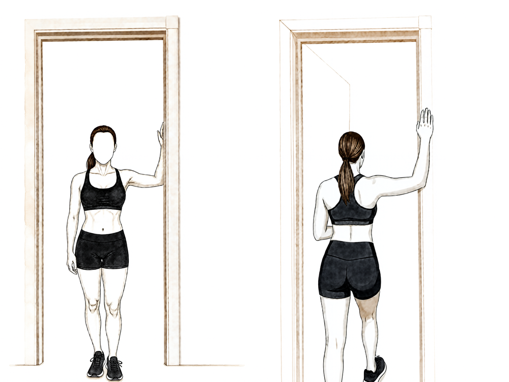
दरवाज़े में छाती का स्ट्रेच
दरवाज़े में खड़े हों, अग्रबाहु चौखट पर टिकी हों और कोहनियां लगभग कंधे की ऊंचाई पर हों। एक पैर आगे बढ़ाएं और धीरे से आगे झुकें जब तक छाती के सामने वाले हिस्से में खिंचाव महसूस न हो, फिर रोककर रखें। इसे हल्का रखें और कभी ज़बरदस्ती न करें।
~20–30 सेकंड तक रोकें, जैसा आपका फिजियोथेरेपिस्ट बताए
Kieran Hirpara 4.0
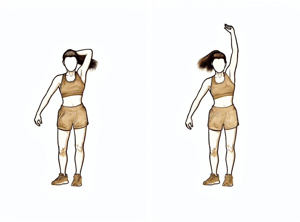
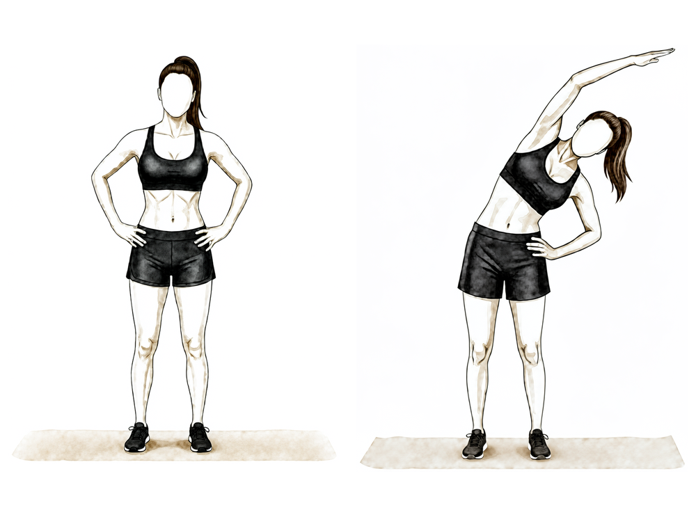
लैटिसिमस (पीठ के बगल वाली मांसपेशी) स्ट्रेच
ऑपरेशन वाली बांह को सिर के ऊपर ले जाएं (ज़रूरत हो तो ठीक वाले हाथ से उसे सहारा दें) और धीरे से दूसरी तरफ़ झुकें जब तक पीठ के बगल में नीचे तक खिंचाव महसूस न हो, फिर रोककर रखें। इसे आरामदायक और अपनी रेंज के भीतर रखें।
~20–30 सेकंड तक रोकें, जैसा आपका फिजियोथेरेपिस्ट बताए
फ्रैक्चर जुड़ जाने और आपकी एक्टिव हरकत लौटने के साथ, ध्यान ताकत दोबारा बनाने पर जाता है। प्रतिरोध वाला काम हल्के से शुरू होता है (रोटेटर कफ़, डेल्टॉइड और कंधे की चपटी हड्डी की मांसपेशियों के लिए इलास्टिक बैंड और हल्के वज़न) और धीरे-धीरे आगे बढ़ता है। साथ में स्ट्रेचिंग जारी रहती है, जिसका लक्ष्य सभी दिशाओं में पूरी हरकत है, जिसमें पीठ के पीछे हाथ ले जाना भी शामिल है। इस चरण के दौरान रोज़मर्रा की सामान्य गतिविधियां काफ़ी हद तक पहले जैसी हो जानी चाहिए, और मनोरंजन की हल्की गतिविधियां आम तौर पर फिर शुरू हो जाती हैं, जैसा आपका फिजियोथेरेपिस्ट बताए।
आपके फिजियोथेरेपिस्ट के लिए:
लक्ष्य
- सामान्य तकनीक के साथ सभी दिशाओं में पूरी एक्टिव रेंज ऑफ मोशन
- रोटेटर कफ़, डेल्टॉइड और स्कैपुलर मांसपेशियों की ताकत और सहनशक्ति को चरणबद्ध तरीके से वापस लाना
प्रबंधन
- रोटेटर कफ़, डेल्टॉइड और स्कैपुलर स्टेबिलाइज़र के लिए आइसोमेट्रिक्स से इलास्टिक बैंड और फिर हल्के फ़्री-वेट (लगभग 0.5–2 किलो) स्ट्रेंथनिंग की ओर बढ़ें: कम भार, ज़्यादा दोहराव (उदाहरण के लिए 2–3 सेट, हर सेट में 8–12 बार), ज़रूरत से ज़्यादा भार से बचने के लिए प्रतिरोध वाले सेशन सप्ताह में लगभग 3 बार
- घुमाने (रोटेशन) की ताकत का काम शुरू में बांह शरीर के बगल में रखकर, कंधे की ऊंचाई से नीचे
- कंधे की चपटी हड्डी के स्थिर आधार के लिए एंटीरियर डेल्टॉइड और ट्रेपेज़ियस–सेराटस एंटीरियर फ़ोर्स कपल पर ज़ोर दें
- सभी दिशाओं में आख़िरी रेंज के लिए लचीलापन प्रोग्राम: पोस्टीरियर कैप्सूल (क्रॉस-बॉडी) स्ट्रेच, पीठ के पीछे इंटरनल रोटेशन, छाती की सामने की दीवार / पेक्टोरैलिस माइनर स्ट्रेच, दरवाज़े में खड़े होकर स्ट्रेच
- रेंज के अनुसार पीठ के पीछे इंटरनल रोटेशन और ग्रेड III–IV मोबिलाइज़ेशन शुरू करें
- कम प्रतिरोध के साथ अपर-बॉडी एर्गोमीटर (हाथों से चलाई जाने वाली साइकिल मशीन); सामान्य एरोबिक कंडीशनिंग
सावधानियां
- ताकत का काम आरामदायक रेंज के भीतर रहे, और उससे ऐसा दर्द नहीं होना चाहिए जो देर तक बना रहे
- इस चरण के दौरान ऑपरेशन वाली बांह से लगभग 4–5 किलो से ज़्यादा वज़न नहीं उठाना
- बांह के ज़रिए ज़्यादा वज़न डालने से बचें (पुश-अप और इस तरह के काम बाद में आते हैं)
अगले चरण में जाने के मानदंड
- बिना किसी क्षतिपूरक तरीके के पूरी एक्टिव रेंज ऑफ मोशन
- ताकत बढ़ाने का प्रोग्राम बिना दर्द भड़के या रेंज घटे सहन हो
चरण IV — पूरी गतिविधि पर लौटना (लगभग 4½–6 महीने से)
आख़िरी चरण भारी वज़न उठाने, शारीरिक मेहनत वाले काम, सिर के ऊपर हाथ ले जाकर किए जाने वाले कामों और खेल पर चरणबद्ध वापसी है। ताकत का काम भारी प्रतिरोध और कई जोड़ों वाली (कंपाउंड) हरकतों की ओर बढ़ता है, और जहां आपके काम या खेल के लिए ज़रूरी हो, वहां तेज़ और ज़्यादा डायनेमिक एक्सरसाइज़ की ओर भी। ज़्यादातर लोग लगभग छह महीने तक अपनी सामान्य गतिविधियों पर लौट आते हैं, हालांकि ताकत और आत्मविश्वास में आम तौर पर एक साल तक सुधार होता रहता है। सही मंज़िल इस पर निर्भर करती है कि आपको बांह से क्या काम लेना है, इसलिए भारी शारीरिक मेहनत वाले काम या संपर्क वाले और सिर के ऊपर हाथ ले जाने वाले खेलों पर वापसी कैलेंडर से तय नहीं होती, बल्कि डॉ. हिरपरा और आपके फिजियोथेरेपिस्ट के साथ मिलकर तय की जाती है।
आपके फिजियोथेरेपिस्ट के लिए:
लक्ष्य
- काम, मनोरंजन और खेल की पूरी गतिविधि पर लौटना
- ऑपरेशन वाली बांह की ताकत दूसरी तरफ़ की बांह के क़रीब पहुंचना
प्रबंधन
- बैंड, फ़्री-वेट और जिम में की जाने वाली कंपाउंड हरकतों के ज़रिए प्रगतिशील प्रतिरोध
- सहन होने के अनुसार पुश-अप की प्रगति (दीवार → बेंच → घुटनों के बल → पूरा) और क्लोज़्ड-चेन स्थिरता का काम
- लगभग 4½ महीने से: जहां ज़रूरी हो, एक्सेंट्रिक लोडिंग, प्लायोमेट्रिक्स (वज़नी गेंद के साथ काम), प्रोप्रियोसेप्टिव और रिदमिक-स्टेबिलाइज़ेशन ड्रिल
- 90° एलिवेशन पर प्रतिरोध के साथ रोटेशन, और ज़रूरत के अनुसार खेल या पेशे से जुड़े अंतराल (इंटरवल) प्रोग्राम
सावधानियां
- आगे बढ़ना लक्षणों के अनुसार ही रहता है: अगर दर्द या रेंज में कमी दोबारा हो, तो थोड़ा पीछे हटें और पहले आरामदायक हरकत वापस लाएं
प्रोग्राम से छुट्टी के मानदंड
- जहां मापा गया हो, वहां ऑपरेशन वाली बांह की ताकत दूसरी तरफ़ की कम से कम लगभग 80% हो
- ताकत के बढ़ते काम के साथ कोई दर्द न हो
- घर पर मेंटेनेंस प्रोग्राम स्वतंत्र रूप से कर सकें
आपके प्रोटोकॉल के बाद
ऊपर दिए गए चरण प्रॉक्सिमल ह्यूमरस फ्रैक्चर को जोड़ने के ऑपरेशन के प्रकाशित रिहैबिलिटेशन प्रोटोकॉल से लिए गए हैं: मैसाचुसेट्स जनरल ब्रिघम स्पोर्ट्स मेडिसिन, ट्विन सिटीज़ ऑर्थोपेडिक्स, यूकॉन मस्कुलोस्केलेटल इंस्टिट्यूट, एनवाईयू लैंगोन ऑर्थोपेडिक सेंटर और साउथ बेंड ऑर्थोपेडिक्स, साथ ही प्रॉक्सिमल ह्यूमरस फ्रैक्चर के बाद रिहैबिलिटेशन की एक सिस्टमैटिक रिव्यू (व्यवस्थित समीक्षा)। इस ऑपरेशन के प्रकाशित प्रोटोकॉल में कंधे की ज़्यादातर सर्जरी की तुलना में ज़्यादा अंतर होता है, क्योंकि सही रफ़्तार इस पर निर्भर करती है कि किसी व्यक्ति के फ्रैक्चर को कैसे जोड़ा गया और वह कैसे जुड़ता है; इसलिए चरणों में आपकी प्रगति डॉ. हिरपरा आपकी फॉलो-अप जांचों में तय करते हैं, और उनके बीच आपका फिजियोथेरेपिस्ट उसमें बदलाव करता है। यह पेज क्लिनिक की ठीक होने से जुड़ी सामान्य सलाह के साथ काम करता है; देखें ऑपरेशन के बाद दर्द को संभालना और घाव की देखभाल। ऑपरेशन के बारे में जानने के लिए देखें प्रॉक्सिमल ह्यूमरस फ़िक्सेशन।
अगर आप इस प्रोटोकॉल के पीछे के प्रमाण पढ़ना चाहते हैं (प्लेट से जोड़ने के बाद जल्दी बनाम देर से हरकत शुरू करने पर शोध, सर्जरी बनाम स्लिंग की बहस, और वे जटिलताएं जिनसे बचने के लिए यह चरणबद्ध प्रगति बनाई गई है), तो पूरे संदर्भों वाला प्रमाणों का सारांश इस पेज के साथ पीडीएफ के रूप में उपलब्ध है।
Evidence & references
This is the clinical evidence summary written for health professionals. It is technical, and it lists the research this page was built from. You do not need to read it to understand your treatment or to make a decision about it.
Proximal Humerus Fracture Fixation (Locking-Plate ORIF) — Post-operative Rehabilitation: Evidence
Topic scope: Post-operative rehabilitation after open reduction and internal fixation (ORIF) of a proximal humerus fracture with a locking (angular-stable) plate and screws. This page covers the evidence behind the phased rehabilitation program — early protected/passive motion, deferred active and resisted motion, and graded strengthening — and the surgical-outcome facts that shape it. It does not cover the separate pathways of non-operative fracture management, intramedullary nailing, hemiarthroplasty or reverse total shoulder arthroplasty, although the operative-versus-non-operative debate is summarised because it frames who is offered this operation at all.
Defining principle of the rehab here (mobilise early, protect the tuberosities): A locking plate is an angular-stable construct — the screws lock into the plate, so the fixation holds even in osteoporotic bone without relying on bone-to-screw friction. Because that stability is present from day one, this protocol now permits early active (and active-assisted) shoulder elevation in a pain-free range for stable fixation, consistent with the early-active-motion RCT evidence (Loew 2025), rather than holding the shoulder passive-only for six weeks. The element still keyed to biological healing is rotation and rotator-cuff loading: where the tuberosity fragments to which the rotator cuff attaches are part of the fracture, active/resisted rotation and cuff work are held back until those fragments unite, whereas where the tuberosities are uninvolved or solidly fixed the surgeon may clear cuff loading sooner. The brake is therefore biological, not mechanical — the plate is strong immediately — and it is now selective (rotation/cuff), not a blanket movement ban. Progression of cuff loading remains governed by fracture stability and radiographic healing, not the calendar. This places the protocol close to the early-active-movement end of the spectrum while still being more tuberosity-aware than a pure debridement/decompression, and far less tuberosity-dependent than a fracture arthroplasty, where healing of the tuberosities to the prosthesis dictates a slower, stricter cuff-loading timetable.
The operation
A proximal humerus fracture is a break of the upper end of the arm bone, near the shoulder. In ORIF the fragments are realigned (reduced) and held with a pre-contoured locking plate on the outer surface of the bone, fixed with multiple locking screws into the humeral head. Where the tuberosities (the bony knobs carrying the rotator-cuff attachments) are part of the fracture, they are reduced and secured to the plate, often reinforced with heavy sutures through the cuff. The plate provides immediate mechanical stability; the rehabilitation then protects the biological healing of the fracture and the tuberosity fragments.
Evidence by theme
1. Modern locking plates permit early active motion — supported by RCT evidence and adopted here for stable fixation
The historical "restrictive" protocol kept the arm immobilised with no active movement for ~6 weeks, with passive limits of flexion to ~90°, external rotation to ~20° and internal rotation to the belly. The rationale for early motion is to prevent the shoulder stiffening (adhesive capsulitis is a recognised complication of these fractures) while still protecting the bone. Because an angular-stable locking plate is mechanically strong from day one, early active elevation can be permitted for stable fixation; the element kept keyed to biological healing is rotation and rotator-cuff loading, because that is what pulls on the tuberosity fragments.
The more aggressive question — can patients move actively from the start? — has now been tested. A prospective randomised controlled trial (Loew et al., J Orthop, 2025) compared a conventional 4-week sling-immobilisation group against an early functional group with no movement or force restrictions after locking-plate ORIF (both groups avoided heavy lifting and impact for 3 months). At 24 months there was no significant difference in DASH or Constant score: Constant score averaged 81.3 (conventional) vs 78.4 (early functional), with relative Constant score 89.8% of the uninjured side in both groups — i.e. early active motion was non-inferior. Moderate (single RCT). This is consistent with the broader signal that early intensive mobilisation yields similar outcomes to conventional later mobilisation after operative treatment. In line with this evidence, Dr Hirpara's protocol now permits early active elevation in a pain-free range for stable fixation; the literature still has no consensus on the optimal regimen, so the one element kept deliberately cautious and keyed to radiographic healing is active/resisted rotation and rotator-cuff loading, because that is the movement that stresses the healing tuberosity fragments.
2. Post-operative protocols are highly heterogeneous, but converge on short immobilisation and early passive ROM
The best summary of practice is a systematic review of 45 cohorts (40 articles, 3,507 patients, 3,519 fractures) (Budharaju et al., Shoulder Elbow, 2024). Across studies:
- Sling immobilisation averaged 3.1 weeks (most commonly 3 weeks; range 0–6).
- Passive ROM began at ~0.9 weeks on average (most commonly at 2 days).
- Active ROM began at ~2.5 weeks on average (most commonly at 3 weeks).
- Strengthening began at ~5.5 weeks on average (most commonly at 6 weeks).
The authors emphasised substantial variability regardless of management, concluded that this heterogeneity limits cross-study comparison, and noted that early mobilisation may produce superior function — supporting consideration of shorter immobilisation. The synthesis page's "~3 weeks in the sling, then wean" reflects this averaged convention, not a trial-proven optimum. Moderate for the descriptive pattern; weak/consensus for any specific timetable.
3. Progression is governed by fracture stability and healing, not the calendar — because of the tuberosities and biological complications
The locking plate is strong immediately; what limits the rehab is the bone. Two facts anchor the "wait-for-healing" rule:
- Tuberosity / cuff loading. When the tuberosities are part of the fracture, loading the rotator cuff (forced/resisted external rotation and resisted cuff work) too early risks displacing fragments before they unite. This is the explicit reason the early phases keep rotation cautious and defer cuff loading until healing, even though early active elevation in a pain-free range is permitted; where the tuberosities are uninvolved or solidly fixed, the surgeon may clear cuff loading sooner.
- The major complications of locking-plate ORIF are largely biological and mechanical, and several are loading- and reduction-sensitive. A systematic review (Thanasas / Brorson-class series) reports the commonest complications as intra-articular screw perforation (~9–12%), varus collapse (~6.8%), loss of reduction, avascular necrosis of the humeral head (~4.6%, reported range 0–15%), subacromial impingement (~5%), adhesive capsulitis (~4%), nonunion (~1.5%) and deep infection (~1.4%). Screw perforation and avascular necrosis frequently coincide, because a head that collapses or undergoes AVN lets fixed-length locked screws migrate into the joint. Moderate (pooled observational series).
This complication profile is why progression waits on radiographs: premature loading risks tipping a borderline reduction into varus collapse or screw cut-out. Some surgeons even advocate early planned plate removal to avoid secondary screw penetration once the head shows AVN/collapse (Dimitriou et al., J Orthop, 2019) — a salvage strategy, not part of routine rehab, but it illustrates how mechanical and biological failure interact.
4. Who is offered ORIF at all — the operative-versus-non-operative debate (PROFHER and after)
The single most influential trial is PROFHER (Rangan et al., JAMA, 2015; 250 patients, 32 UK centres, displaced fractures involving the surgical neck): surgery showed no important difference in Oxford Shoulder Score versus sling-based non-operative care over 2 years, and was more expensive. The 5-year follow-up (Handoll/Keding et al., Bone Joint J, 2017) confirmed no significant difference in shoulder function or quality of life persisting to 5 years. A smaller RCT in displaced 3-part fractures in the elderly (Fjalestad et al., J Shoulder Elbow Surg, 2012) similarly found no functional advantage to internal fixation over non-operative care in that group, and a systematic review and meta-analysis (Beks et al., J Shoulder Elbow Surg, 2018) found no clear superiority of operative treatment across observational and randomised data combined. Strong (multiple RCTs + SR-MA).
The clinical upshot — and the reason this matters to a rehab page — is that ORIF is selectively indicated, typically in younger patients, in fractures where reduction and stable fixation are achievable and worthwhile, and where the alternative (non-operative care or arthroplasty) is judged less favourable. The decision is individualised; "difficulty in decision-making" for displaced fractures is itself documented as affecting outcomes (Okike et al., J Shoulder Elbow Surg, 2018). Patients should understand that being offered ORIF is a considered judgement, not an automatic consequence of the fracture.
5. Adjacent rehabilitation evidence (non-operative immobilisation duration)
Although it concerns non-operatively treated fractures, a relevant randomised controlled trial (Tanji et al., J Bone Joint Surg Am, 2021) compared 1 versus 3 weeks of immobilisation and supports the broad theme that earlier movement is at least as good as longer immobilisation for many proximal humerus fractures. It does not directly govern the post-ORIF protocol but reinforces the same direction-of-travel away from prolonged slings. Moderate (RCT, non-operative population).
Phased post-operative timeline (consistent with the synthesis page)
| Phase | Window | Sling | Shoulder motion | Strengthening | Governing rule |
|---|---|---|---|---|---|
| I — Protection & early passive motion | Weeks 0–6 | Full-time ~3 weeks, then weaned; off for exercises/hygiene; not in bed | Early active-assisted/active elevation permitted in a pain-free range from the outset (supine-start → upright); passive as needed; pendulums; elbow/wrist/hand active. ER kept to a gentle ~30–40° default; rotation cautious — defer active/resisted cuff work until tuberosities heal (surgeon-guided) | None at the shoulder (deltoid/periscapular isometrics as comfort allows) | Protect fixation + tuberosities; settle pain/swelling |
| II — Regaining active movement | Weeks 6–12 | Discarded by the 6-week review at the latest | Active-assisted → active in all planes (supine-start elevation); submaximal cuff/deltoid isometrics once healing confirmed | Light elbow/scapular work; no resisted cuff work until union (typically not before 8–12 weeks) | Radiographic healing at the ~6-week review gates active motion |
| III — Strengthening | ~Months 3–4½ | Off | Full active ROM in all planes, including behind-the-back | Graded bands → light free weights (~0.5–2 kg) for cuff, deltoid, scapula | Union confirmed; symptom-guided load progression |
| IV — Return to full activity | ~4½–6 months | Off | Full, with dynamic/overhead drills as relevant | Heavier resistance, compound + sport/work-specific | Return-to-task agreed with surgeon/physio, not the calendar |
The phase boundaries are individualised at Dr Hirpara's reviews on the basis of X-ray healing — this is the operative consequence of the "stability- and healing-governed, not calendar-governed" principle.
Key controversies / evidence quality
-
Early active versus delayed (protected) mobilisation after ORIF. A single RCT (Loew 2025) found unrestricted early active motion non-inferior to 4-week immobilisation at 24 months, and the broader literature leans toward shorter immobilisation. The protocol here aligns with that early-active-motion evidence: early active and active-assisted elevation in a pain-free range is permitted for stable fixation, rather than holding the shoulder passive-only for six weeks. Because there is still no consensus and no large confirmatory trial (protocols remain heterogeneous — Budharaju 2024), the protocol keeps one deliberate, evidence-aware caution: active/resisted rotation and cuff loading is keyed to radiographic tuberosity healing rather than released wholesale on day one. Moderate evidence, unsettled.
-
Operative versus non-operative treatment of displaced fractures. PROFHER (2015) and its 5-year follow-up (2017), plus an elderly-3-part RCT (Fjalestad 2012) and a meta-analysis (Beks 2018), found no clear functional benefit of surgery on average — which is why ORIF is selectively, not routinely, offered. The trials enrolled broad/older populations; the subgroup most likely to benefit from fixation (younger patients, good bone, reconstructable head-preserving fractures) is exactly where this operation is concentrated. Strong evidence overall; subgroup benefit remains debated.
-
The post-operative rehab protocol itself is consensus/expert. No high-level RCT defines the optimal phase structure, ROM limits or strengthening onset after ORIF. The timings here are drawn from published surgeon protocols and the systematic-review averages, individualised at review. Weak/consensus.
Evidence-strength flags (summary)
- STRONG (RCT / SR-MA): operative versus non-operative equivalence on average for displaced fractures (PROFHER 2-yr JAMA 2015 + 5-yr Bone Joint J 2017; Fjalestad 2012 RCT; Beks 2018 SR-MA).
- MODERATE (single RCT / pooled series): early active motion non-inferior to immobilisation after ORIF at 24 months (Loew 2025 RCT); 1-vs-3-week immobilisation non-operatively (Tanji 2021 RCT); locking-plate complication profile — screw perforation, varus collapse, AVN, etc. (pooled observational series); descriptive practice pattern of short sling + early passive ROM (Budharaju 2024 SR of 45 cohorts).
- WEAK / CONSENSUS: the specific phased rehabilitation timetable after ORIF (no defining rehab RCT; published surgeon protocols + systematic-review averages; progression individualised by radiographic healing).
Citations
RAG corpus (180,000+ Orthopaedic articles) — real DOIs
- Five-year follow-up results of the PROFHER trial comparing operative and non-operative treatment of adults with a displaced fracture of the proximal humerus. Bone Joint J. 2017. DOI: 10.1302/0301-620x.99b3.bjj-2016-1028
- Operative versus nonoperative treatment of proximal humeral fractures: a systematic review, meta-analysis, and comparison of observational studies and randomized controlled trials. J Shoulder Elbow Surg. 2018. DOI: 10.1016/j.jse.2018.03.009
- Internal fixation versus nonoperative treatment of displaced 3-part proximal humeral fractures in elderly patients: a randomized controlled trial. J Shoulder Elbow Surg. 2012. DOI: 10.1016/j.jse.2010.12.018
- One Versus 3-Week Immobilization Period for Nonoperatively Treated Proximal Humeral Fractures: a randomized controlled trial. J Bone Joint Surg Am. 2021. DOI: 10.2106/jbjs.20.02137
- Contemporary Management of Proximal Humeral Fractures. J Am Acad Orthop Surg. 2024. DOI: 10.5435/jaaos-d-24-01073
- The Use of Precontoured Humeral Locking Plates in the Management of Displaced Proximal Humerus Fracture. J Am Acad Orthop Surg. 2009. DOI: 10.5435/00124635-200909000-00005
- Use of locking plates in the treatment of proximal humerus fractures. J Shoulder Elbow Surg. 2010. DOI: 10.1016/j.jse.2010.01.001
- Functional results and unfavorable events after treatment of proximal humerus fractures using a new locking plate system. BMC Musculoskelet Disord. 2023. DOI: 10.1186/s12891-023-06176-5
- Fracture site augmentation with calcium phosphate cement reduces screw penetration after open reduction–internal fixation of proximal humeral fractures. J Shoulder Elbow Surg. 2012. DOI: 10.1016/j.jse.2011.09.017
- Difficulty in decision making in the treatment of displaced proximal humerus fractures: the effect of uncertainty on surgical outcomes. J Shoulder Elbow Surg. 2018. DOI: 10.1016/j.jse.2017.09.033
Literature (URLs)
- Loew M, et al. Postoperative treatment of proximal humerus fractures with an early active motion protocol: a prospective randomized controlled trial. J Orthop. 2025. https://www.sciencedirect.com/science/article/pii/S1058274625001867 (Constant 81.3 conventional vs 78.4 early-functional at 24 months; early active motion non-inferior)
- Budharaju A, Hones KM, Hao KA, et al. Rehabilitation protocols in proximal humerus fracture management: a systematic review. Shoulder Elbow. 2024;16(4):449–458. https://pmc.ncbi.nlm.nih.gov/articles/PMC11437559/ (45 cohorts; sling 3.1 wk, passive 0.9 wk, active 2.5 wk, strengthening 5.5 wk; early mobilisation may improve function)
- Rangan A, et al. (PROFHER). Surgical vs nonsurgical treatment of adults with displaced fractures of the proximal humerus: the PROFHER randomized clinical trial. JAMA. 2015;313(10):1037–1047. https://pubmed.ncbi.nlm.nih.gov/25756440/ (250 patients; no important difference in Oxford Shoulder Score at 2 years)
- Complications associated with locking plate of proximal humerus fractures (systematic review of complication rates). PMC. https://pmc.ncbi.nlm.nih.gov/articles/PMC5858203/ (screw perforation ~9–12%, varus collapse ~6.8%, AVN ~4.6%)
- Avascular necrosis and posttraumatic arthritis after proximal humerus fracture internal fixation: evaluation and management. PMC. https://pmc.ncbi.nlm.nih.gov/articles/PMC9889581/ (AVN reported range 0–15%)
- Late screw-related complications in locking plating of proximal humerus fractures: a systematic review. Injury. https://www.sciencedirect.com/science/article/abs/pii/S0020138319306989
- Dimitriou D, et al. Early locking plate removal following ORIF of proximal humeral fractures could prevent secondary implant-related complications. J Orthop. 2019;17:106–109. https://pmc.ncbi.nlm.nih.gov/articles/PMC6919395/
Published rehabilitation protocols (patient-guidance — basis for the phase structure)
- Massachusetts General Brigham Sports Medicine. Rehabilitation Protocol for Proximal Humeral Fracture Open Reduction Internal Fixation (ORIF). https://www.massgeneral.org/assets/MGH/pdf/orthopaedics/sports-medicine/physical-therapy/rehabilitation-protocol-for-proximal-humeral-fracture-with-ORIF.pdf
- LaPrade CM. Post-Surgical Physical Therapy Protocol: Proximal Humerus Fracture ORIF. Twin Cities Orthopedics. https://tcomn.com/wp-content/uploads/2024/08/CML_Proximal-Humerus-ORIF-PT_10-2024.pdf
- Coyner KJ. ORIF Proximal Humerus Fractures Protocol. UConn Musculoskeletal Institute. https://www.drcoyner.com/pdf/orif-proximal-humerus-fractures-protocol.pdf
- Jazrawi LM. Rehabilitation Protocol: Proximal Humerus Open Reduction & Internal Fixation (ORIF). NYU Langone Orthopedic Center. https://www.newyorkortho.com/pdf/proximal-humerus-fracture-orif-post-op-instructions-and-rehab.pdf
- South Bend Orthopaedics. ORIF Proximal Humerus Fracture Rehab Protocol. https://www.sbortho.com/wp-content/uploads/2023/09/br-pt-fracture-orif-proximal-humerus.pdf