Patients › Hand
संक्रमित फ्लेक्सर शीथ (पायोजेनिक फ्लेक्सर टेनोसाइनोवाइटिस)
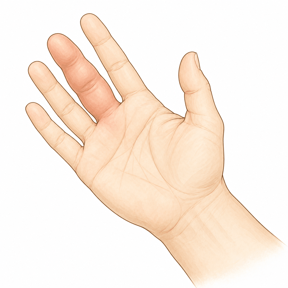
आपको क्या महसूस होता है
संक्रमित फ्लेक्सर शीथ (उंगली मोड़ने वाले टेंडन का आवरण) उस सुरंग का संक्रमण (इन्फेक्शन) है जिसमें से उंगली को मोड़ने वाला टेंडन (कंडरा) सरकता है। अक्सर पूरी उंगली किसी एक दुखती जगह पर नहीं, बल्कि अपनी पूरी लंबाई में एक जैसी सूज जाती है। उंगली थोड़ी मुड़ी हुई रह सकती है और उसे सीधा करना मुश्किल हो सकता है। उसे सीधा करने पर, या जब कोई और धीरे से उसे आपके लिए सीधा करे, आमतौर पर सबसे ज़्यादा दर्द होता है, ख़ासकर उंगली की जड़ के पास, जहां सूजन दिख सकती है।
दर्द किसी एक जोड़ पर नहीं, बल्कि टेंडन की लाइन के साथ-साथ होता है, उंगली की जड़ से नीचे हथेली की ओर। यह आमतौर पर हर समय बना रहता है और उंगली को धकेलकर सीधा करने पर बढ़ जाता है। उंगली को थोड़ा मोड़ने से अक्सर आराम मिलता है, इसीलिए उंगली मुड़कर रहती है। सूजन और दर्द की वजह से रोज़ के काम मुश्किल हो सकते हैं: केतली पकड़ना, चाबी घुमाना, बटन लगाना, पेन पकड़ना या रेलिंग पकड़ना।
यह संक्रमण तेज़ी से बढ़ सकता है, और यह ऐसा संक्रमण है जिसमें इंतज़ार नहीं करना चाहिए। अगर आपकी उंगली या हाथ गर्म, लाल, सूजा हुआ और दर्दनाक हो जाए, ख़ासकर बुखार के साथ, तो उसी दिन इमरजेंसी विभाग जाएं। इसके लिए पहले जीपी (आपके फैमिली डॉक्टर) के रेफ़रल की ज़रूरत नहीं है। अगर आपकी उंगली फीकी (सफ़ेद पड़ी हुई), ठंडी, सफ़ेद या नीली पड़ जाए, या उसमें अचानक महसूस करने की क्षमता या हिलने-डुलने की ताकत चली जाए, तो भी यही करें।
असल में क्या हो रहा है
हर उंगली की निचली (हथेली वाली) सतह के साथ-साथ एक मोड़ने वाला टेंडन चलता है, एक मज़बूत डोरी जो उंगली के सिरे को हथेली की ओर खींचती है। यह टेंडन एक बंद सुरंग के अंदर सरकता है जिसे फ्लेक्सर शीथ कहते हैं। इस सुरंग के अंदर एक चिकनी परत होती है जो टेंडन को पोषण देती है, उसे चिकना रखती है और आसानी से सरकने देती है, कुछ-कुछ ग्रीस लगी आस्तीन में से गुज़रते केबल की तरह।
जब बैक्टीरिया उस आस्तीन के अंदर पहुंच जाते हैं, आमतौर पर किसी कट, चुभन वाले घाव या दांत से काटे जाने (बाइट) के रास्ते, तो वह बंद जगह मवाद से भर जाती है। क्योंकि आस्तीन बंद होती है, इसलिए मवाद ठीक उसी सतह पर दबाव के साथ जमा रहता है जिस पर टेंडन को सरकना होता है। यह दबाव टेंडन तक ख़ून पहुंचाने वाले रास्ते को दबा देता है और टेंडन और उसकी परत के बीच निशान वाला ऊतक (स्कार टिश्यू) बनना शुरू कर देता है। इसीलिए पूरी उंगली एक जैसी सूज जाती है, इसीलिए वह मुड़कर रहती है, और इसीलिए उसे सीधा करने पर इतना दर्द होता है: सुरंग के हर हिस्से में एक साथ सूजन (इन्फ्लेमेशन) होती है।
संक्रमण फैल सकता है। अंगूठे की सुरंग हथेली में तरल से भरी एक जगह से जुड़ी होती है जिसे रेडियल बर्सा कहते हैं, और छोटी उंगली की सुरंग एक दूसरी जगह से जुड़ी होती है जिसे अल्नर बर्सा कहते हैं। बहुत से लोगों में ये जगहें कलाई की ओर ऊपर तक जाती हैं, इसलिए एक उंगली का संक्रमण हथेली की ओर, या उस पार अंगूठे या छोटी उंगली तक भी फैल सकता है, और मवाद का ऐसा जमाव बना सकता है जिसे हॉर्सशू एब्सेस (घोड़े की नाल के आकार का फोड़ा) कहते हैं।
सब कुछ समय पर निर्भर करता है। टेंडन का काम सरकने पर टिका है, और उसके सामने फंसा मवाद जितनी देर रहता है, उतना ही स्थायी नुकसान करता है। जल्दी इलाज होने पर ज़्यादातर उंगलियां अच्छी तरह ठीक हो जाती हैं। बहुत देर तक छोड़ देने पर ख़ुद टेंडन मर सकता है, उंगली निशान वाले ऊतक की वजह से जकड़ सकती है, और गंभीर मामलों में उंगली खोनी पड़ सकती है। तुरंत एंटीबायोटिक और सर्जरी के बावजूद भी बाद में उंगली में कुछ जकड़न रहना आम है, क्योंकि भरता हुआ ऊतक सरकने के बजाय चिपक जाता है।
मवाद कितनी जल्दी निकालना है, यह तय करने से पहले आपके सर्जन जांचेंगे कि संक्रमण कितना फैल चुका है।
हम इसके लिए क्या कर सकते हैं
डॉ. कीरन हिरपरा, जो माटर प्राइवेट हॉस्पिटल, रॉकहैम्पटन में ऊपरी अंग (कंधे से हाथ तक) के सर्जन हैं, संक्रमित फ्लेक्सर शीथ को इमरजेंसी मानते हैं और तुरंत इलाज शुरू करते हैं। यह संक्रमण इंतज़ार नहीं करता, और आपको भी इंतज़ार नहीं करना चाहिए। मक़सद संक्रमण को उससे पहले साफ़ करना है, जब तक वह उस टेंडन को नुकसान न पहुंचा दे जिससे आपकी उंगली मुड़ती है।
पहला कदम आपकी बांह में ड्रिप के ज़रिए दी जाने वाली एंटीबायोटिक है। एंटीबायोटिक मवाद निकालने के साथ मिलकर काम करती है, उसकी जगह नहीं लेती। क्योंकि शीथ एक बंद सुरंग है, इसलिए अकेले एंटीबायोटिक से अक्सर संक्रमण साफ़ नहीं हो पाता। मवाद निकालने से टेंडन पर से दबाव हटता है और एंटीबायोटिक पूरे हिस्से तक पहुंच पाती है।
ज़्यादातर मामलों में शीथ से मवाद निकालने के लिए सर्जरी की ज़रूरत होती है। हम छोटे चीरे लगाते हैं, यानी त्वचा में छोटे कट, ताकि सुरंग को खोलकर मवाद को धोकर निकाला जा सके। शीथ को जल्दी धोकर साफ़ करने से टेंडन की वह सरकने वाली सतह बची रहती है जिसकी उसे ज़रूरत है। सर्जरी ठीक कब और किस तरह की होगी, यह इस पर निर्भर करता है कि संक्रमण कितना फैल चुका है और आप कितनी जल्दी पहुंचे हैं।
सर्जरी के बाद आपकी उंगली को आराम देने के लिए स्प्लिंट की ज़रूरत पड़ सकती है। सर्जरी के बाद आपकी हैंड थेरेपी एक्सटेंड रिहैबिलिटेशन में रूबी डूलन के साथ होती है। रूबी हैंड थेरेपिस्ट (हाथ की विशेषज्ञ थेरेपिस्ट) हैं: वे आपके व्यायामों में मार्गदर्शन करती हैं और आपको जिस भी स्प्लिंट की ज़रूरत हो, उसे बनाती हैं। उनका ध्यान टेंडन को फिर से सरकने लायक बनाने पर रहता है, क्योंकि टेंडन और उसकी परत के बीच बना निशान वाला ऊतक ही बाद में हिलने-डुलने को सीमित करता है।
कुछ संक्रमण असामान्य कीटाणुओं से होते हैं, जैसे माइकोबैक्टीरिया या फंगस (फफूंद), जिनके लिए लंबे कोर्स में ख़ास एंटीबायोटिक या एंटीफंगल (फंगस-रोधी) इलाज की ज़रूरत होती है। अगर संक्रमण ने टेंडन को बुरी तरह नुकसान पहुंचाया है, तो उसे दोबारा बनाना एक अलग ऑपरेशन है, जो संक्रमण के पूरी तरह शांत हो जाने के बाद किया जा सकता है।
नतीजा बहुत हद तक समय पर निर्भर करता है। जल्दी आना ही वह एक चीज़ है जो आपकी उंगली को सबसे ज़्यादा बचाती है, इसलिए अगर आपकी उंगली गर्म, लाल, सूजी हुई और दर्दनाक है, ख़ासकर बुखार के साथ, तो उसी दिन इमरजेंसी विभाग जाएं।
क्या उम्मीद करें
संक्रमित फ्लेक्सर शीथ का नतीजा ज़्यादातर इस पर निर्भर करता है कि उसका इलाज कितनी जल्दी होता है। जल्दी आने से आपकी उंगली को फिर से सामान्य रूप से मुड़ने का सबसे अच्छा मौका मिलता है। इंतज़ार करने से मवाद दबाव के साथ टेंडन के सामने जमा रहता है, और वह दबाव ऐसा नुकसान करता है जिसे बाद में ठीक नहीं किया जा सकता।
तुरंत इलाज होने पर ज़्यादातर उंगलियां शांत हो जाती हैं और अगले सप्ताहों और महीनों में फिर से हिलने लगती हैं। फिर भी, बाद में कुछ जकड़न रहना आम है, उन लोगों में भी जो वैसे स्वस्थ हैं और जिन्हें जल्दी एंटीबायोटिक और सर्जरी मिली। भरता हुआ ऊतक सरकने के बजाय चिपक जाता है, इसलिए हो सकता है कि उंगली पहले की तरह आसानी से न मुड़े या सीधी न हो। हैंड थेरेपी इस पर काम करती है, लेकिन इसमें समय और लगातार मेहनत लगती है।
अगर संक्रमण गंभीर हो या उसे बहुत देर तक छोड़ दिया जाए, तो नतीजा ज़्यादा मुश्किल होता है। टेंडन के चारों ओर की सुरंग नष्ट हो सकती है, और निशान वाले ऊतक की वजह से उंगली हमेशा के लिए जकड़ी रह सकती है। कुछ मामलों में सूजन इतनी गंभीर हो जाती है कि वह ख़ुद उंगली तक ख़ून पहुंचाने वाले रास्ते को दबा देती है; इस समस्या को कम्पार्टमेंट सिंड्रोम कहते हैं। सबसे गंभीर संक्रमणों में उंगली मर सकती है और उसे काटकर निकालना (एम्प्यूटेशन) पड़ सकता है, तब भी जब इलाज समय पर और पूरी तरह हुआ हो।
कुछ कम आम चीज़ें भी हो सकती हैं। कभी-कभी संक्रमण पूरी तरह साफ़ नहीं होता और दूसरी बार धुलाई की ज़रूरत पड़ती है। बहुत कम मामलों में, इलाज के सप्ताहों बाद उंगली में कोई असामान्य कीटाणु सामने आ सकता है, जिसके लिए आगे और इलाज की ज़रूरत होती है।
आप जिस एक चीज़ पर नियंत्रण रख सकते हैं, वह है समय। अगर आपकी उंगली या हाथ गर्म, लाल, सूजा हुआ और दर्दनाक हो जाए, ख़ासकर बुखार के साथ, तो उसी दिन इमरजेंसी विभाग जाएं। इसके लिए पहले जीपी के रेफ़रल की ज़रूरत नहीं है। अगर आपकी उंगली फीकी (सफ़ेद पड़ी हुई), ठंडी, सफ़ेद या नीली पड़ जाए, या उसमें अचानक महसूस करने की क्षमता या हिलने-डुलने की ताकत चली जाए, तो भी यही करें।
डॉक्टर को कब दिखाएं
यह संक्रमण एक इमरजेंसी है, और यह तेज़ी से बढ़ता है। अगर आपकी उंगली या हाथ गर्म, लाल, सूजा हुआ और दर्दनाक हो जाए, ख़ासकर बुखार के साथ, तो उसी दिन इमरजेंसी विभाग जाएं। अगर आपकी उंगली फीकी (सफ़ेद पड़ी हुई), ठंडी, सफ़ेद या नीली पड़ जाए, या उसमें अचानक महसूस करने की क्षमता या हिलने-डुलने की ताकत चली जाए, तो भी यही करें। इसके लिए पहले जीपी के रेफ़रल की ज़रूरत नहीं है। अगर आप क्लिनिक से संपर्क नहीं कर पाते, उदाहरण के लिए क्लिनिक के समय के बाद या सप्ताहांत में, तो अपने सबसे नज़दीकी इमरजेंसी विभाग जाएं।
चेतावनी वाले लक्षण इस संक्रमण के स्वरूप से मेल खाते हैं: पूरी उंगली एक जैसी सूज जाती है, वह मुड़ी हुई रहती है, टेंडन की लाइन के साथ-साथ दर्द होता है, और उसे सीधा करने पर बहुत दर्द होता है। जांच करवाने से पहले इन सभी लक्षणों का होना ज़रूरी नहीं है। जिस उंगली में इनमें से सिर्फ़ कुछ लक्षण हों, उसमें भी यह संक्रमण हो सकता है।
अगर लक्षण शांत नहीं हो रहे हैं, सप्ताहों में बिगड़ते जा रहे हैं, या आपको हाथ इस्तेमाल करने से रोकते हैं, तो अपने जीपी को दिखाएं या विशेषज्ञ से जांच के लिए कहें।
विस्तार से
Advanced reading: the deeper science (optional)
यह हिस्सा आपके अपने इलाज के फ़ैसलों के लिए ज़रूरी जानकारी से आगे जाता है। संक्रमित फ्लेक्सर शीथ के बारे में अतिरिक्त पढ़ना फ़ायदेमंद है, क्योंकि यह हाथ का वह एक संक्रमण है जो सचमुच इमरजेंसी है, और क्योंकि इसका अंत कैसा होगा यह सबसे ज़्यादा तय करने वाली दो बातें आपके अस्पताल पहुंचने से पहले ही तय हो चुकी होती हैं, उनमें से एक तो सालों पहले।
निदान 1930 के दशक के चार लक्षणों पर टिका है, जिनकी कभी ठीक से जांच नहीं हुई
एलन कैनावेल ने संक्रमित फ्लेक्सर शीथ के चार लक्षण बताए थे: उंगली का उसकी पूरी लंबाई में सूजा होना, न कि एक जगह पर, उसका थोड़ा मुड़ा हुआ रहना, टेंडन शीथ की पूरी लंबाई में दबाने पर दर्द, न कि सिर्फ़ एक जोड़ पर, और, चारों में सबसे काम का, जब कोई धीरे से उंगली को सीधा करे तो तेज़ दर्द होना [1]।
लगभग सौ साल बाद भी, यही चार लक्षण निदान का तरीका हैं, और इनकी संवेदनशीलता, विशिष्टता और अलग-अलग जांचने वालों के बीच विश्वसनीयता कभी ठीक से स्थापित नहीं की गई [1]। ऐसे निदान के लिए यह एक बड़ी कमी है, जिसमें देरी की क़ीमत उंगलियां खोकर चुकानी पड़ती है।
इसका व्यावहारिक नतीजा वह हिस्सा है जो याद रखने लायक है: हर मामले में चारों लक्षण नहीं दिखते, ख़ासकर बच्चों में, और एक या ज़्यादा लक्षणों के न होने से निदान को ख़ारिज नहीं किया जा सकता [1]। जो उंगली कैनावेल के सिर्फ़ एक टेस्ट में खरी न उतरे, उसे साफ़ मान लेना सही नहीं है।
मवाद कैसे निकाला जाता है, इससे बाद में उंगली के हिलने पर फ़र्क पड़ता है
निदान हो जाने के बाद शीथ का दबाव हटाना ज़रूरी होता है, और इसके दो मुख्य तरीके हैं: सर्जरी से शीथ को खोलना, या उसमें एक पतला कैथेटर (नली) डालकर बहुत छोटे घाव के रास्ते उसे धोना।
763 मरीज़ों को शामिल करने वाली एक सिस्टमैटिक समीक्षा में पाया गया कि कैथेटर से धुलाई से खुली धुलाई की तुलना में जोड़ ज़्यादा हिल-डुल पाया (रेंज ऑफ मोशन), और यह कि सिर्फ़ मवाद निकालने पर निर्भर रहने के बजाय इलाज के हिस्से के रूप में एंटीबायोटिक देने से भी हिलना-डुलना बेहतर हुआ [2]।
ध्यान दें कि यह फेलन (उंगली के पोर का गहरा संक्रमण) के मामले के बिल्कुल उलट है, जहां ठीक से मवाद निकाल दिए गए पोर के फोड़े में किसी एंटीबायोटिक की ज़रूरत नहीं होती। फ़र्क बनावट का है: फेलन एक बंद थैली है जिसे आप पूरी तरह खाली कर सकते हैं, जबकि शीथ एक लंबी नली है जिसके अंदर वही सतह होती है जिस पर टेंडन को सरकना होता है। आप उस चीज़ को नुकसान पहुंचाए बिना इसे खुरचकर साफ़ नहीं कर सकते जिसे आप बचाने की कोशिश कर रहे हैं, इसलिए एंटीबायोटिक वह काम करती है जो सर्जरी नहीं कर सकती।
उंगली खोने का जोखिम ज़्यादातर मरीज़ की अपनी सेहत पर निर्भर करता है
यह सबसे असहज नतीजा है और सबसे काम का भी। मिलाकर देखे गए मामलों में, उंगली काटने की दर सर्जरी की तकनीक के बजाय मुख्य रूप से पहले से मौजूद सेहत से तय हुई: डायबिटीज़ वाले मरीज़ों में 39%, किडनी फ़ेल होने वालों में 64%, और हाथ-पैर की खून की नलियों की बीमारी (पेरिफ़ेरल वैस्कुलर डिज़ीज़) वालों में 71%, और ये सभी आंकड़ों के हिसाब से महत्वपूर्ण थे [2]।
ये संख्याएं उस बीमारी से अलग एक बीमारी को दिखाती हैं जो किसी स्वस्थ व्यक्ति को फांस चुभने की चोट से होती है। इसी वजह से डायबिटीज़ या कमज़ोर खून के बहाव वाले व्यक्ति में संक्रमित शीथ का इलाज ज़्यादा जल्दी किया जाता है और दोबारा धुलाई का फ़ैसला जल्दी लिया जाता है, और इसी वजह से "क्या मेरी उंगली बचेगी" का ईमानदार जवाब ऑपरेशन थिएटर में होने वाली किसी भी चीज़ से ज़्यादा इस सवाल के जवाब पर निर्भर करता है कि "आपकी सेहत में और क्या चल रहा है"।
देरी लगभग किसी भी और चीज़ से ज़्यादा क्यों मायने रखती है
शीथ एक बंद जगह है जिसमें खून का बहाव कम होता है और इसके अंदर एक टेंडन होता है जो सरकने पर निर्भर करता है। उस जगह के अंदर दबाव में भरा मवाद एक साथ दो काम करता है: यह टेंडन की खून की आपूर्ति का गला घोंटता है, और उन चिपकावों (एडहीज़न) के बीज बोता है जो बाद में हिलना-डुलना सीमित करते हैं। दोनों समय पर निर्भर हैं, इसीलिए जल्दी इलाज से लगातार बेहतर नतीजे मिलते हैं [2] और इसीलिए इस ख़ास संक्रमण के लिए आम सलाह जीपी अपॉइंटमेंट के बजाय इमरजेंसी विभाग जाने की है [3]।
सब कुछ सही और समय पर किए जाने पर भी बाद में जकड़न आम है। कितनी जकड़न होगी, यह ज़्यादातर इस बात से तय होता है कि शीथ कितनी देर दबाव में रहा, और यही वह एक चीज़ है जिस पर मरीज़ असर डाल सकता है, जल्दी आकर।
संदर्भ
[1] Kennedy CD, Huang JI, Hanel DP. In brief: Kanavel's signs and pyogenic flexor tenosynovitis. Clin Orthop Relat Res. 2016;474(1):280-284. https://doi.org/10.1007/s11999-015-4367-x
[2] Giladi AM, Malay S, Chung KC. A systematic review of the management of acute pyogenic flexor tenosynovitis. J Hand Surg Eur Vol. 2015;40(7):720-728. https://doi.org/10.1177/1753193415570248
[3] Goyal K, Speeckaert AL. Pyogenic flexor tenosynovitis: evaluation and management. Hand Clin. 2020;36(3):323-329. https://doi.org/10.1016/j.hcl.2020.03.005
Evidence & references
This is the clinical evidence summary written for health professionals. It is technical, and it lists the research this page was built from. You do not need to read it to understand your treatment or to make a decision about it.
Overview
- Pyogenic flexor tenosynovitis is an uncommon closed-space infection of the hand [2].
- Pyogenic flexor tenosynovitis can result in severe stiffness and other sequela [2].
- Acute flexor tendon sheath infections are associated with significant morbidity [3].
- 43% of patients with acute flexor tendon sheath infections have a poor result [3].
- The consequences of pyogenic flexor tenosynovitis can include destruction of the tendon sheath [6].
- The consequences of pyogenic flexor tenosynovitis can include scarring and inflammation causing oedema of the soft tissues of the finger [6].
- The consequences of pyogenic flexor tenosynovitis can include resultant finger stiffness [6].
- The consequences of pyogenic flexor tenosynovitis can occasionally include compartment syndrome [6].
- Sonographic evidence of a swollen tendon and fluid in the flexor sheath correlates strongly with the diagnosis of acute suppurative tenosynovitis [4].
- Sonographic evidence of a swollen tendon and fluid in the flexor sheath should prompt early surgical drainage [4].
- Any suspicion of flexor tendon sheath phlegmon should lead to urgent surgical exploration [10].
- Any suspicion of flexor tendon sheath phlegmon should not lead to blind antibiotic prescription [10].
- The absence of pain on passive extension in advanced stages is a grave prognostic sign indicating tissue necrosis rather than improvement [7].
Anatomy & Pathophysiology
Flexor Tendon Anatomy and Sheath Structure
- The extrinsic finger flexors consist of the flexor digitorum profundus and the flexor digitorum superficialis [13].
- The flexor digitorum profundus inserts on the proximal volar aspect of the distal phalanx, flexing the distal interphalangeal joint as well as the proximal interphalangeal and metacarpophalangeal joints [13].
- The flexor digitorum superficialis acts as a flexor of the proximal interphalangeal and metacarpophalangeal joints [13].
- As flexor tendons pass distal to the metacarpal neck, they enter the fibroosseous tunnel, or digital flexor sheath [24].
- The fibroosseous tunnel extends distally to the proximal aspect of the distal phalanx [24].
- The tendinous sheath consists of annular pulleys, which provide mechanical stability, and cruciate pulleys, which provide flexibility [24].
- The first, third, and fifth annular pulleys (A1, A3, and A5) are located over the metacarpophalangeal, proximal interphalangeal, and distal interphalangeal joints, respectively [24].
- The second and fourth pulleys (A2 and A4) are situated over the middle portion of the proximal and middle phalanges [24].
- The A2 and A4 pulleys are the most essential in maintaining the mechanical advantage of the flexor tendons [24].
- The tenosynovium that lines the fibroosseous tunnel supplies both nutrition and lubrication to the poorly vascularized flexor tendons [24].
- Within the sheath, tendon vascularity is supplied via the vincula system: the vinculum longus and brevis [24].
- The tenosynovial sheath of the flexor pollicis longus is continuous with the radial bursa [24].
- The tenosynovial sheath to the little finger is continuous with the ulnar digital bursa [24].
- In some patients, the radial and ulnar bursae communicate, allowing a so-called horseshoe abscess to spread between the thumb and little finger if infection occurs in the flexor tendon sheath of either one of these digits [24].
Pathophysiology of Pyogenic Flexor Tenosynovitis
- Pyogenic flexor tenosynovitis (PFT) is an uncommon closed-space infection of the hand that can result in severe stiffness and other sequela [2].
- Acute flexor tendon sheath infections are associated with significant morbidity, with 43% of patients having a poor result [3].
- The consequences of pyogenic flexor tenosynovitis can be destruction of the tendon sheath, scarring and inflammation causing oedema of the soft tissues of the finger with resultant finger stiffness or, occasionally, compartment syndrome [6].
- Sonographic evidence of a swollen tendon and fluid in the flexor sheath correlates strongly with the diagnosis of acute suppurative tenosynovitis and should prompt early surgical drainage [4].
- Any suspicion of flexor tendon sheath phlegmon should lead to urgent surgical exploration, not blind antibiotic prescription [10].
- Although unusual, nonsuppurative tenosynovitis secondary to foreign body migration may occasionally account for an otherwise unexplained mild but persistent tenosynovitis [11].
Classification
- Nonsuppurative tenosynovitis secondary to foreign body migration may occasionally account for an otherwise unexplained mild but persistent tenosynovitis [11].
Investigations
- A careful physical examination is essential to direct care and future testing if indicated [12].
- Diagnostic tests such as imaging and serum laboratory studies are useful in the determination of pathology but can be expensive, time consuming, and often nonspecific [12].
Treatment
Operative
- A minimally invasive, ultrasound-guided, percutaneous flexor tendon sheath lavage technique was effective in 65% of cases [1].
- A minimally invasive, ultrasound-guided, percutaneous flexor tendon sheath lavage technique was safe in 100% of cases [1].
- Any suspicion of flexor tendon sheath phlegmon should not be managed with blind antibiotic prescription [10].
- Placement of irrigation catheters in ulnar or radial bursitis may be facilitated by passing an irrigation catheter over a previously placed guide wire [9].
Outcomes and Prognosis
- No patient had a recurrence of the infection following closed tendon sheath irrigation for pyogenic flexor tenosynovitis [5].
- Use of a protocol for the treatment of severe infections of the hand resulted in a shorter hospital stay compared with patients treated before the protocol's institution [8].
- Use of a protocol for the treatment of severe infections of the hand resulted in faster healing compared with patients treated before the protocol's institution [8].
- Use of a protocol for the treatment of severe infections of the hand resulted in fewer complications, including recrudescence of infection, reoperation, stiffness, arthritis, and osteomyelitis, compared with patients treated before the protocol's institution [8].
Complications
- Pyogenic flexor tenosynovitis is an uncommon closed-space infection of the hand that can result in severe stiffness and other sequela [2].
- Complications associated with severe hand infections include recrudescence of infection, reoperation, stiffness, arthritis, and osteomyelitis [8].
Recovery
- The consequences of pyogenic flexor tenosynovitis can be destruction of the tendon sheath [6].
- The consequences of pyogenic flexor tenosynovitis can be scarring and inflammation causing oedema of the soft tissues of the finger with resultant finger stiffness [6].
- The consequences of pyogenic flexor tenosynovitis can occasionally be compartment syndrome [6].
- Use of a treatment protocol resulted in a shorter hospital stay compared with patients treated before the protocol's institution [8].
- Use of a treatment protocol resulted in faster healing compared with patients treated before the protocol's institution [8].
- Use of a treatment protocol resulted in fewer complications, including recrudescence of infection, reoperation, stiffness, arthritis, and osteomyelitis, compared with patients treated before the protocol's institution [8].
- No patient had a recurrence of the infection following closed tendon sheath irrigation [5].
Key Evidence
- [L5] Our results have shown that this minimally invasive, ultrasound-guided, percutaneous flexor tendon sheath lavage technique was effective in 65% of cases and safe in 100% of cases. [1] (10.1016/j.hansur.2018.12.001)
- [L5] Pyogenic flexor tenosynovitis (PFT) is an uncommon closed-space infection of the hand that can result in severe stiffness and other sequela. [2] (10.2106/jbjs.rvw.26.00015)
- [L4] Acute flexor tendon sheath infections are associated with significant morbidity, with 43% of patients having a poor result. [3] (10.1016/0363-5023(90)90064-x)
- [L4] Sonographic evidence of a swollen tendon and fluid in the flexor sheath correlates strongly with the diagnosis of acute suppurative tenosynovitis and should prompt early surgical drainage. [4] (10.1016/0363-5023(89)90027-0)
- [L4] No patient had a recurrence of the infection. [5] (10.1016/s0363-5023(78)80141-5)
- [L5] The consequences of pyogenic flexor tenosynovitis can be destruction of the tendon sheath, scarring and inflammation causing oedema of the soft tissues of the finger with resultant finger stiffness or, occasionally, compartment syndrome. [6] (10.1016/j.jhsb.2006.02.019)
- [L3] The absence of pain on passive extension in advanced stages is a grave prognostic sign indicating tissue necrosis rather than improvement. [7] (10.1016/j.injury.2026.113631)
- [L3] Use of the protocol resulted in a shorter hospital stay, faster healing, and fewer complications (recrudescence of infection, reoperation, stiffness, arthritis, and osteomyelitis) when compared with 107 patients who were treated before institution of the protocol. [8] (10.1016/s0363-5023(88)80060-1)
- [L5] Placement of irrigation catheters in other closed space infections such as in ulnar or radial bursitis may also be facilitated by passing an irrigation catheter over a previously placed guide wire. [9] (10.1016/s0266-7681(97)80290-2)
- [L4] Any suspicion of flexor tendon sheath phlegmon should lead to urgent surgical exploration, not blind antibiotic prescription. [10] (10.1016/j.main.2011.10.025)
- [L5] Although unusual, the situation described may occasionally account for an otherwise unexplained mild but persistent tenosynovitis. [11] (10.1016/s0363-5023(83)80176-2)
References
[1] Designing a minimally-invasive, ultrasound-guided, percutaneous flexor tendon sheath lavage technique: a cadaver study. Hand Surgery and Rehabilitation. 2019. DOI: 10.1016/j.hansur.2018.12.001
[2] Management of Pyogenic Flexor Tenosynovitis. JBJS Reviews. 2026. DOI: 10.2106/jbjs.rvw.26.00015
[3] Acute flexor tendon sheath infections. The Journal of Hand Surgery. 1990. DOI: 10.1016/0363-5023(90)90064-x
[4] Use of sonography in the early detection of suppurative flexor tensosynovitis. The Journal of Hand Surgery. 1989. DOI: 10.1016/0363-5023(89)90027-0
[5] Closed tendon sheath irrigation for pyogenic flexor tenosynovitis. The Journal of Hand Surgery. 1978. DOI: 10.1016/s0363-5023(78)80141-5
[6] MRSA Pyogenic Flexor Tenosynovitis Leading to Digital Ischaemic Necrosis and Amputation. Journal of Hand Surgery. 2006. DOI: 10.1016/j.jhsb.2006.02.019
[7] Predictive Factors for Outcomes in Pyogenic Flexor Tenosynovitis: A 10-Year Analysis of 341 Patients. Injury. 2026. DOI: 10.1016/j.injury.2026.113631
[8] A protocol for the treatment of severe infections of the hand. The Journal of Hand Surgery. 1988. DOI: 10.1016/s0363-5023(88)80060-1
[9] Passage of an Irrigation Catheter with the Aid of a Guide Wire for Pyogenic Flexor Tenosynovitis. Journal of Hand Surgery. 1997. DOI: 10.1016/s0266-7681(97)80290-2
[10] Phlegmons des gaines des tendons fléchisseurs des doigts : étude de 120 cas. Chirurgie de la Main. 2011. DOI: 10.1016/j.main.2011.10.025
[11] Nonsuppurative tenosynovitis secondary to foreign body migration. The Journal of Hand Surgery. 1983. DOI: 10.1016/s0363-5023(83)80176-2
[12] Orthopaedic Knowledge Update 13 Ebook Without Multimedia. Anatomy, Evaluation, Clinical Examination, and Imaging > Evaluation and Clinical Examination: Current Concepts.
[13] A Lange Medical Book Current Diagnosis Treatment In Orthopedics Fifth Edition. 9Hand Surgery > Image DISORDERS OF THE MUSCULATURE OF THE HAND.
[24] A Lange Medical Book Current Diagnosis Treatment In Orthopedics Fifth Edition. 9Hand Surgery > FLEXOR TENDON INJURY.