Patients › Elbow
Rekonstruksyon ng Ligamento sa Siko (Stabilisasyon)
Bakit iminungkahi ang operasyong ito
Si Dr Kieran Hirpara, isang upper-limb surgeon sa Mater Private Hospital Rockhampton, ay itinutugma ang gamutan sa iyong partikular na pinsala. Ang mga pasyente ay karaniwang nirerefer sa aming klinika ng kanilang GP; kung iminungkahi ng isang physiotherapist na magpatingin sa amin, kakailanganin mo pa rin ng referral mula sa iyong GP upang maging eligible para sa Medicare rebate. Sa iyong appointment, kumukuha kami ng history, sinusuri ang iyong siko, at nag-aayos ng mga scan kung kinakailangan upang malaman kung ano ang problema.
Ang elbow ligament reconstruction ay isang operasyon na muling bumubuo sa isang gasgas o punit na ligament gamit ang isang piraso ng donor tendon, upang panatilihing matatag ang joint. Karaniwan namin itong iminumungkahi kapag nararamdaman mo pa ring bumibigay ang iyong siko, o hindi nananatili sa puwesto, matapos ang ibang gamutan na hindi nagbigay ng sapat na pagbuti. Inaalok din ito kapag ang siko ay mapapanatili lamang sa joint sa pamamagitan ng paghawak dito nang halos diretso, o kapag ang isang fracture sa paligid ng joint ay nag-iwan dito na unstable. Ang layunin ay isang matatag na siko na maaari mong igalaw, gamitin, at asahan nang walang sakit. Ang allograft reconstruction ay nagbabalik ng stability ng siko sa humigit-kumulang 85% ng mga siko na may posterolateral rotatory instability. Pag-uusapan namin ito nang detalyado kasama ka at magpapasya nang magkasama kung ito ay angkop para sa iyong siko at sa iyong mga layunin.
Bago ang operasyon
Sa mga linggo bago ang operasyon, tinatapos namin ang pagpaplano gamit ang mga scan gaya ng X-ray, MRI (isang scan na nagpapakita ng mga soft tissue tulad ng mga ligament), o ultrasound. Sa araw ng operasyon, itigil ang pagkain at pag-inom pitong oras bago ito. Humihingi kami ng pitong oras upang maaaring mapabilis ang iyong operasyon kung maagang matapos ang listahan sa theatre; kumpirmasyon ng iyong surgeon ang eksaktong oras ng iyong pag-aayuno. Maaaring kailanganin mong itigil muna ang ilang gamot bago ang operasyon, at bibigyan ka namin ng malinaw na mga tagubilin kung alin sa mga ito at kung gaano katagal. Magdala ng nakasulat na listahan ng lahat ng iyong iniinom o ginagamit, kabilang ang mga tablet, patak, at cream. Mag-ayos ng taong maghahatid sa iyo pauwi pagkatapos. Magsuot ng maluwag at komportableng damit na may mga manggas na madaling maiangat lampas sa iyong siko. Kung mayroon kang iba pang kondisyong medikal, maaaring kailanganin mo rin ng mga blood test o pagsusuri kasama ang anaesthetist (ang doktor na nagbibigay ng anaesthetic).
Sa araw ng operasyon
Darating ka sa surgical admissions unit ng ospital, kung saan ka ire-rehistro at ihahanda para sa theatre. Makikilala mo doon ang anaesthetist. Ang operasyong ito ay ginagawa sa ilalim ng general anaesthetic. Minsan ay nagdaragdag ng regional nerve block para sa pagpapaginhawa ng sakit pagkatapos ng operasyon; tatalakayin ito ng anaesthetist sa iyo sa araw na iyon. Pagkatapos ay dadalhin ka sa operating theatre, kung saan isasagawa ang operasyon. Kapag natapos na ito, magigising ka sa recovery area, kung saan babantayan ka ng mga nurse habang nawawala ang bisa ng anaesthetic. Kapag stable ka na, pupunta ka sa ward o uuwi na, depende sa procedure at sa iyong paggaling.
Simple lang ang layunin ng araw na ito: ibalik ang sapat na stability sa iyong siko upang maaari itong simulang igalaw agad pagkatapos ng surgery. Ang pagpapanatiling hindi gumagalaw ng siko sa loob ng mahabang panahon pagkatapos ay may tendensiyang magdulot ng paninigas nito, kaya ang maagang paggalaw ay bahagi ng plano.
Ano ang kinapapalooban ng operasyon
Ang mga eksaktong hakbang ay nakadepende sa kung aling mga istruktura sa paligid ng iyong siko ang napinsala, at pinaplano namin ito mula sa iyong mga scan bago ang araw ng operasyon. Kung ang bali (fracture) ay bahagi ng problema, aayusin muna ng iyong surgeon ang mga nabaling piraso ng buto gamit ang mga screw o isang maliit na plate. Kung ang isang nabaling piraso ng buto sa itaas ng forearm ay hindi na maaaring kumpunihin, maaari itong palitan ng isang metal implant. Kapag secure na ang buto, ang mga napunit na ligament ay kumpunihin o muling bubuuin.
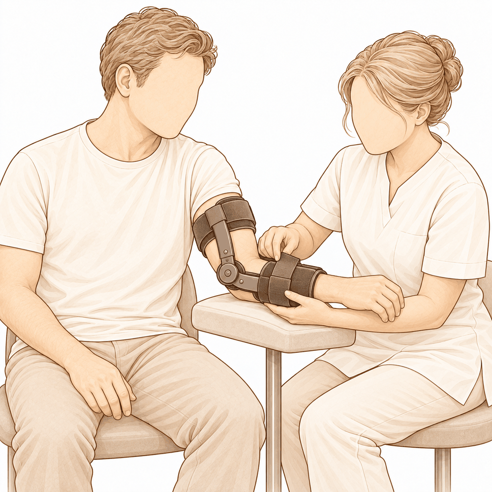
Ang muling pagbuo ng ligament ay nangangahulugang paggawa ng bago mula sa isang piraso ng tendon. Maaari itong manggaling sa donor tissue, o mula sa iyong sariling katawan. May mga maliliit na anchor na humahawak sa bagong ligament sa buto sa mga tamang posisyon, at sinusuri ng iyong surgeon na ang siko ay nananatili sa joint sa buong range of movement nito bago tapusin. Minsan, isang matibay na suture tape ang idinaragdag kasabay ng pagkumpuni upang magsilbing internal brace, na sumusuporta sa ligament habang ito ay gumagaling. Kung ang siko ay hindi pa rin nananatiling matatag, maaaring lagyan ng pansamantalang hinge upang mapanatili ito sa posisyon habang naghihilom ang lahat.
Ang operasyon ay madalas na maaaring gawin sa pamamagitan ng maliliit na hiwa sa halip na isang mahabang bukasan. Para sa ilang ligament rebuilds, sapat na ang hiwa na may laki na mga 2 hanggang 3 cm. Ang pagtatrabaho sa pamamagitan ng maliliit na hiwa ay nagpoprotekta sa mga malulusog na tendon at muscle sa paligid ng joint, at umiiwas sa pag-abala sa mismong joint lining.
Kapag tapos na ang pagkumpuni, ang mga hiwa ay sasara gamit ang mga tahi at tatakpan ng dressing. Ang layunin ng buong operasyon ay isang matatag na siko na maaaring magsimulang gumalaw agad pagkatapos, dahil ang pagpapanatili nito na hindi gumagalaw nang masyadong matagal ay may tendensiyang mag-iwan dito ng paninigas (stiffness).
Pagkatapos ng operasyon
Magigising ka sa recovery area, kung saan babantayan ka nang maigi ng mga nurse habang nawawala ang bisa ng anaesthetic. Ang iyong braso ay ilalagay sa isang sling o susuportahan ng mga unan, na may dressing sa ibabaw ng mga sugat. Bibigyan ka namin ng pain relief at titiyakin na ito ay gumagana bago ka gumalaw. Dapat may kasama ka sa unang 24 oras pagkatapos mong umuwi. Sasabihin sa iyo ng iyong team kung uuwi ka sa araw ring iyon o mananatili ng isang gabi sa ospital. Hahayaan naming nakalagay ang dressing sa loob ng humigit-kumulang 10 araw; pakiusap na huwag itong tanggalin bago ang panahong iyon maliban kung sinabi namin sa iyo. Papalitan o tatanggalin namin ito kapag nakita ka na namin. Karamihan ng mga tao ay maaaring maglakad-lakad at gumawa ng mga magagaan na gawain agad, ngunit panatilihing nakapahinga ang iyong siko sa sling kapag ikaw ay nakatayo at kumikilos.
Paggaling
Sa unang ilang araw, ang iyong siko ay magiging masakit at mamamaga. Unti-unti itong huhupa. Ang pahinga, pagpapanatiling nakataas ng iyong braso, at ang pain relief na ibibigay namin sa iyo ay magpapabawas ng discomfort. Ang ilang pananakit kapag nagsimula mo nang igalaw ang joint ay normal at bumubuti habang lumilipas ang mga linggo.
Uuwi ka na ang iyong braso ay nasa sling. Maaari kang maglakad-lakad at gumawa ng mga magaang gawain agad, ngunit panatilihing nakapahinga ang iyong siko sa sling kapag ikaw ay gumagalaw. Ang iyong kamay at pulso ay maaaring igalaw nang maaga, at ang mga banayad na ehersisyo para sa mga ito ay karaniwang nagsisimula sa loob ng ilang araw. Ang hand therapy pagkatapos ng operasyon ay kay Ruby Doolan sa Extend Rehabilitation. Si Ruby ay isang hand therapist: gagabayan niya ang iyong mga ehersisyo at gagawa ng anumang splint na iyong kakailanganin. Ang layunin ng therapy ay matatag at maagang paggalaw, dahil ang pagpapanatiling hindi gumagalaw ng siko nang masyadong matagal ay may tendensiyang mag-iwan nito na matigas (stiff).
Habang humuhupa ang pamamaga, mas gagamitin mo na ang iyong braso: pagkain, pagsusulat, at mga magaang gawaing bahay. Kapag kuntento na ang iyong surgeon na secure ang repair, tatanggalin na ang sling at magsisimula ka nang itiklop at ituwid ang siko mismo. Ang paggalaw ay madalas na bumubuti nang mabilis sa simula, pagkatapos ay mas mabagal. Kapag kaya mo nang humawak at kumapit sa mga bagay nang walang sakit, ang mga pang-araw-araw na aktibidad ay magiging mas madali. Ang pagmamaneho ay hindi ligtas habang ang iyong braso ay nasa sling, at kakailanganin mong maging may kakayahang humawak sa manibela gamit ang dalawang kamay at rumesponde sa isang emergency stop, nang walang iniinom na matapang na pain medication. Ang aming gabay sa pagmamaneho pagkatapos ng upper-limb surgery ay nagpapaliwanag kung kailan ka maaaring bumalik.
Ang bawat isa ay gumagaling sa sarili nilang bilis. Maaaring magkaiba ang iyong timeline, at gagabayan ka ng iyong surgeon at therapist sa prosesong ito.
Ano ang maaaring maging problema
Karamihan sa mga pasyente ay gumagaling nang maayos, ngunit paminsan-minsan ay maaaring magkaroon ng mga problema. Binabantayan kayo nang maigi ng inyong surgeon at ng team upang maagapan ang anumang isyu.
Ang nerve na dumadaan sa loob ng inyong siko ay maaaring mairita pagkatapos ng operasyon. Maaari kayong makaramdam ng pangingilig, parang tinutusok ng karayom, o pamamanhid sa inyong ring finger at kalingkingan. Kadalasan ay nawawala ito nang kusa, ngunit ipaalam sa amin kung hindi ito humuhupa, o kung nanghihina ang mga daliri.
Ang siko ay maaaring manatiling maluwag o pakiramdam na muling bumibigay. Kung ang inyong siko ay nagsisimulang maging hindi matatag, o nalalaglag mula sa posisyon nito, makipag-ugnayan agad sa klinika.
Ang siko ay maaari ring tumigas. Maaaring mahirapan kayong ituwid o itiklop nang lubos ang braso, at ang paggalaw ay maaaring maramdamang nakabara sa halip na masakit. Banggitin ito sa inyong review, dahil ang karagdagang therapy o iba pang gamutan ay maaaring makatulong.
Ang impeksyon ay hindi karaniwan ngunit seryoso. Bantayan ang malalim at pumupulsong sakit na hindi humuhupa sa simpleng painkiller, pamumula na kumakalat mula sa sugat, o pagtagas ng likido mula rito. Kung mapansin ang mga ito, tawagan ang klinika sa mismong araw na iyon, o pumunta sa emergency department kung kayo ay nilalagnat o masama ang pakiramdam.
Ang maliliit na metal anchor o screw na ginamit upang hawakan ang repair ay maaaring magdulot ng iritasyon, o ang isang pansamantalang hinge ay maaaring lumuwag. Maaari kayong makaramdam ng bagong pag-click, pag-catch, o bukol sa ilalim ng balat. Banggitin ito sa inyong susunod na review.
Kung saan ginamit ang isang bahagi ng inyong sariling tendon upang muling buuin ang ligament, ang bahaging iyon ay maaaring manatiling maselan o kumikirot sa loob ng ilang panahon. Ipaalam sa amin kung ito ay lumalala sa halip na bumubuti.
Ang buto ay maaaring mabuo kung saan hindi ito dapat, sa paligid ng joint o sa loob nito. Maaari nitong limitahan ang paggalaw o magdulot ng pag-catch at pag-grind. Kung ang inyong siko ay huminto sa pag-unlad, banggitin ito sa inyong review.
Ang peklat mismo ay maaaring paminsan-minsang bumuka o maglabas ng likido. Kung ang sugat ay bumukas, lalong namumula, o nagsimulang magkaroon ng discharge, makipag-ugnayan sa amin sa halip na maghintay.
Ang table ng mga komplikasyon sa pahinang ito ay naglilista ng mga tipikal na rate kung nais ninyo ang mga detalye.
Kailan dapat tumawag sa amin
Karamihan sa mga problema ay lumalabas nang maaga, at mas gusto naming malaman ang mga ito nang mas maaga kaysa huli na. Tumawag sa amin kung ikaw ay may lagnat, kung ang sugat ay lalong namumula o nagsisimulang maglabas ng likido, o kung ang iyong sakit ay patuloy na lumalala sa halip na humupa. Pumunta sa emergency kung ikaw ay may pamamaga o sakit sa iyong binti (calf), o biglaang pagkapos ng hininga. Tumawag sa amin agad kung ang iyong ring finger at kalingkingan ay namamanhid, kung ang iyong kamay ay nanghihina, o kung hindi mo na maigalaw ang iyong braso. Kung ang iyong siko ay nalukot o pakiramdam mo ay muling bumibigay, makipag-ugnayan sa klinika sa mismong araw na iyon.
Evidence & references
This is the clinical evidence summary written for health professionals. It is technical, and it lists the research this page was built from. You do not need to read it to understand your treatment or to make a decision about it.
Overview
- Surgical ligament reconstruction or repair is the most favorable treatment option for restoration of normal elbow function in posterolateral rotatory instability [4].
- Repair and reconstruction of the lateral ulnar collateral ligament offers practical and often successful solutions for patients with posterolateral rotatory instability of the elbow [10].
- Improving arthroscopic techniques provide a satisfactory alternative in stabilizing the elbow [7].
- The gravity varus test has biomechanical plausibility and confirms that pronation stabilizes the lateral elbow [1].
- An all-arthroscopic reconstruction technique for the lateral ulnar collateral ligament is reproducible and avoids residual instability [2].
- Open posterolateral ligament plication and lateral ulnar collateral ligament repair using an all-suture construct allows for complete posterolateral stabilization of the elbow with a single implant and bone preservation [3].
- Suture-augmented lateral ulnar collateral ligament and radial collateral ligament reconstruction provides a reproducible, anatomically based construct that restores posterolateral elbow stability and addresses the complex spectrum of lateral-sided injuries observed in posterolateral rotatory instability [5].
- Further follow-up and wider experience of the Surgilig/LockDown technique is required to assess its long-term functional outcome in comparison with more established surgical techniques for stabilization of posterolateral elbow instability [6].
- Arthroscopic lateral ulnar collateral ligament plication/reconstruction with augmented lateral collateral ligament imbrication allows effective management of elbow instability while promoting quicker patient recovery and long-term functional restoration [8].
- Acute repair of the lateral ulnar collateral ligament with a cortical button resulted in satisfactory clinical outcomes and successful restoration of elbow stability [9].
- The use of suture button fixation for repair of the lateral ulnar collateral ligament in terrible triad injuries has not been previously described [11].
- Corrective open-wedge osteotomy with structural bone allograft restored radial head alignment, eliminated dynamic subluxation, and achieved stable elbow function without the need for ligament reconstruction in complex posterolateral rotatory instability due to malunited radius fracture [12].
- Reconstruction of the lateral ulnar collateral ligament with a tendon graft offers an alternative that restores stability through a dynamic “sling effect” rather than rigid constraint [13].
Anatomy & Pathophysiology
Bony Anatomy
- The elbow is a trocho-ginglymoid joint consisting of medial and lateral articulations that provide bony stability [22].
- The ulnohumeral joint is formed by the articulation of the trochlea with the ulna within the greater sigmoid notch [22].
- The ulnohumeral joint provides highly congruent anatomy through almost 180° of articular contact, except for a bare area of the greater sigmoid notch devoid of cartilage [22].
- The radiocapitellar joint is formed by the articulation of the capitellum and the radial head [22].
- The radial head is a concave elliptical structure covered with articular cartilage along the radiocapitellar joint and approximately 270° of the articular margin [22].
- The radius is held in close approximation to the ulna at the proximal radioulnar joint by the annular ligament [22].
- The distal humeral articulation is angled 30° from the longitudinal axis of the humerus [22].
- The axis of rotation is angulated 5° to 7° in the coronal plane relative to the epicondylar axis, with the medial side more distal than the lateral side [22].
- The articular surface of the distal humerus is angled 30 degrees anterior to the humeral shaft axis [28].
- In full extension, 60% of axial load is transmitted through the radiocapitellar joint [28].
- The medial column of the distal humerus diverges from the humeral shaft at a 45-degree angle in the coronal plane [30].
- The lateral column of the distal humerus diverges from the humeral shaft at a 20-degree angle in the coronal plane [30].
- The trochlea is covered by articular cartilage anteriorly, inferiorly, and posteriorly, creating an arc of almost 270 degrees [33].
- The olecranon fossa and coronoid fossa are separated by a thin bony septum which is occasionally absent [33].
- The ulna medially bends approximately 8° at 8 cm from the tip of the olecranon [22].
- The articulation to the tip of the coronoid is approximately 30° from the long axis of the ulna in the sagittal plane [22].
Ligamentous Anatomy
- The lateral collateral ligament complex consists of the radial collateral ligament, the lateral ulnar collateral ligament (LUCL), and the annular ligament [33].
- The radial collateral ligament originates from an isometric point on the lateral epicondyle and fans out to attach to the annular ligament [33].
- The LUCL arises from an isometric point on the lateral epicondyle and attaches to the crista supinatoris of the proximal ulna [33].
- The annular ligament attaches to the anterior and posterior margins of the lesser sigmoid notch [33].
- The LUCL origin center is located 10.7 mm from the lateral epicondyle [21].
- The LUCL insertion is located 3.3 mm from the apex of the supinator crest [21].
- The LUCL acts as a posterior buttress for the radial head and the ulna to prevent subluxation [17].
- The LUCL is considered the principal constraint of the elbow joint against posterolateral rotatory instability (PLRI) [17].
- The lateral collateral ligament contributes 14% of varus stability of the elbow with the joint in full extension [37].
- The lateral collateral ligament contributes 9% of varus stability of the elbow with the joint in 90 degrees of flexion [37].
- The medial ulnar collateral ligament (MUCL) is the primary restraint to valgus stress within functional elbow range of motion [28].
- The MUCL originates on the posterior medial epicondyle and inserts on the sublime tubercle of the medial coronoid process [28].
- The anterior bundle of the MUCL is the primary restraint to valgus stress within functional elbow range of motion [28].
- The posterior bundle of the MUCL is the primary restraint to valgus stress with the elbow in maximal flexion [28].
- The common extensor tendon is a secondary stabilizer of the lateral elbow [16].
- The joint capsule is a secondary stabilizer of the elbow [19].
- The radiocapitellar articulation is a secondary stabilizer of the elbow [19].
Biomechanics & Pathophysiology
- Elbow stability is determined by primary and secondary stabilizers, with injury to these structures causing instability [19].
- The three primary stabilizers of the elbow are the ulnohumeral articulation, the MUCL, and the LUCL complex [19].
- Secondary stabilizers of the elbow include the radiocapitellar articulation, common flexor tendon, common extensor tendon, and joint capsule [19].
- Tensile forces are present at the medial elbow while compressive forces are present at the lateral elbow [23].
- The capsule allows maximum distension at approximately 70 to 80 degrees of flexion [23].
- The anterior capsule attaches at a point approximately 6 mm distal to the tip of the coronoid [23].
- Pronation stabilizes the lateral elbow [1].
- The LUCL is taut in both flexion and extension [37].
- Disruption of the LUCL results in posterolateral rotatory instability [37].
- Failure of primary stabilizers results in increased stress on secondary stabilizers, leading to capsular contractures, chondromalacia, osteophytes, and loose bodies [37].
- Ulnar nerve symptoms may develop from traction, scarring, or osteophyte impingement following primary stabilizer failure [37].
- The typical injury pattern for traumatic elbow dislocation involves a valgus internal rotation force of the forearm on a slightly flexed extremity [37].
- In traumatic elbow dislocation, structures are disrupted on the lateral side first, progressing medially as more force is applied [37].
- Posterolateral structures are most commonly affected when recurrence or persistence of instability results from traumatic dislocation [37].
- A coronoid fracture in association with disruption of the posterior band of the ulnar collateral ligament can result in symptomatic posteromedial instability [37].
- The normal elbow has a range of motion from 0° to 140° from extension to flexion [19].
- The normal elbow has a range of motion of 75° and 85° in pronation and supination respectively [19].
- A functional arc for elbow flexion and extension is 100° [19].
- A functional arc for forearm rotation is 100° [19].
- The normal valgus carrying angle of the elbow is 5 to 10 degrees for men and 10 to 15 degrees for women [28].
- The overall valgus angle of the elbow in extension, including the ulna, is 10 to 17 degrees [33].
- The distal humerus articular surface is internally rotated 3 to 8 degrees axially [33].
- As the elbow flexes, it internally rotates resulting in slight varus alignment [33].
Classification
- The lateral ulnar collateral ligament (LUCL) acts as a posterior buttress for the radial head and the ulna to prevent subluxation [17].
- Pronation stabilizes the lateral elbow in a cadaveric model of posterolateral rotatory instability [1].
Clinical Presentation
- The gravity varus test has biomechanical plausibility for evaluating posterolateral rotatory instability of the elbow [1].
- Complex posterolateral rotatory instability can be caused by a malunited radius fracture [12].
- Four-dimensional computed tomography scan can be used to diagnose complex posterolateral rotatory instability due to malunited radius fracture [12].
- Dynamic subluxation is a feature of complex posterolateral rotatory instability associated with malunited radius fracture [12].
Investigations
Physical Examination
- Pronation stabilizes the lateral elbow during the gravity varus test [1].
- Elbow stability is determined by primary stabilizers, which include the ulnohumeral articulation, the medial ulnar collateral ligament (MUCL), and the lateral ulnar collateral ligament (LUCL) complex [19].
- Secondary stabilizers of the elbow include the radiocapitellar articulation, the common flexor tendon, the common extensor tendon, and the joint capsule [19].
- The physical examination for elbow pathology is directed by the history and the location of patient pain in the anterior, posterior, medial, or lateral aspect of the elbow [19].
- The LUCL is considered the principal constraint of the elbow joint against posterolateral rotatory instability [17].
- The lateral ulnar collateral ligament injury is the cause of recurrent instability following elbow dislocation [41].
Imaging
- Plain radiographs remain the hallmark and best screening test for elbow evaluation [19].
- Standard radiographic views for elbow evaluation include AP, lateral, and oblique views [36].
- CT is helpful when assessing for malunion architecture and the location and pattern of osteophytes or loose bodies [36].
- Three-dimensional CT is used to check for heterotopic ossification [36].
- CT is not necessary when elbow stiffness is entirely soft-tissue related [36].
- CT is beneficial if any joint incongruity or abnormal bony anatomy is present [36].
- MRI can be used to evaluate ligaments and tendons, but it is rarely indicated for elbow stiffness [36].
- Magnetic resonance evaluation of the elbow includes imaging of ligament complexes [20].
- MRI findings in acute elbow dislocation provide insight into the mechanism of injury [20].
- MR evaluation of instability patterns emphasizes the soft-tissue lesions that result from dislocation [20].
- Four-dimensional computed tomography scan can be used for diagnosis in complex posterolateral rotatory instability due to malunited radius fracture [12].
Treatment
Operative Techniques and Constructs
- Surgical ligament reconstruction or repair is the most favorable treatment option for restoration of normal elbow function [4].
- Repair and reconstruction of the lateral ulnar collateral ligament (LUCL) offers practical and often successful solutions for patients with posterolateral rotatory instability (PLRI) of the elbow [10].
- An all-arthroscopic reconstruction technique for the LUCL is reproducible and achieves reconstruction while avoiding residual instability [2].
- Open posterolateral ligament plication and LUCL repair using an all-suture construct allows for complete posterolateral stabilization of the elbow with a single implant and bone preservation [3].
- A suture-augmented LUCL and radial collateral ligament reconstruction method provides a reproducible, anatomically based construct that restores posterolateral elbow stability and addresses the complex spectrum of lateral-sided injuries observed in PLRI [5].
- Acute repair of the LUCL with a cortical button resulted in satisfactory clinical outcomes and successful restoration of elbow stability [9].
- Arthroscopic LUCL plication/reconstruction with augmented lateral collateral ligament imbrication is a minimally invasive method that allows effective management of elbow instability while promoting quicker patient recovery and long-term functional restoration [8].
- Arthroscopic-assisted LUCL reconstruction provides less insult and dissection to the soft tissue at the lateral side of the elbow while being an excellent tool to diagnose any concomitant intraarticular pathologies [15].
- A knotless, onlay technique for LUCL reconstruction performs reconstruction with a tendon graft without violation of the extensor origin and soft tissue envelop [16].
- The use of knotless anchors and an onlay technique shortens operative time, reduces the required surgical exposure, and removes the risk of tunnel osteolysis or fracture and resultant graft failure while maintaining a broad bone surface for graft incorporation [16].
- A minimally-invasive dissection prevents iatrogenic injury to the common extensor origin, an important secondary stabilizer of the lateral elbow, and the articular capsule [16].
- Remaining extracapsular with a minimally-invasive technique avoids plication of the capsular structures or risk of formation of intra-articular adhesions, theoretically reducing the risk of any postoperative loss of range of motion [16].
Outcomes and Considerations
- The docking technique originally described by Jones et al. in 2012 is the most common method in use in contemporary practice for LUCL reconstruction [16].
- The docking technique has shown recurrent instability rates as high as 25% [16].
- A minimally-invasive approach allows for earlier rehabilitation and return of range of motion, reduced postoperative pain, and reduced operative time as a repair of the common extensor origin is not required following LUCL reconstruction [16].
- Fluoroscopic guidance can help to reduce injuries to unintended structures that could be foreseen due to a limited exposure [16].
- Postoperative stiffness is a known complication of LUCL reconstruction and occurs not uncommonly [16].
Non-Ligamentous Interventions
- Corrective open-wedge osteotomy with structural bone allograft restored radial head alignment, eliminated dynamic subluxation, and achieved stable elbow function without the need for ligament reconstruction in cases of complex PLRI due to malunited radius fracture [12].
Instability and Recurrence
- The docking technique for LUCL reconstruction has shown recurrent instability rates as high as 25% [16].
- The knotless, onlay technique for LUCL reconstruction removes the risk of tunnel osteolysis or fracture and resultant graft failure [16].
- The arthroscopic technique for LUCL reconstruction achieves reconstruction and avoids residual instability [2].
- The all-suture construct for posterolateral ligament plication and LUCL repair allows for complete posterolateral stabilization of the elbow [3].
- The suture-augmented LUCL and radial collateral ligament reconstruction method restores posterolateral elbow stability [5].
- Acute repair of the LUCL with a cortical button resulted in successful restoration of elbow stability [9].
- Repair and reconstruction of the LUCL offers practical and often successful solutions for patients with PLRI of the elbow [10].
- Reconstruction of the lateral ulnar collateral ligament with a tendon graft restores stability through a dynamic “sling effect” rather than rigid constraint [13].
Stiffness and Range of Motion
- The minimally-invasive, extracapsular technique for LUCL reconstruction theoretically reduces the risk of postoperative loss of range of motion by avoiding plication of capsular structures or risk of formation of intra-articular adhesions [16].
- The minimally-invasive method for managing elbow instability promotes quicker patient recovery and long-term functional restoration [8].
Iatrogenic Injury and Soft Tissue
- The minimally-invasive dissection for LUCL reconstruction prevents iatrogenic injury to the common extensor origin, an important secondary stabilizer of the lateral elbow [16].
- The minimally-invasive dissection for LUCL reconstruction prevents iatrogenic injury to the articular capsule [16].
- The arthroscopic-assisted LUCL reconstruction provides less insult and dissection to the soft tissue at the lateral side of the elbow [15].
Long-Term Outcomes
Recovery
- The minimally invasive arthroscopic method allows effective management of elbow instability while promoting quicker patient recovery and long-term functional restoration [8].
- The all-arthroscopic reconstruction technique is reproducible and achieves the reconstruction of the lateral ulnar collateral ligament as well as avoids residual instability [2].
- The open posterolateral ligament plication and lateral ulnar collateral ligament repair technique allows for complete posterolateral stabilization of the elbow with a single implant and bone preservation [3].
- The suture-augmented lateral ulnar collateral ligament and radial collateral ligament reconstruction method provides a reproducible, anatomically based construct that restores posterolateral elbow stability and addresses the complex spectrum of lateral-sided injuries observed in posterolateral rotatory instability [5].
- Reconstruction of the lateral ulnar collateral ligament with a tendon graft offers an alternative, restoring stability through a dynamic “sling effect” rather than rigid constraint [13].
- Clinical practice supports formal reconstruction of the lateral ulnar collateral ligament for chronic late presenting injury with posterolateral rotatory instability in the patient with open physes, without concern for causing significant growth arrest [14].
Key Evidence
- [L5] These findings support the biomechanical plausibility of the test and confirm that pronation stabilizes the lateral elbow. [1] (10.1016/j.jseint.2026.101792)
- [L5] The presented arthroscopic technique is reproducible and achieves the reconstruction of the LUCL of the elbow as well as avoids residual instability. [2] (10.1016/j.eats.2024.103096)
- [L5] The technique allows for complete posterolateral stabilization of the elbow with a single implant and bone preservation. [3] (10.1016/j.eats.2024.103172)
- [L4] Surgical ligament reconstruction or repair is the most favorable treatment option for restoration of normal elbow function. [4] (10.1016/s1058-2746(03)00091-0)
- [L5] The described method provides a reproducible, anatomically based construct that restores posterolateral elbow stability and addresses the complex spectrum of lateral-sided injuries observed in PLRI. [5] (10.1016/j.eats.2025.103797)
- [L4] Further follow-up and wider experience of the technique is required to assess its long-term functional outcome in comparison with more established surgical techniques for stabilization of posterolateral elbow instability. [6] (10.1097/bte.0000000000000025)
- [L5] Improving arthroscopic techniques provide a satisfactory alternative in stabilizing the elbow. [7] (10.1016/s0278-5919(05)70246-5)
- [L5] This minimally invasive method allows effective management of elbow instability while promoting quicker patient recovery and long-term functional restoration. [8] (10.1016/j.eats.2025.103529)
- [L4] The study found that acute repair of the LUCL with a cortical button resulted in satisfactory clinical outcomes and successful restoration of elbow stability. [9] (10.1097/bte.0000000000000178)
- [L5] Repair and reconstruction of the LUCL now offers practical and often successful solutions for patients with PLRI of the elbow. [10] (10.1016/j.csm.2004.06.010)
- [L4] The use of suture button fixation for repair of lateral ulnar collateral ligament has not been previously described. [11] (10.1016/j.eats.2023.10.004)
- [L5] Corrective open-wedge osteotomy with structural bone allograft restored radial head alignment, eliminated dynamic subluxation, and achieved stable elbow function without the need for ligament reconstruction. [12] (10.1016/j.xrrt.2026.100840)
- [L5] Reconstruction of the lateral ulnar collateral ligament with a tendon graft offers an alternative, restoring stability through a dynamic “sling effect” rather than rigid constraint. [13] (10.1002/atn2.70037)
- [L5] Clinical practice supports formal reconstruction of the LUCL for chronic late presenting injury with PLRI in the patient with open physes, without concern for causing significant growth arrest. [14] (10.1097/bth.0b013e3181dd88f3)
- [L5] It provides less insult and dissection to the soft tissue at the lateral side of the elbow while being an excellent tool to diagnose any concomitant intraarticular pathologies. [15] (10.1016/j.eats.2024.103101)
- [L5] [16] (10.1002/atn2.70135)
- [L5] On the basis of these morphological features, the LUCL might act a posterior buttress for the radial head and the ulna to prevent its subluxation, and therefore it is considered to be the principal constraint of the elbow joint against PLRI. [17] (10.1177/1558944716660555mm)
References
[1] Biomechanical Evaluation of the Gravity Varus Test in a Cadaveric Model of Posterolateral Rotatory Instability of the Elbow. JSES International. 2026. DOI: 10.1016/j.jseint.2026.101792
[2] Posterolateral Elbow Dislocation: An All‐Arthroscopic Reconstruction of the Lateral Ulnar Collateral Ligament. Arthroscopy Techniques. 2024. DOI: 10.1016/j.eats.2024.103096
[3] Open Posterolateral Ligament Plication and Lateral Ulnar Collateral Ligament Repair in Posterolateral Rotatory Instability of the Elbow Using an All‐Suture Construct. Arthroscopy Techniques. 2024. DOI: 10.1016/j.eats.2024.103172
[4] Surgical reconstruction for posterolateral rotatory instability of the elbow. Journal of Shoulder and Elbow Surgery. 2003. DOI: 10.1016/s1058-2746(03)00091-0
[5] Suture‐Augmented Lateral Ulnar Collateral Ligament and Radial Collateral Ligament Reconstruction for Subacute and Chronic Posterolateral Rotatory Instability. Arthroscopy Techniques. 2025. DOI: 10.1016/j.eats.2025.103797
[6] Surgical Repair of Posterolateral Rotatory Instability of the Elbow Using Surgilig/LockDown. Techniques in Shoulder & Elbow Surgery. 2014. DOI: 10.1097/bte.0000000000000025
[7] Posterolateral Rotatory Instability of the Elbow. Clinics in Sports Medicine. 2001. DOI: 10.1016/s0278-5919(05)70246-5
[8] Arthroscopic Lateral Ulnar Collateral Ligament Plication/Reconstruction With Augmented Lateral Collateral Ligament Imbrication. Arthroscopy Techniques. 2025. DOI: 10.1016/j.eats.2025.103529
[9] Clinical Outcomes of Lateral Ulnar Collateral Ligament Repair of the Elbow Using a Cortical Button. Techniques in Shoulder & Elbow Surgery. 2019. DOI: 10.1097/bte.0000000000000178
[10] PLRI: posterolateral rotatory instability of the elbow. Clinics in Sports Medicine. 2004. DOI: 10.1016/j.csm.2004.06.010
[11] Suture Button Repair for Lateral Ulnar Collateral Ligament in Terrible Triad Injuries: Surgical Technique. Arthroscopy Techniques. 2024. DOI: 10.1016/j.eats.2023.10.004
[12] Complex posterolateral rotatory instability due to malunited radius fracture: diagnosis with four-dimensional computed tomography scan and corrective osteotomy. JSES Reviews, Reports, and Techniques. 2026. DOI: 10.1016/j.xrrt.2026.100840
[13] Arthroscopic Lateral Collateral Ligament Reconstruction With Tendon Graft in High‐Grade Atraumatic Posterolateral Rotatory Instability in Elbows. Arthroscopy Techniques. 2026. DOI: 10.1002/atn2.70037
[14] Surgical Treatment of Posterolateral Rotatory Instability of the Elbow in Children. Techniques in Hand & Upper Extremity Surgery. 2010. DOI: 10.1097/bth.0b013e3181dd88f3
[15] Arthroscopic‐Assisted Lateral Ulnar Collateral Ligament Reconstruction for Posterolateral Rotatory Instability of the Elbow: A Technical Note. Arthroscopy Techniques. 2024. DOI: 10.1016/j.eats.2024.103101
[16] Lateral Ulnar Collateral Ligament Reconstruction Through a Mini‐Invasive Approach. Arthroscopy Techniques. 2026. DOI: 10.1002/atn2.70135
[17] Morphology of the Lateral Ulnar Collateral Ligament of the Elbow. HAND. 2016. DOI: 10.1177/1558944716660555mm
[19] Orthopaedic Knowledge Update 13 Ebook Without Multimedia. Anatomy, Biomechanics, Physical Examination, and Imaging of the Elbow > Summary and Conclusions.
[20] Orthopaedic Knowledge Update Sports Medicine 6. Magnetic Resonance Imaging of the Elbow > Annotated References.
[21] Orthopaedic Knowledge Update 13 Ebook Without Multimedia. Anatomy, Biomechanics, Physical Examination, and Imaging of the Elbow > Annotated References.
[22] Orthopaedic Knowledge Update 13 Ebook Without Multimedia. Anatomy, Biomechanics, Physical Examination, and Imaging of the Elbow > Anatomy > Bony Anatomy.
[23] Miller S Review Of Orthopaedics. SECTION 16 PATELLAR TRACKING IN TOTAL KNEE ARTHROPLASTY > TABLE 2.3 Shoulder Spaces.
[28] Miller S Review Of Orthopaedics. ELBOW.
[30] A Lange Medical Book Current Diagnosis Treatment In Orthopedics Fifth Edition. 2Musculoskeletal Trauma Surgery > INJURIES AROUND THE ELBOW.
[33] Rockwood And Green S Fractures In Adults. 29: Principles of Nonunion and Bone Defect Treatment > Pathoanatomy and Applied Anatomy Related to Distal Humerus Fractures.
[36] Aaos Comprehensive Orthopaedic Review 3. Elbow Stiffness* > IV. Evaluation.
[37] Campbell S Operative Orthopaedics 4 Volume Set. POSTERIOR SURGICAL APPROACH FOR QUADRILATERAL SPACE SYNDROME > MCLAUGHLIN PROCEDURE > ARTHROSCOPIC SURGERY.
[41] A Lange Medical Book Current Diagnosis Treatment In Orthopedics Fifth Edition. 2Musculoskeletal Trauma Surgery > ELBOW DISLOCATION.