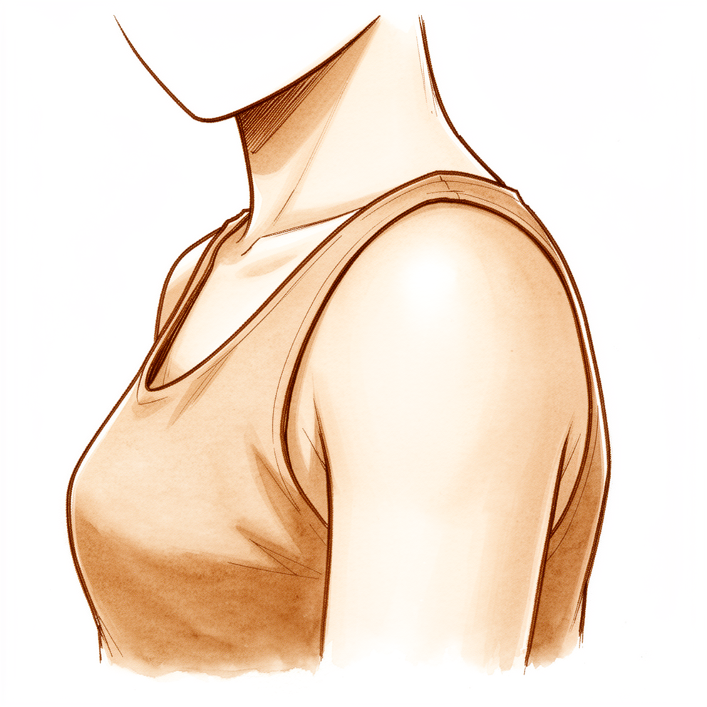
Những triệu chứng bạn đang gặp phải
Hội chứng lạo xạo xương bả vai có nghĩa là bạn nghe thấy hoặc cảm thấy tiếng lạo xạo, tiếng lục cục hoặc tiếng bật phát ra từ xương bả vai khi xương này chuyển động trên thành ngực. Đôi khi các bác sĩ gọi đây là “hội chứng tấm ván giặt”, vì âm thanh này có thể có cảm giác như một bánh xe lăn qua tấm ván giặt. Bản thân tiếng kêu này rất phổ biến. Khoảng một phần ba số người hoàn toàn không có triệu chứng gì vẫn có chút tiếng lạo xạo ở xương bả vai. Nếu chỉ có tiếng kêu mà không đau, tình trạng này không cần điều trị.
Điều thường khiến mọi người đi khám là cơn đau. Cơn đau nằm quanh hoặc bên dưới xương bả vai, thường ở gần bờ trong, phía gần cột sống nhất. Cơn đau thường nặng hơn khi làm các hoạt động đưa tay qua đầu, với tay lên cao hoặc với ngang qua người, và có thể biểu hiện bằng việc giảm thành tích khi chơi thể thao. Cơn đau bắt nguồn từ túi hoạt dịch bị viêm, tức lớp đệm mỏng chứa dịch giúp xương bả vai trượt êm trên các xương sườn, hoặc từ chính hình dạng của xương. Thành ngực hoạt động như một hộp cộng hưởng, vì vậy tiếng kêu bạn nghe thấy to hơn so với chuyển động gây ra nó.
Cơn đau có thể khiến các công việc hằng ngày trở nên khó khăn. Nhấc một chiếc chảo nặng lên kệ cao, phơi quần áo lên dây, hoặc với tay lấy dây an toàn đều có thể gây đau. Một số người thấy đau nhiều nhất sau khi hoạt động chứ không phải trong lúc hoạt động. Cơn đau có thể nặng đến mức làm gián đoạn giấc ngủ hoặc khiến bạn không thể sử dụng cánh tay bình thường.
Nếu các triệu chứng của bạn không thuyên giảm, ngày càng nặng hơn qua nhiều tuần, khiến bạn thức giấc vào ban đêm, hoặc khiến bạn không thể làm việc hay sử dụng cánh tay, hãy đến gặp bác sĩ đa khoa hoặc đề nghị được bác sĩ chuyên khoa khám.
Điều gì đang thực sự xảy ra
Xương bả vai của bạn được thiết kế để trượt. Xương này nằm trên thành ngực và được ngăn cách với thành ngực bằng một lớp mô mỡ mỏng và trơn, hoạt động như một miếng đệm kín giữa hai bề mặt chuyển động. Bốn khớp phối hợp với nhau để cử động vai, và khoảng hai phần ba chuyển động đến từ chính khớp chỏm cầu – ổ chảo, một phần ba còn lại đến từ việc xương bả vai trượt trên các xương sườn. Khi chuyển động trượt này êm ái, bạn sẽ không bao giờ để ý đến nó.
Vấn đề trong hội chứng lạo xạo xương bả vai là có điều gì đó làm xáo trộn chuyển động trượt này. Đôi khi đó là hình dạng của xương: bờ trong của xương bả vai có thể gập góc hoặc cong về phía các xương sườn nhiều hơn bình thường, nên xương nằm sát thành ngực hơn mức cần thiết. Đôi khi đó là một khối xương thừa nhỏ trên xương bả vai. Thường gặp là các cơ quanh xương bả vai hoạt động không đều, khiến xương bả vai không di chuyển êm theo đúng đường khi bạn cử động. Bất kỳ nguyên nhân nào trong số này cũng có thể khiến xương bả vai cọ xát hoặc bị vướng thay vì trượt êm.
Sự cọ xát này gây kích ứng lớp đệm giữa xương bả vai và các xương sườn. Lớp đệm đó, gọi là túi hoạt dịch, là một túi mỏng chứa dịch, hoạt động như một bộ phận giảm chấn giữa xương và mô mềm. Khi bị viêm, túi này dày lên và đau khi ấn vào, và đó chính là cơn đau bạn cảm thấy ở bên dưới hoặc quanh xương bả vai. Sự ma sát cũng có thể làm chính mô đó dày lên theo kiểu phản ứng, tức là phản ứng của cơ thể trước việc bị cọ xát lặp đi lặp lại.
Điều này giải thích các triệu chứng đã mô tả ở trên. Tiếng kêu phát ra từ các bề mặt bị thô ráp cọ vào nhau, và thành ngực khuếch đại nó như một hộp cộng hưởng. Cơn đau đến từ lớp đệm bị viêm chứ không phải từ bản thân tiếng kêu. Đó là lý do hai vấn đề này được điều trị khác nhau, và vì sao việc điều trị tập trung vào làm dịu tình trạng viêm và tập luyện lại các cơ kiểm soát xương bả vai.
Những biện pháp chúng tôi có thể áp dụng
Bác sĩ Kieran Hirpara, bác sĩ phẫu thuật chi trên tại Bệnh viện tư nhân Mater Rockhampton, sẽ bắt đầu bằng các phương pháp ít xâm lấn nhất phù hợp với tình trạng của bạn. Thông thường, bệnh nhân được bác sĩ đa khoa giới thiệu đến phòng khám chúng tôi; nếu vật lý trị liệu viên khuyên bạn nên đến gặp chúng tôi, bạn vẫn cần có giấy giới thiệu từ bác sĩ đa khoa để được hưởng mức hoàn trả từ chương trình Medicare. Tại phòng khám, chúng tôi sẽ hỏi bệnh sử, khám vai và chỉ định chụp hình ảnh khi cần để tìm ra nguyên nhân gây ra tiếng kêu và cơn đau.
Ở hầu hết mọi người, tình trạng sẽ ổn định mà không cần phẫu thuật, vì vậy việc điều trị bắt đầu bằng các biện pháp không phẫu thuật. Bước đầu tiên là một chương trình vật lý trị liệu nhằm tăng cường sức mạnh cho các cơ kiểm soát xương bả vai, kèm theo kéo giãn các cơ đã bị co cứng. Thay đổi các hoạt động gây ra triệu chứng cũng giúp ích. Hãy kiên trì thử phương pháp này một cách nghiêm túc trong 3 đến 6 tháng trước khi nghĩ đến bất kỳ biện pháp nào khác.
Nếu cơn đau vẫn còn, thuốc có thể giúp làm dịu tình trạng viêm. Thuốc kháng viêm dạng viên, cùng loại mà nhiều người vẫn dùng khi đau nhức cơ, là một phần của giai đoạn này. Chúng tôi cũng có thể tiêm steroid và thuốc gây tê tại chỗ vào túi hoạt dịch bên dưới xương bả vai, dưới hướng dẫn hình ảnh. Mũi tiêm này có hai tác dụng: làm dịu lớp đệm bị viêm, và xác nhận rằng đây chính là nơi gây ra cơn đau của bạn.
Phẫu thuật chỉ được cân nhắc khi cơn đau vẫn tiếp diễn sau vài tháng áp dụng các phương pháp điều trị này và chúng không mang lại đủ sự cải thiện. Ca mổ là phẫu thuật nội soi, được thực hiện qua các vết rạch nhỏ với một camera. Ca mổ loại bỏ túi hoạt dịch bị viêm nằm giữa xương bả vai và các xương sườn. Góc trên trong của xương bả vai chỉ được mài bớt nếu trong lúc mổ phát hiện góc này nhô ra. Ở hầu hết mọi người, cơn đau cải thiện đáng kể sau phẫu thuật, nhưng tiếng lục cục thường vẫn còn. Chúng tôi sẽ cùng bạn trao đổi về ý nghĩa của phẫu thuật đối với bạn và cùng quyết định xem đó có phải là bước phù hợp hay không.
Những điều bạn có thể mong đợi
Ở hầu hết mọi người, tình trạng sẽ ổn định mà không cần phẫu thuật. Việc điều trị bắt đầu bằng vật lý trị liệu để tăng cường sức mạnh cho các cơ kiểm soát xương bả vai, thuốc kháng viêm và thay đổi các hoạt động gây ra triệu chứng. Hãy kiên trì thử các biện pháp này một cách nghiêm túc trong 3 đến 6 tháng. Nhiều người thấy cơn đau dịu đi trong khoảng thời gian này và không bao giờ cần phẫu thuật.
Nếu cơn đau vẫn còn sau vài tháng áp dụng các phương pháp điều trị này, phẫu thuật có thể được cân nhắc. Phẫu thuật nội soi loại bỏ túi hoạt dịch bị viêm nằm giữa xương bả vai và các xương sườn, và chỉ mài bớt góc trên trong của xương bả vai nếu phát hiện góc này nhô ra. Ở hầu hết mọi người, cơn đau cải thiện đáng kể sau phẫu thuật. Tiếng lục cục thường vẫn còn, vì các phương pháp điều trị giúp giảm đau chủ yếu khắc phục cơn đau chứ không khắc phục tiếng kêu. Việc hết hoàn toàn mọi triệu chứng không phải là kết quả thường gặp.
Nếu không được điều trị, các triệu chứng có thể kéo dài và có thể nặng đến mức làm gián đoạn giấc ngủ hoặc khiến bạn không thể sử dụng cánh tay bình thường. Tình trạng này có thể khó nhận biết và khó điều trị, vì vậy bạn nên sớm có chẩn đoán rõ ràng thay vì chờ nó tự khỏi.
Nếu các triệu chứng của bạn không thuyên giảm, ngày càng nặng hơn qua nhiều tuần, khiến bạn thức giấc vào ban đêm, hoặc khiến bạn không thể làm việc hay sử dụng cánh tay, hãy đến gặp bác sĩ đa khoa hoặc đề nghị được bác sĩ chuyên khoa khám.
Khi nào nên đi khám bác sĩ
Chỉ riêng tiếng kêu thì không phải là lý do để lo lắng. Khoảng một phần ba số người hoàn toàn không có triệu chứng gì vẫn có chút tiếng lạo xạo ở xương bả vai, và nếu chỉ có tiếng kêu thì không cần điều trị. Điều quan trọng là cơn đau. Hãy đến gặp bác sĩ đa khoa hoặc đề nghị được bác sĩ chuyên khoa khám nếu bạn bị đau quanh hoặc bên dưới xương bả vai mà không thuyên giảm, ngày càng nặng hơn qua nhiều tuần, khiến bạn thức giấc vào ban đêm, hoặc khiến bạn không thể làm việc hay sử dụng cánh tay. Cũng hãy đề nghị được khám nếu cơn đau nặng đến mức làm gián đoạn giấc ngủ, nếu thành tích thể thao của bạn giảm sút, hoặc nếu các hoạt động đưa tay qua đầu như với tay lên cao hay với ngang qua người gây ra cơn đau. Hội chứng lạo xạo xương bả vai có thể khó nhận biết, vì vậy việc sớm có chẩn đoán rõ ràng là điều đáng làm.
Phân tích chi tiết hơn
Advanced reading: the deeper science (optional)
Phần này đi sâu hơn những gì bạn cần biết để đưa ra quyết định điều trị cho bản thân. Hội chứng lạo xạo xương bả vai đáng để đọc thêm vì tiếng kêu và cơn đau là hai vấn đề khác nhau, và các phương pháp điều trị có hiệu quả chủ yếu khắc phục vấn đề thứ hai chứ không phải vấn đề thứ nhất.
Tiếng kêu thì phổ biến; cơn đau mới là vấn đề
Tiếng lạo xạo từ xương bả vai, nếu chỉ có vậy, không phải là dấu hiệu của bệnh. Một bài tổng quan kinh điển về tình trạng này dẫn lại một nghiên cứu ban đầu ghi nhận tiếng lạo xạo xương bả vai ở 31% trong số 100 người hoàn toàn không có triệu chứng [1]. Điều biến tiếng kêu thành một bệnh lý là cơn đau, thường do túi hoạt dịch bị viêm: lớp mô mỏng có lót dịch giúp xương bả vai trượt trên các xương sườn.
Hình dạng xương cũng đóng một vai trò. Trong một nghiên cứu hình ảnh học, những người có xương bả vai cong về phía trước dọc theo bờ trong (hình dạng lõm, với “góc thân xương bả vai phía trong” dương) có nguy cơ cao gấp 12 lần bị hội chứng lạo xạo xương bả vai [2]. Phép đo đó hóa ra kém hữu ích hơn so với lúc đầu: một nghiên cứu sau đó phát hiện cùng góc này thay đổi đáng kể tùy theo hướng nhìn, nên nó không đủ tin cậy để tự mình chẩn đoán tình trạng này [3]. Trong một mô hình trên tử thi, đặc điểm xương quan trọng nhất là phần bên trong của xương bả vai nằm nhô về phía trước, hướng về các xương sườn, xa đến mức nào [4].
Hầu hết mọi người ổn định mà không cần phẫu thuật
Trong một loạt ca gồm 30 xương bả vai ở 28 bệnh nhân được điều trị bằng một chương trình tăng cường sức mạnh cơ chuyên biệt, 14 (50%) đạt kết quả tốt chỉ với các bài tập và 6 (21%) chỉ cần dùng thêm thuốc kháng viêm, vì vậy 71% đạt kết quả tốt hoặc tốt hơn theo thang phân loại riêng của nghiên cứu mà không cần phẫu thuật [5].
Thời gian theo dõi dài nhất đến từ Phần Lan. Mười hai người được phẫu thuật mở và 9 người được điều trị không phẫu thuật đã được đánh giá lại sau trung bình 22 năm. Mức đau khi gắng sức thấp ở cả hai nhóm: 0,8 trên 10 sau phẫu thuật và 1,5 trên 10 khi không phẫu thuật, một khác biệt quá nhỏ để có thể chắc chắn là có thật (p = 0,357) [6]. Cơn đau cải thiện nhanh sau phẫu thuật ở 7 trên 12 người, nhưng về lâu dài cả hai cách điều trị đều dẫn đến kết quả gần như nhau.
Mũi tiêm thuốc gây tê tại chỗ và steroid vào túi hoạt dịch có hai tác dụng: điều trị tình trạng viêm và kiểm tra xem túi hoạt dịch có thực sự là nguồn gây đau hay không. Trong một quy trình điều trị tại Anh, 74 trên 94 bệnh nhân vẫn còn khó chịu sau vật lý trị liệu và được tiêm dưới hướng dẫn siêu âm; 55 người có đáp ứng ban đầu tốt, và đáp ứng đó kéo dài ở 17 người [7]. Trong số 42 bệnh nhân được phẫu thuật và được theo dõi, những người đã đáp ứng tốt với mũi tiêm cải thiện nhiều hơn sau phẫu thuật so với những người không đáp ứng [7]. Một loạt ca lớn hơn tại Mỹ, được theo dõi ít nhất năm năm, không tìm thấy mối liên hệ tương tự [8], vì vậy đáp ứng kém với mũi tiêm không loại trừ khả năng phẫu thuật.
Phẫu thuật đạt được gì, và không đạt được gì
Loạt ca dài hạn lớn nhất đã theo dõi 66 người trong 5 đến 15 năm sau phẫu thuật nội soi cắt bỏ túi hoạt dịch và mài bớt góc của xương bả vai. Điểm số vai trung bình tăng từ 56,7 lên 87,2 trên 100, mức đau giảm từ 5,2 xuống 1,4 trên 10, và 77,6% đạt mức cải thiện đủ lớn để cảm nhận được. Trong số 42 người chơi thể thao, 38 (90,5%) đã quay lại chơi và 31 (73,8%) trở lại ở mức bằng hoặc gần bằng trước đây. Tám (12,1%) người cần phẫu thuật thêm [8]. Một loạt ca khác gồm 74 xương bả vai có tỷ lệ phẫu thuật thêm tương tự (8 trên 74) và nhận thấy những bệnh nhân lớn tuổi hơn, những người có triệu chứng lâu hơn, và những người có điểm sức khỏe tinh thần thấp hơn trước phẫu thuật thường có kết quả kém hơn [9].
Khỏi hoàn toàn là trường hợp ngoại lệ. Một tổng quan hệ thống về 203 vai được phẫu thuật cho thấy 42 (20,7%) khỏi hoàn toàn, 139 (68,5%) cải thiện nhưng vẫn còn một số triệu chứng, và 22 (10,8%) có kết quả kém [10]. Một tổng quan năm 2026 trên 464 bệnh nhân đưa ra cùng kết luận chung: cả điều trị không phẫu thuật lẫn phẫu thuật đều giúp giảm đau, nhưng các triệu chứng cơ học, tức tiếng lục cục và lạo xạo, thường không biến mất hoàn toàn [11].
Có đáng cắt bỏ phần xương không?
Mài bớt góc trên trong của xương bả vai là phần truyền thống của ca mổ, vì vậy thật đáng ngạc nhiên khi bước này có thể không mang lại thêm nhiều lợi ích. Một trung tâm đã so sánh 24 người chỉ được cắt bỏ túi hoạt dịch với 32 người được mài bớt cả góc xương. Điểm đau cuối cùng (3,4 và 2,8 trên 10, p = 0,351) và điểm số vai (75,8 và 76,5, p = 0,895) là như nhau, và tiếng lục cục vẫn còn ở 58% và 59% [12]. Ca mổ ngắn hơn khi không can thiệp vào xương: 37,5 so với 61,2 phút [12]. Các nhóm được theo dõi trong những khoảng thời gian khác nhau và không được phân nhóm ngẫu nhiên, vì vậy đây chưa phải là kết luận cuối cùng, nhưng đó là lý do quyết định mài bớt xương được đưa ra trong lúc mổ chứ không phải từ trước.
Tài liệu tham khảo
[1] Kuhn JE, Plancher KD, Hawkins RJ. Symptomatic Scapulothoracic Crepitus and Bursitis. J Am Acad Orthop Surg. 1998;6(5):267-73. https://doi.org/10.5435/00124635-199809000-00001 [2] Spiegl UJ, Petri M, Smith SW, Ho CP, Millett PJ. Association between scapula bony morphology and snapping scapula syndrome. J Shoulder Elbow Surg. 2015;24(8):1289-95. https://doi.org/10.1016/j.jse.2014.12.034 [3] Percin B, Featherall J, Tashjian RZ, Chalmers PN, Joyce CD, Mortensen AJ, et al. The influence of scapular orientation on the medial scapula corpus angle in snapping scapula syndrome. JSES Int. 2025;9(1):70-8. https://doi.org/10.1016/j.jseint.2024.08.179 [4] Tahal DS, Katthagen JC, Marchetti DC, Mikula JD, Montgomery SR, Brady A, et al. A Cadaveric Model Evaluating the Influence of Bony Anatomy and the Effectiveness of Partial Scapulectomy on Decompression of the Scapulothoracic Space in Snapping Scapula Syndrome. Am J Sports Med. 2017;45(6):1276-82. https://doi.org/10.1177/0363546516687755 [5] Groh G, Simoni M, Allen T, Dwyer T, Heckman M, Rockwood C. Treatment of snapping scapula with a periscapular muscle strengthening program. J Shoulder Elbow Surg. 1996;5(2):S6. https://doi.org/10.1016/s1058-2746(96)80089-9 [6] Vastamäki M, Vastamäki H. Open Surgical Treatment for Snapping Scapula Provides Durable Pain Relief, but so Does Nonsurgical Treatment. Clin Orthop Relat Res. 2016;474(3):799-805. https://doi.org/10.1007/s11999-015-4614-1 [7] Tytherleigh-Strong G, Gill J, Griffiths E, Al-Hadithy N. Prognosis After Arthroscopic Superior Medial Scapuloplasty for Snapping Scapula Syndrome Improves After a Transient Beneficial Response With an Ultrasound-Guided Subscapular Cortisone Injection. Arthroscopy. 2020;36(12):2965-72. https://doi.org/10.1016/j.arthro.2020.07.024 [8] Rupp M, Geissbuhler AR, Rutledge JC, Amendola R, Hanson JA, Doan KC, et al. Minimum 5-Year Clinical and Return-to-Sport Outcomes After Primary Arthroscopic Scapulothoracic Bursectomy and Partial Scapulectomy for Snapping Scapula Syndrome. Am J Sports Med. 2024;52(6):1449-56. https://doi.org/10.1177/03635465241243072 [9] Menge TJ, Horan MP, Tahal DS, Mitchell JJ, Katthagen JC, Millett PJ. Arthroscopic Treatment of Snapping Scapula Syndrome: Outcomes at Minimum of 2 Years. Arthroscopy. 2016;33(4):726-32. https://doi.org/10.1016/j.arthro.2016.08.029 [10] Memon M, Kay J, Simunovic N, Ayeni OR. Arthroscopic management of snapping scapula syndrome improves pain and functional outcomes, although a high rate of residual symptoms has been reported. Knee Surg Sports Traumatol Arthrosc. 2017;26(1):221-39. https://doi.org/10.1007/s00167-017-4693-5 [11] Wallace K, Mitchell JK, Lauck BJ, Reed J, Conlon S, Kamath GV. Snapping Scapula Syndrome: A Systematic Review of Treatment Options and Outcomes. J Shoulder Elbow Surg. 2026. https://doi.org/10.1016/j.jse.2026.05.023 [12] Featherall J, Christensen GV, Mortensen AJ, Wheelwright JC, Chalmers PN, Tashjian RZ. Arthroscopic scapulothoracic bursectomy with and without superomedial angle scapuloplasty: a comparison of patient-reported outcomes. J Shoulder Elbow Surg. 2023;32(9):1945-52. https://doi.org/10.1016/j.jse.2023.03.020
Evidence & references
This is the clinical evidence summary written for health professionals. It is technical, and it lists the research this page was built from. You do not need to read it to understand your treatment or to make a decision about it.
Overview
- Snapping scapula syndrome is caused by either osseous lesions or scapulothoracic bursitis [3].
- The condition is also referred to as “washboard syndrome” and is attributed to bony and soft tissue abnormalities [9].
- Snapping scapula represents the clinical expression of scapulothoracic impingement due to mechanical factors and/or muscle imbalance [7].
- Osteochondroma of the scapula is a rare entity that may present with snapping scapula syndrome when it alters scapulothoracic mechanics [6].
- Snapping scapula syndrome can be a debilitating disorder [2].
- Diagnosis and management of snapping scapula syndrome remains challenging despite recent advances [1].
- The condition can be difficult to recognize and treat [3].
- Four-dimensional computed tomography scans facilitate preoperative planning by defining pathology and improving assessment of the amount and location of scapular bone and soft tissue causing symptoms [18].
Treatment
- Treatment of patients with snapping scapula syndrome begins with nonoperative methods [5].
- When nonoperative treatment fails, several surgical options exist [5].
- Arthroscopic management of snapping scapula syndrome yields improvement in pain, crepitus, and range of motion in a majority of patients [4].
- Most patients experience residual symptoms following arthroscopic management [4].
- Arthroscopic scapulothoracic treatment provides significant pain relief and functional improvement for painful snapping scapula symptoms not responding to non-surgical treatment modalities [8].
- Arthroscopic treatment for snapping scapula syndrome resulted in significant improvement in pain and function with high patient satisfaction at a mean follow-up of 3.4 years [14].
- Arthroscopic scapuloplasty leads to significant improvement in pain and function in patients with snapping scapula syndrome refractory to conservative treatment [15].
- Arthroscopic surgery is an effective treatment for snapping scapula syndrome in both primary and revision cases, showing significant improvements in all postoperative outcome scores at a mean of 3.4 years [17].
- Arthroscopic resection of the superomedial corner of the scapula is a useful technique in patients whose pain and clicking can be clinically localized and who have failed to respond to conservative measures [19].
- Arthroscopic resection of the medial superior hook formation in combination with partial bursectomy provided good results with significant gains with respect to the WORC score in 90 % of the patients troubled by painful snapping scapula for a long time and without relief by exercise-based rehabilitation [35].
- At the end of 1 year, all patients in a case series had a constant score above 90 and were completely relieved of snapping around the scapula having full pain-free movements at the shoulder and scapulothoracic joints [11].
- A patient remained asymptomatic with full range of motion and no further complaints of pain or snapping at twenty-six months postoperatively [12].
- At 2 years 6 months after the operation, snapping and pain were completely gone and the patient was satisfied with the results [13].
Anatomy & Pathophysiology
Etiology and Mechanism
- Snapping scapula is the clinical expression of scapulothoracic impingement due to mechanical factors and/or muscle imbalance [7].
- The condition is attributed to bony and soft tissue abnormalities [9].
- Osteochondroma of the scapula may present with snapping scapula syndrome when it alters scapulothoracic mechanics [6].
- Subscapular elastofibroma is a reactive pseudotumor resulting from mechanical friction of the scapula on the chest wall [86].
Bony Anatomy and Morphology
- The scapula is separated from the chest wall by thin gliding fibro-fatty tissue, allowing its smooth excursion over the chest wall [53].
- Anterior angulation of the medial scapula in the axial plane was associated with snapping scapula syndrome [34].
- Patients with a concave-shaped scapula and a positive medial scapula corpus angle may be at risk for snapping scapula syndrome [47].
- The anterior offset of the scapula appeared to be the most important bony parameter to consider during preoperative planning and the evaluation of scapulothoracic space decompression with partial scapulectomy [46].
- The weakest area of the circumference of the biomechanical body of the scapula is the connection of the scapular spine and the medial border of the scapula, known as the spinomedial angle [53].
- The superior angle and the adjacent part of the supraspinous fossa form an appendage that serves as a surface of insertion or origin of muscles but does not transmit compressive forces from the glenoid [53].
Soft Tissue and Neurovascular Anatomy
- Neurologic structures around the scapula vary significantly between the neutral arm position and the chicken-wing position commonly used in the arthroscopic treatment of snapping scapula syndrome [85].
- The scapula is attached to the axial skeleton by the clavicle, specifically by the acromioclavicular and sternoclavicular joints [53].
- The scapula serves for attachment of a number of muscles originating from the axial skeleton that control its motion [53].
Classification
- Mauclaire classified scapulothoracic crepitus into three types: froissement (a gentle friction sound thought to be physiologic), frottement (a louder sound with grating, which is usually pathologic), and craquement (a loud snapping sound, which is always pathologic) [26].
- Frottement may suggest a soft-tissue lesion or bursitis, while craquement may suggest an osseous lesion as the source of symptomatic scapulothoracic crepitus [26].
- The snapping scapula syndrome is caused by either osseous lesions or scapulothoracic bursitis [3].
- Snapping scapula is the clinical expression of the scapulothoracic impingement due to mechanical factors and/or muscle imbalance [7].
- The snapping scapula, also called “washboard syndrome,” is a controversial condition attributed to bony and soft tissue abnormalities [9].
- Rib fractures at the posterior angle could cause snapping scapula syndrome [20].
Clinical Presentation
- Snapping scapula syndrome is an uncommon condition that is likely underdiagnosed [42].
- The condition can produce severe shoulder pain and dysfunction [24].
- Patients often present with decreased athletic performance and increased pain with overhead activities [42].
- Crepitus is a reported symptom in patients with snapping scapula syndrome [42].
- High clinical suspicion is necessary in patients presenting with medial scapular border tenderness, palpable crepitus, and audible snapping [40].
- Potential factors causing snapping scapula syndrome include bony prominence at the superomedial scapular angle, imbalance of the periscapular muscles, and scapulothoracic bursitis [42].
- Scapulothoracic crepitus is not necessarily a pathologic condition, as it was found in 31% of 100 normal asymptomatic persons [26].
- Scapulothoracic crepitus is considered pathologic if it is associated with pain, winging, or other disorders of the scapulothoracic articulation [26].
- The condition has been referred to by various names, including snapping scapula, washboard syndrome, scapulothoracic syndrome, rolling scapula, grating scapula, and scapulocostal syndrome [26].
- Mauclaire classified scapulothoracic crepitus into three types: froissement (gentle friction sound), frottement (louder sound with grating), and craquement (loud snapping sound) [26].
- Scapular noises arise from anatomic changes in the tissue interposed between the scapula and the chest wall and/or an incongruent scapulothoracic articulation [26].
- The air-filled thoracic cavity acts as a resonating chamber that amplifies scapulothoracic crepitus [26].
- A mass composed of alternating strands of fatty and fibrous tissue located at the inferior pole of the scapula is pathognomonic for elastofibroma dorsi [36].
- Scapular osteochondroma is a rarely reported cause of scapular pseudo-winging and should be considered in the differential diagnosis when winging and a mass are present [43].
- The differential diagnosis for causes of snapping scapula includes idiopathic causes, scapulothoracic bursitis, osteochondromas, and elastofibromas [42].
- Four-dimensional computed tomography scans facilitate preoperative planning by defining pathology and assessing the amount and location of scapular bone and soft tissue causing symptoms [18].
- Plain radiographs and CT scans provide detailed information about osseous abnormalities in snapping scapula syndrome [42].
- MRI characterizes bursal and other soft-tissue pathologies in snapping scapula syndrome [42].
Investigations
- Symptomatic scapulothoracic crepitus is considered pathologic when associated with pain, winging, or other disorders of the scapulothoracic articulation [26].
- Scapulothoracic crepitus is attributed to anatomic changes in the tissue interposed between the scapula and the chest wall or an incongruent scapulothoracic articulation [26].
- The air-filled thoracic cavity acts as a resonating chamber that amplifies scapulothoracic crepitus noises [26].
- Frottement (a louder sound with grating) usually suggests a soft-tissue lesion or bursitis, while craquement (a loud snapping sound) always suggests an osseous lesion [26].
- Four-dimensional computed tomography (4D CT) scans define pathology well and improve the assessment of the amount and location of scapular bone and soft tissue causing symptoms in snapping scapula syndrome [18].
- Fluoroscopy provides a real-time method for evaluating and documenting abnormal osseous mechanics of the scapulothoracic bursal region [31].
- Anterior angulation of the medial scapula in the axial plane is associated with snapping scapula syndrome [34].
- The anterior offset of the scapula is the most important bony parameter to consider during preoperative planning and evaluation of scapulothoracic space decompression with partial scapulectomy [46].
- Imaging-guided injections provide an alternative means of diagnosis in cases where direct imaging is usually not successful [48].
- Rib fractures at the posterior angle can cause snapping scapula syndrome [20].
Treatment
Non-Operative Management
- In most cases, nonoperative treatment is curative and includes physical therapy for scapular muscle strengthening and nonsteroidal anti-inflammatory medications [29].
- Nonsurgical management remains the first treatment option, with surgery recommended after 6 months of nonsurgical treatment with no improvement [42].
- Nonoperative management of snapping scapula syndrome in the form of analgesia, physiotherapy, local corticosteroid injection, and/or extracorporeal shockwave therapy should be initiated for 3 to 6 months before considering surgical management [40].
- Strengthening weak periscapular muscle groups combined with simultaneous stretching of contracted muscles and training of antagonistic muscle groups can yield good clinical results [42].
- Physical therapy, injections with local anesthetics and/or steroids, and NSAIDs can be useful in nonsurgical management [42].
- Patients treated nonsurgically achieved results that were not distinguishable from the surgically treated group in a small series with a minimum of 16 years follow-up [24].
- At a minimum of 16 years follow-up, patients treated nonsurgically reported little pain, some crepitus (usually painless), and a consistent ability to return to work [24].
Operative Management
- When nonoperative treatment fails, several surgical options exist for snapping scapula syndrome [5].
- Most patients experience residual symptoms after arthroscopic management of snapping scapula syndrome [4].
- For patients who fail conservative management, arthroscopic bursectomy and partial scapulectomy is a reasonable option [23].
- Arthroscopic bursectomy and partial scapulectomy is technically demanding and requires an in-depth understanding of the complex anatomy of the scapulothoracic region [23].
- Improvement in functional outcomes and pain relief are reliably achieved after surgical treatment of snapping scapula syndrome [28].
- Poorer outcomes can be seen in workers' compensation patients as well as those with residual symptomatic shoulder pathology following surgical treatment [28].
- Operative time is shorter without scapuloplasty when performing arthroscopic scapulothoracic bursectomy [30].
- Open surgical treatment for snapping scapula provides durable pain relief [24].
- Patients treated surgically for snapping scapula report little pain, some crepitus (usually painless), and a consistent ability to return to work at a minimum of 16 years follow-up [24].
- At the end of 1 year, all patients in a case series of arthroscopic management for recalcitrant scapulothoracic bursitis had a constant score above 90 and were completely relieved of snapping around the scapula [11].
- Patients in a case series of arthroscopic management for recalcitrant scapulothoracic bursitis had full pain-free movements at the shoulder and scapulothoracic joints at 1 year [11].
- A patient with snapping scapula syndrome remained asymptomatic with full range of motion and no further complaints of pain or snapping at twenty-six months postoperatively [12].
- Snapping and pain were completely gone and the patient was satisfied with the results at 2 years 6 months after operation for abnormal angulation of the superior angle of the scapula [13].
- Arthroscopic access to the scapulothoracic space is simple and reproducible, with bursal tissue clearance optimizing visualization [25].
- Arthroscopic treatment of snapping scapula follows 8 key steps to ensure a safe and effective procedure [10].
- A technique for arthroscopic management of snapping scapula syndrome includes scapulothoracic bursectomy and superomedial partial scapulectomy, which has been refined over nearly two decades of experience [16].
- Most authors have recommended partial scapulectomy or resection of the superomedial angle of the scapula in patients with scapular crepitus and pain caused by bony incongruity [81].
- The decision for and timing of operative management should be individualized for each patient [81].
- Operative options include partial scapulectomy and open versus arthroscopic bursectomy [81].
Complications
- Arthroscopic management of snapping scapula syndrome results in most patients experiencing residual symptoms [4].
- Snapping scapula syndrome is difficult to recognize and treat [3].
Recovery
Non-Operative
- Nonoperative treatment for snapping scapula syndrome includes physical therapy for scapular muscle strengthening and nonsteroidal anti-inflammatory medications [29].
- In most cases, nonoperative treatment is curative [29].
Operative Outcomes
- Improvement in functional outcomes and pain relief are reliably achieved after surgical treatment [28].
- Poorer outcomes can be seen in workers' compensation patients as well as those with residual symptomatic shoulder pathology [28].
- At the end of 1 year, all the patients had a constant score above 90 and were completely relieved of snapping around the scapula having full pain-free movements at the shoulder and scapulothoracic joints [11].
- The patient remained asymptomatic with full range of motion and no further complaints of pain or snapping at twenty-six months postoperatively [12].
- A year after excision, the patient has not suffered recurrent pain in the left shoulder [90].
Surgical Technique and Planning
- Operative time is shorter without scapuloplasty [30].
- The 4D CT scan images defined pathology well in patients with snapping scapula syndrome and improved assessment of the amount and location of the scapular bone and soft tissue causing symptoms [18].
Key Evidence
- [L5] Diagnosis and management of snapping scapula syndrome remains challenging despite recent advances. [1] (10.5435/jaaos-21-04-214)
- [L4] Snapping scapula syndrome can be a debilitating disorder. [2] (10.1016/j.arthro.2012.05.889)
- [L5] The snapping scapula syndrome is caused by either osseous lesions or scapulothoracic bursitis and can be difficult to recognize and treat. [3] (10.1155/2013/635628)
- [L1] Arthroscopic management of snapping scapula syndrome yields improvement in pain, crepitus, and range of motion in a majority of patients; however, most patients experience residual symptoms. [4] (10.1007/s00167-017-4693-5)
- [L5] Treatment of patients with snapping scapula syndrome begins with nonoperative methods; when nonoperative treatment fails, several surgical options exist. [5] (10.1177/0363546504268790)
- [Case_report] Osteochondroma of the scapula is a rare entity that may present with snapping scapula syndrome when it alters scapulothoracic mechanics. [6] (10.1016/j.otsr.2017.01.019)
- [L4] Snapping scapula is the clinical expression of the scapulothoracic impingement due to mechanical factors and/or muscle imbalance. [7] (10.1016/s1058-2746(96)80466-6)
- [L4] Arthroscopic scapulothoracic treatment provides significant pain relief and functional improvement for painful snapping scapula symptoms not responding to non-surgical treatment modalities. [8] (10.2174/1874325001711010785)
- [L5] The snapping scapula, also called “washboard syndrome” is a controversial condition attributed to bony and soft tissue abnormalities. [9] (10.11138/mltj/2013.3.2.080)
- [L5] Arthroscopic treatment of snapping scapula follows 8 key steps to ensure a safe and effective procedure. [10] (10.1016/j.eats.2025.103671)
- [L4] At the end of 1 year, all the patients had a constant score above 90 and were completely relieved of snapping around the scapula having full pain-free movements at the shoulder and scapulothoracic joints. [11] (10.13107/jocr.2025.v15.i02.5290)
- [Case_report] The patient remained asymptomatic with full range of motion and no further complaints of pain or snapping at twenty-six months postoperatively. [12] (10.2106/00004623-198264060-00020)
- [L5] At 2 years 6 months after the operation, snapping and pain were completely gone and the patient was satisfied with the results. [13] (10.1016/s1058-2746(03)00178-2)
- [L3] Arthroscopic treatment for snapping scapula syndrome resulted in significant improvement in pain and function with high patient satisfaction at a mean follow-up of 3.4 years. [14] (10.1177/2325967116s00097)
- [L3] Arthroscopic scapuloplasty leads to significant improvement in pain and function in patients with snapping scapula syndrome refractory to conservative treatment. [15] (10.1016/j.arthro.2020.07.024)
- [L5] This Technical Note presents a technique for arthroscopic management of snapping scapula syndrome that includes scapulothoracic bursectomy and superomedial partial scapulectomy, which has been refined over nearly two decades of experience. [16] (10.1016/j.eats.2022.02.028)
- [L5] The editorial commentary notes that the referenced study by Menge et al. concludes that arthroscopic surgery is an effective treatment for snapping scapula syndrome in both primary and revision cases, showing significant improvements in all postoperative outcome scores at a mean of 3.4 years. [17] (10.1016/j.arthro.2016.11.017)
- [L5] The 4D CT scan images defined pathology well in patients with snapping scapula syndrome and improved assessment of the amount and location of the scapular bone and soft tissue causing symptoms. [18] (10.1016/j.jse.2014.09.020)
- [L4] Arthroscopic resection of the superomedial corner of the scapula is a useful technique in patients whose pain and clicking can be clinically localized and who have failed to respond to conservative measures. [19] (10.1016/s1058-2746(99)90056-3)
- [L5] It should be noted that rib fractures at the posterior angle could cause snapping scapula syndrome. [20] (10.1016/s1058-2746(03)00055-7)
- [Paper] For those patients who fail conservative management, arthroscopic bursectomy and partial scapulectomy is a reasonable option, but is technically demanding and requires an in-depth understanding of the complex anatomy of the scapulothoracic region. [23] (10.1016/j.csm.2014.06.003)
- [L3] [24] (10.1007/s11999-015-4614-1)
- [Paper] Arthroscopic access to the scapulothoracic space is simple and reproducible, with bursal tissue clearance optimizing visualization. [25] (10.1016/j.eats.2015.07.002)
- [L4] [26] (10.5435/00124635-199809000-00001)
- [L4] Improvement in functional outcomes and pain relief are reliably achieved after surgical treatment, although poorer outcomes can be seen in workers' compensation patients as well as those with residual symptomatic shoulder pathology. [28] (10.1177/2325967113505739)
- [L5] In most cases, nonoperative treatment is curative and includes physical therapy for scapular muscle strengthening and nonsteroidal anti-inflammatory medications. [29] (10.1016/j.arthro.2008.12.022)
- [L3] Operative time is shorter without scapuloplasty. [30] (10.1016/j.jse.2023.03.020)
- [L5] Fluoroscopy provides a realtime method of evaluating and documenting abnormal osseous mechanics of the scapulothoracic bursal region. [31] (10.1007/s11420-007-9047-2)
- [L3] Anterior angulation of the medial scapula in the axial plane was associated with SSS. [34] (10.1016/j.jse.2014.12.034)
- [L4] In this study, it was found that arthroscopic resection of the medial superior hook formation in combination with partial bursectomy provided good results with significant gains with respect to the WORC score in 90 % of the patients troubled by painful snapping scapula for a long time and without relief by exercise-based rehabilitation. [35] (10.1007/s00590-012-1154-1)
- [L4] A mass composed of alternating strands of fatty and fibrous tissue is pathognomonic if it is located at the inferior pole of the scapula. [36] (10.1016/j.jbspin.2004.04.006)
- [L4] [40] (10.1177/19417381211029211)
- [Case_report] Scapular osteochondroma is a rarely reported cause of scapular pseudo-winging, and should be considered in the differential diagnosis when winging and a mass are present. [43] (10.1007/s11552-014-9659-1)
- [L5] The anterior offset of the scapula appeared to be the most important bony parameter to consider during preoperative planning and the evaluation of scapulothoracic space decompression with partial scapulectomy. [46] (10.1177/0363546516687755)
- [L3] Those patients with a concave-shaped scapula and a positive MSCA may be at risk for SSS. [47] (10.1177/2325967114s00063)
- [L4] [48] (10.2214/ajr.181.5.1811232)
- [L5] [81] (10.1177/1941738109338359)
- [L5] Neurologic structures around the scapula vary significantly between the neutral arm position and the chicken-wing position commonly used in the arthroscopic treatment of SSS. [85] (10.1016/j.jse.2022.03.029)
- [L4] Subscapular elastofibroma is a reactive pseudotumor resulting from mechanical friction of the scapula on the chest wall. [86] (10.1016/s1058-2746(05)80008-4)
- [L5] A year after excision, the patient has not suffered recurrent pain in the left shoulder. [90] (10.1016/1058-2746(93)90078-u)
References
[1] Snapping Scapula Syndrome: Diagnosis and Management. Journal of the American Academy of Orthopaedic Surgeons. 2013. DOI: 10.5435/jaaos-21-04-214
[2] Technique and Outcomes of Arthroscopic Scapulothoracic Bursectomy and Partial Scapulectomy. Arthroscopy. 2012. DOI: 10.1016/j.arthro.2012.05.889
[3] Scapulothoracic Anatomy and Snapping Scapula Syndrome. Anatomy Research International. 2013. DOI: 10.1155/2013/635628
[4] Arthroscopic management of snapping scapula syndrome improves pain and functional outcomes, although a high rate of residual symptoms has been reported. Knee Surgery, Sports Traumatology, Arthroscopy. 2017. DOI: 10.1007/s00167-017-4693-5
[5] Nonoperative and Operative Management of Snapping Scapula. The American Journal of Sports Medicine. 2004. DOI: 10.1177/0363546504268790
[6] The unusual osteochondroma: A case of snapping scapula syndrome and review of the literature. Orthopaedics & Traumatology: Surgery & Research. 2017. DOI: 10.1016/j.otsr.2017.01.019
[7] Surgical management of snapping scapula. Journal of Shoulder and Elbow Surgery. 1996. DOI: 10.1016/s1058-2746(96)80466-6
[8] Outcome of Scapulothoracic Arthroscopy for Painful Snapping Scapula. The Open Orthopaedics Journal. 2017. DOI: 10.2174/1874325001711010785
[9] Snapping scapula syndrome: current concepts review in conservative and surgical treatment. Muscles, Ligaments and Tendons Journal. 2013. DOI: 10.11138/mltj/2013.3.2.080
[10] Full‐House Arthroscopic Treatment of Snapping Scapula Syndrome: Bursectomy, Levator Scapulae Release, and Partial Scapulectomy. Arthroscopy Techniques. 2025. DOI: 10.1016/j.eats.2025.103671
[11] Arthroscopic Management of Snapping Scapular Syndrome due to Recalcitrant Scapulothoracic Bursitis – A Case Series. Journal of Orthopaedic Case Reports. 2025. DOI: 10.13107/jocr.2025.v15.i02.5290
[12] The snapping scapula syndrome. A case report.. The Journal of Bone & Joint Surgery. 1982. DOI: 10.2106/00004623-198264060-00020
[13] Snapping scapula caused by abnormal angulation of the superior angle of the scapula. Journal of Shoulder and Elbow Surgery. 2004. DOI: 10.1016/s1058-2746(03)00178-2
[14] Two-Year Outcomes following Arthroscopic Treatment for Snapping Scapula Syndrome. Orthopaedic Journal of Sports Medicine. 2016. DOI: 10.1177/2325967116s00097
[15] Prognosis After Arthroscopic Superior Medial Scapuloplasty for Snapping Scapula Syndrome Improves After a Transient Beneficial Response With an Ultrasound‐Guided Subscapular Cortisone Injection. Arthroscopy. 2020. DOI: 10.1016/j.arthro.2020.07.024
[16] Arthroscopic Treatment of Snapping Scapula Syndrome With Scapulothoracic Bursectomy and Partial Scapulectomy. Arthroscopy Techniques. 2022. DOI: 10.1016/j.eats.2022.02.028
[17] Editorial Commentary: Arthroscopy of the Scapulothoracic Joint for Treatment of Snapping Scapula Syndrome—Minimum 2‐Year Results in a Large Series, a Long‐Awaited Publication. Arthroscopy. 2017. DOI: 10.1016/j.arthro.2016.11.017
[18] Four-dimensional computed tomography scans facilitate preoperative planning in snapping scapula syndrome. Journal of Shoulder and Elbow Surgery. 2015. DOI: 10.1016/j.jse.2014.09.020
[19] Arthroscopic partial resection of the scapula for snapping scapula: A new technique. Journal of Shoulder and Elbow Surgery. 1999. DOI: 10.1016/s1058-2746(99)90056-3
[20] Snapping scapula syndrome due to malunion of rib fractures: a case report. Journal of Shoulder and Elbow Surgery. 2004. DOI: 10.1016/s1058-2746(03)00055-7
[23] Snapping Scapula Syndrome in the Military. Clinics in Sports Medicine. 2014. DOI: 10.1016/j.csm.2014.06.003
[24] Open Surgical Treatment for Snapping Scapula Provides Durable Pain Relief, but so Does Nonsurgical Treatment. Clinical Orthopaedics & Related Research. 2016. DOI: 10.1007/s11999-015-4614-1
[25] Arthroscopic Scapulothoracic Decompression for Snapping Scapula Syndrome. Arthroscopy Techniques. 2015. DOI: 10.1016/j.eats.2015.07.002
[26] Symptomatic Scapulothoracic Crepitus and Bursitis. Journal of the American Academy of Orthopaedic Surgeons. 1998. DOI: 10.5435/00124635-199809000-00001
[28] Functional Outcomes After Arthroscopic Scapulothoracic Bursectomy and Partial Superomedial Angle Scapulectomy. Orthopaedic Journal of Sports Medicine. 2013. DOI: 10.1177/2325967113505739
[29] The Snapping Scapula: Diagnosis and Treatment. Arthroscopy. 2009. DOI: 10.1016/j.arthro.2008.12.022
[30] Arthroscopic scapulothoracic bursectomy with and without superomedial angle scapuloplasty: a comparison of patient-reported outcomes. Journal of Shoulder and Elbow Surgery. 2023. DOI: 10.1016/j.jse.2023.03.020
[31] Imaging-Guided Treatment of Scapulothoracic Bursitis. HSS Journal®: The Musculoskeletal Journal of Hospital for Special Surgery. 2007. DOI: 10.1007/s11420-007-9047-2
[34] Association between scapula bony morphology and snapping scapula syndrome. Journal of Shoulder and Elbow Surgery. 2015. DOI: 10.1016/j.jse.2014.12.034
[35] Arthroscopic treatment for snapping scapula: a prospective case series. European Journal of Orthopaedic Surgery & Traumatology. 2013. DOI: 10.1007/s00590-012-1154-1
[36] Imaging study findings in elastofibroma dorsi. Joint Bone Spine. 2004. DOI: 10.1016/j.jbspin.2004.04.006
[40] Diagnosis and Treatment of Snapping Scapula Syndrome: A Scoping Review. Sports Health: A Multidisciplinary Approach. 2021. DOI: 10.1177/19417381211029211
[42] Chapter 5 Adhesive Capsulitis, Cartilage Lesions, Nerve Compression Disorders, and Snapping Scapula. 2019.
[43] Pseudo-Winging of the Scapula Caused by Scapular Osteochondroma: Review of Literature and Case Report. HAND. 2014. DOI: 10.1007/s11552-014-9659-1
[46] A Cadaveric Model Evaluating the Influence of Bony Anatomy and the Effectiveness of Partial Scapulectomy on Decompression of the Scapulothoracic Space in Snapping Scapula Syndrome. The American Journal of Sports Medicine. 2017. DOI: 10.1177/0363546516687755
[47] Significant Association Between Snapping Scapula Syndrome and Anterior Angulation of the Superomedial Scapular Angle. Orthopaedic Journal of Sports Medicine. 2014. DOI: 10.1177/2325967114s00063
[48] Fluoroscopically Guided Scapulothoracic Injections. American Journal of Roentgenology. 2003. DOI: 10.2214/ajr.181.5.1811232
[53] Rockwood And Green S Fractures In Adults. 29: Principles of Nonunion and Bone Defect Treatment > Applied Anatomy Related to Scapular Fractures.
[81] Clinical Management of Scapulothoracic Bursitis and the Snapping Scapula. Sports Health: A Multidisciplinary Approach. 2010. DOI: 10.1177/1941738109338359
[85] Anatomic safe zones for arthroscopic snapping scapula surgery: quantitative anatomy of the superomedial scapula and associated neurovascular structures and the effects of arm positioning on safety. Journal of Shoulder and Elbow Surgery. 2022. DOI: 10.1016/j.jse.2022.03.029
[86] Subscapular elastofibroma: A reactive pseudotumor. Journal of Shoulder and Elbow Surgery. 1996. DOI: 10.1016/s1058-2746(05)80008-4
[90] Snapping scapula: Formation of an erosive, subscapular bursal cyst after partial scapulectomy. Journal of Shoulder and Elbow Surgery. 1993. DOI: 10.1016/1058-2746(93)90078-u