Lý do phẫu thuật này được đề xuất
Bác sĩ Kieran Hirpara, bác sĩ phẫu thuật chi trên tại Bệnh viện tư nhân Mater Rockhampton, sẽ lựa chọn phương pháp điều trị phù hợp với chấn thương cụ thể của bạn. Thông thường, bệnh nhân được bác sĩ đa khoa giới thiệu đến phòng khám chúng tôi; nếu vật lý trị liệu viên khuyên bạn nên đến gặp chúng tôi, bạn vẫn cần có giấy giới thiệu từ bác sĩ đa khoa để được hưởng mức hoàn trả chi phí từ Medicare. Trong buổi khám, chúng tôi sẽ hỏi bệnh sử, khám khuỷu tay và chỉ định các xét nghiệm hình ảnh nếu cần thiết để xác định nguyên nhân vấn đề.
Phẫu thuật tái tạo dây chằng khuỷu tay là thủ thuật dùng một đoạn gân hiến tặng để tái tạo lại dây chằng bị mòn hoặc rách, giúp ổn định khớp. Chúng tôi thường đề xuất phương pháp này khi khuỷu tay của bạn vẫn có cảm giác “trôi” hoặc không giữ được tư thế ổn định sau khi các phương pháp điều trị khác chưa mang lại hiệu quả đáng kể. Thủ thuật này cũng được áp dụng khi khuỷu tay chỉ có thể giữ được trạng thái ổn định khi gần như duỗi thẳng, hoặc khi gãy xương quanh khớp khiến nó mất ổn định. Mục tiêu là giúp khuỷu tay trở nên vững chắc, cho phép bạn vận động, sử dụng mà không đau đớn. Phương pháp tái tạo bằng mô hiến tặng giúp khôi phục độ ổn định cho khoảng 85% các trường hợp khuỷu tay bị mất ổn định kiểu xoay sau ngoài. Chúng tôi sẽ trao đổi kỹ với bạn và cùng nhau quyết định xem phương pháp này có phù hợp với tình trạng khuỷu tay cũng như mục tiêu điều trị của bạn hay không.
Trước khi phẫu thuật
Trong những tuần lễ trước khi phẫu thuật, chúng tôi hoàn tất kế hoạch điều trị bằng các phương pháp chẩn đoán hình ảnh như chụp X-quang, chụp MRI (phương pháp cho thấy các mô mềm như dây chằng) hoặc siêu âm. Vào ngày phẫu thuật, bạn cần nhịn ăn uống trong 7 giờ trước đó. Chúng tôi yêu cầu thời gian nhịn ăn 7 giờ để có thể sắp xếp phẫu thuật sớm hơn nếu lịch mổ có sự thay đổi; bác sĩ phẫu thuật sẽ xác nhận chính xác thời gian nhịn ăn dành cho bạn. Có thể bạn cần ngưng dùng một số loại thuốc trước khi phẫu thuật; chúng tôi sẽ cung cấp hướng dẫn rõ ràng về loại thuốc nào và trong bao lâu. Hãy mang theo danh sách viết tay ghi rõ tất cả các loại thuốc bạn đang dùng, bao gồm viên uống, thuốc nhỏ mắt và kem bôi. Hãy sắp xếp người đưa đón bạn về nhà sau phẫu thuật. Mặc quần áo rộng rãi, thoải mái, có tay áo dễ dàng kéo qua khuỷu tay. Nếu bạn mắc các bệnh lý khác, có thể bạn sẽ cần làm xét nghiệm máu hoặc được bác sĩ gây mê (bác sĩ chuyên về gây mê) khám lại.
Vào ngày phẫu thuật
Bạn sẽ đến khu tiếp nhận bệnh nhân phẫu thuật của bệnh viện, nơi bạn được làm thủ tục nhập viện và chuẩn bị cho ca mổ. Tại đây, bạn sẽ gặp bác sĩ gây mê. Ca phẫu thuật này được thực hiện dưới gây mê toàn thân. Đôi khi người ta còn tiêm thuốc gây tê vùng thần kinh để giảm đau sau mổ; bác sĩ gây mê sẽ trao đổi với bạn về vấn đề này vào ngày hôm đó. Sau đó, bạn sẽ được đưa vào phòng mổ để tiến hành phẫu thuật. Khi ca mổ kết thúc, bạn tỉnh dậy tại khu hồi sức, nơi các điều dưỡng sẽ theo dõi tình trạng của bạn cho đến khi thuốc mê hết tác dụng. Khi sức khỏe ổn định, tùy vào loại phẫu thuật và mức độ hồi phục, bạn sẽ được chuyển về phòng bệnh hoặc xuất viện về nhà.
Mục tiêu của ngày hôm đó rất đơn giản: khôi phục độ ổn định cần thiết cho khớp khuỷu tay để bạn có thể bắt đầu vận động ngay sau phẫu thuật. Việc giữ khuỷu tay bất động trong thời gian dài sau mổ thường khiến khớp bị cứng; vì vậy việc vận động sớm cũng là một phần trong kế hoạch điều trị.
Quy trình phẫu thuật
Các bước cụ thể sẽ phụ thuộc vào các cấu trúc xung quanh khuỷu tay bị tổn thương; chúng tôi lên kế hoạch chi tiết dựa trên các hình ảnh chụp chiếu trước ngày phẫu thuật. Nếu có gãy xương, bác sĩ phẫu thuật sẽ cố định các mảnh xương bằng ốc vít hoặc một tấm kim loại nhỏ. Trong trường hợp mảnh xương ở phần trên cẳng tay không thể phục hồi, chúng tôi có thể thay thế bằng một mảnh ghép kim loại. Sau khi xương đã ổn định, các dây chằng bị rách sẽ được khâu lại hoặc tái tạo.
Việc tái tạo dây chằng là quá trình tạo ra một dây chằng mới từ một đoạn gân; đoạn gân này có thể lấy từ mô hiến tặng hoặc từ chính cơ thể bệnh nhân. Các móc nhỏ được dùng để cố định dây chằng mới vào đúng vị trí trên xương; bác sĩ phẫu thuật sẽ kiểm tra xem khuỷu tay có thể vận động trong toàn bộ phạm vi chuyển động hay không trước khi kết thúc ca mổ. Đôi khi, một dải chỉ khâu chắc chắn sẽ được thêm vào bên cạnh vết khâu để đóng vai trò như nẹp nội tại, hỗ trợ dây chằng trong quá trình lành lại. Nếu khuỷu tay vẫn không giữ được ổn định, có thể phải gắn một khớp nối tạm thời để giữ nó ở đúng vị trí cho đến khi hồi phục.
Thông thường, phẫu thuật có thể thực hiện qua những vết mổ nhỏ thay vì một đường rạch dài. Đối với một số ca tái tạo dây chằng, vết mổ dài khoảng 2–3 cm là đủ. Việc mổ qua các vết mổ nhỏ giúp bảo vệ các gân và cơ lành mạnh xung quanh khớp, đồng thời hạn chế ảnh hưởng đến lớp màng bao quanh khớp.
Sau khi hoàn tất việc sửa chữa, các vết mổ sẽ được khâu lại bằng chỉ và băng vết thương. Mục tiêu của toàn bộ ca phẫu thuật là tạo ra một khuỷu tay ổn định để bệnh nhân có thể bắt đầu vận động sớm; việc giữ khuỷu tay bất động quá lâu thường dẫn đến tình trạng cứng khớp.
Sau khi phẫu thuật
Bạn sẽ tỉnh dậy tại khu vực hồi sức, nơi các điều dưỡng sẽ theo dõi sát sao tình trạng của bạn cho đến khi thuốc gây mê hết tác dụng. Cánh tay của bạn sẽ được đặt trong đai treo hoặc kê trên gối, đồng thời các vết thương được băng bó cẩn thận. Chúng tôi sẽ cho bạn dùng thuốc giảm đau và kiểm tra xem thuốc có phát huy tác dụng hay không trước khi bạn di chuyển. Trong 24 giờ đầu sau khi về nhà, cần có người ở bên cạnh bạn. Đội ngũ y tế sẽ thông báo cho bạn biết là bạn có thể về nhà ngay hay phải ở lại bệnh viện qua đêm. Chúng tôi giữ băng gạc trên vết thương khoảng 10 ngày; xin đừng tháo băng ra trước thời hạn đó trừ khi có chỉ định từ chúng tôi. Chúng tôi sẽ thay hoặc gỡ băng gạc khi bạn đến tái khám. Hầu hết mọi người có thể đi lại và thực hiện các công việc nhẹ ngay sau phẫu thuật; tuy nhiên, khi di chuyển, hãy luôn giữ khuỷu tay nằm trong đai treo.
Quá trình hồi phục
Trong vài ngày đầu, khuỷu tay của bạn sẽ cảm thấy đau nhức và sưng tấy. Tình trạng này sẽ dần dịu đi. Việc nghỉ ngơi, giữ tay ở tư thế cao và dùng thuốc giảm đau sẽ giúp giảm cảm giác khó chịu. Việc hơi đau nhức khi bắt đầu vận động khớp là hiện tượng bình thường; tình trạng này sẽ cải thiện dần theo thời gian.
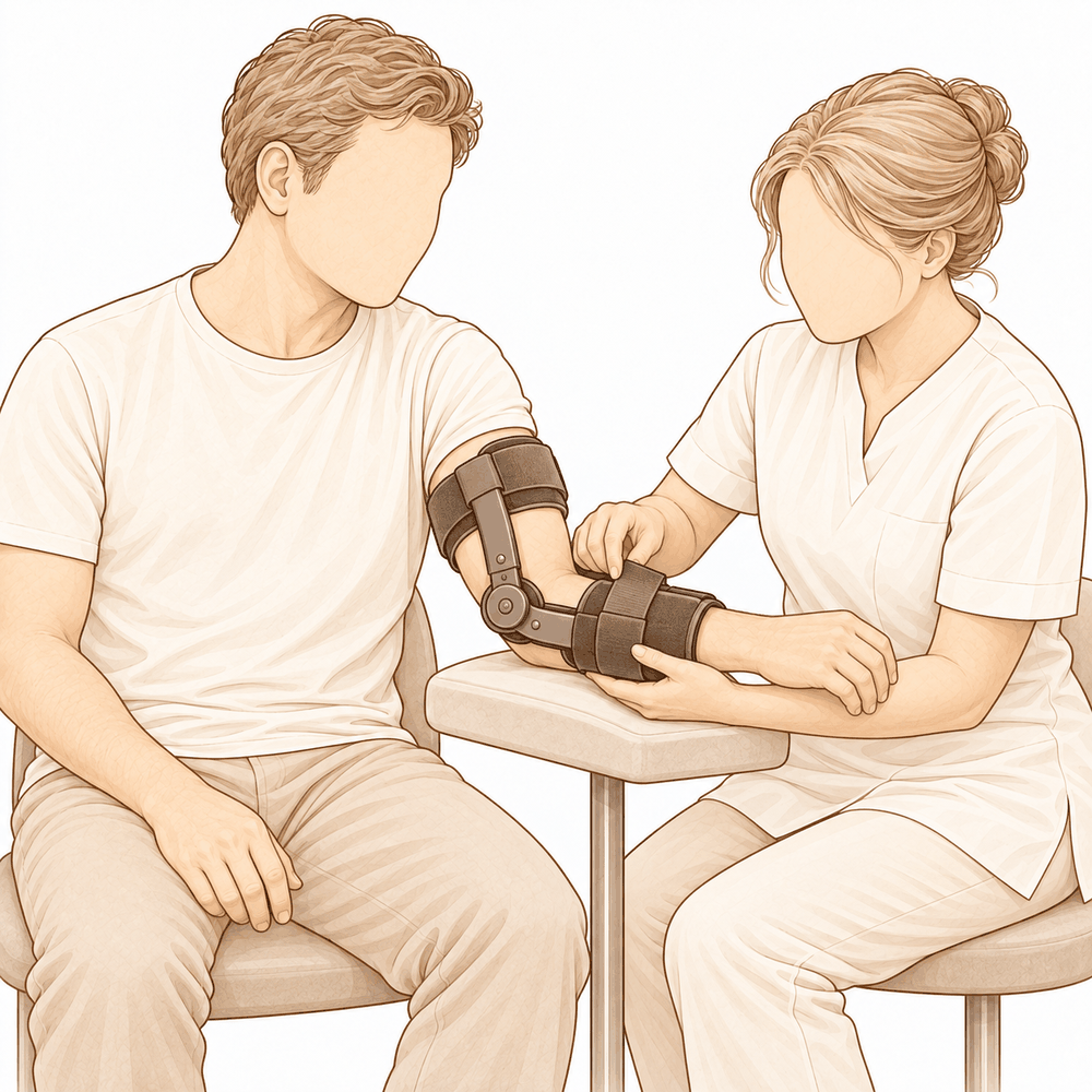
Sau phẫu thuật, bạn sẽ về nhà với cánh tay được đeo nẹp treo. Bạn có thể đi lại và thực hiện các công việc nhẹ ngay lập tức, nhưng hãy để khuỷu tay nghỉ ngơi trong nẹp khi đang vận động. Bàn tay và cổ tay có thể cử động sớm; các bài tập nhẹ dành cho chúng thường được bắt đầu trong vài ngày đầu. Các buổi trị liệu tay sau phẫu thuật sẽ do Ruby Doolan tại Extend Rehabilitation phụ trách. Ruby là chuyên gia trị liệu tay; cô ấy sẽ hướng dẫn các bài tập và chế tạo nẹp nếu cần thiết. Mục tiêu của việc trị liệu là giúp vận động sớm và đều đặn, vì việc để khuỷu tay bất động quá lâu có thể gây cứng khớp.
Khi tình trạng sưng giảm bớt, bạn sẽ sử dụng cánh tay nhiều hơn: ăn uống, viết lách và thực hiện các công việc nhà nhẹ nhàng. Khi bác sĩ phẫu thuật xác nhận rằng vết khâu đã ổn định, nẹp treo sẽ được tháo ra và bạn bắt đầu gập, duỗi khuỷu tay. Ban đầu, khả năng vận động thường cải thiện nhanh chóng, sau đó chậm dần. Khi bạn có thể nắm và giữ các vật thể mà không cảm thấy đau, các hoạt động hàng ngày sẽ trở nên dễ dàng hơn. Việc lái xe là không an toàn khi cánh tay vẫn đang đeo nẹp; bạn cần phải có khả năng nắm vô lăng bằng cả hai tay và phản ứng nhanh trong các tình huống dừng khẩn cấp mà không phụ thuộc vào thuốc giảm đau mạnh. Hướng dẫn của chúng tôi về việc lái xe sau phẫu thuật chi trên sẽ giúp bạn biết khi nào có thể quay lại lái xe.
Mỗi người có tốc độ hồi phục riêng. Lộ trình phục hồi của bạn có thể khác đi một chút; bác sĩ phẫu thuật và chuyên viên trị liệu sẽ hỗ trợ bạn trong suốt quá trình này.
Những biến chứng có thể xảy ra
Hầu hết bệnh nhân đều hồi phục tốt, nhưng đôi khi vẫn có thể gặp phải các vấn đề. Bác sĩ phẫu thuật và đội ngũ y tế sẽ theo dõi sát sao để phát hiện sớm bất kỳ bất thường nào.
Dây thần kinh chạy dọc phía trong khuỷu tay có thể bị kích ứng sau phẫu thuật. Bạn có thể cảm thấy tê, kiến bò hoặc mất cảm giác ở ngón áp út và ngón út. Thông thường tình trạng này tự khỏi; tuy nhiên hãy báo cho chúng tôi nếu triệu chứng không thuyên giảm hoặc ngón tay trở nên yếu đi.
Đôi khi khuỷu tay vẫn còn lỏng lẻo hoặc có cảm giác dễ bị trật khớp. Nếu khuỷu tay trở nên không vững, hoặc có dấu hiệu trật ra khỏi vị trí, hãy liên hệ ngay với phòng khám.
Khuỷu tay cũng có thể bị cứng lại. Bạn có thể gặp khó khăn khi duỗi thẳng hoặc gập cánh tay hoàn toàn; cảm giác cử động bị hạn chế thay vì đau nhức. Hãy đề cập vấn đề này trong lần tái khám, vì vật lý trị liệu bổ sung hoặc điều trị thêm có thể giúp ích.
Nhiễm trùng là tình trạng hiếm gặp nhưng rất nghiêm trọng. Hãy chú ý nếu có cơn đau âm ỉ, dữ dội không giảm khi dùng thuốc giảm đau thông thường; vùng da quanh vết mổ bị đỏ lan rộng; hoặc có dịch chảy ra từ vết thương. Nếu gặp các dấu hiệu này, hãy gọi cho phòng khám ngay trong ngày; nếu có sốt hoặc cảm thấy mệt mỏi, hãy đến phòng cấp cứu.
Các móc kim loại hoặc ốc vít nhỏ dùng để cố định vết khâu đôi khi gây kích ứng; khớp nối tạm thời cũng có thể bị lỏng. Bạn có thể cảm nhận được tiếng lục cục, cảm giác vướng víu hoặc có khối u nhỏ dưới da. Hãy nói với bác sĩ trong lần tái khám tới.
Trong trường hợp dùng một đoạn gân tự thân để tái tạo dây chằng, vùng đó có thể còn đau nhức trong một thời gian. Hãy báo cho chúng tôi nếu cơn đau ngày càng tăng thay vì thuyên giảm.
Đôi khi xương có thể mọc ở những nơi không nên, quanh khớp hoặc bên trong khớp. Điều này có thể hạn chế vận động hoặc gây cảm giác vướng víu, tiếng kêu lạo xạo khi cử động. Nếu khả năng vận động của khuỷu tay không tiến triển, hãy đề cập vấn đề này trong lần tái khám.
Vết sẹo đôi khi có thể bị bong tróc hoặc tiết dịch. Nếu vết thương hở ra, trở nên đỏ hơn hoặc có dịch chảy ra, hãy liên hệ chúng tôi ngay thay vì chờ đợi.
Bảng các biến chứng dưới đây liệt kê tỷ lệ xảy ra của từng tình trạng nếu bạn muốn biết thông tin cụ thể.
Khi nào nên gọi cho chúng tôi
Hầu hết các vấn đề đều xuất hiện từ sớm; vì vậy chúng tôi mong nhận được thông tin càng sớm càng tốt. Hãy gọi cho chúng tôi nếu bạn bị sốt, vết thương trở nên đỏ hơn hoặc có dịch chảy ra, hoặc cơn đau ngày càng tăng thay vì giảm dần. Hãy đến phòng cấp cứu nếu bạn bị sưng hoặc đau ở vùng bắp chân, hoặc đột ngột khó thở. Hãy gọi ngay cho chúng tôi nếu ngón áp út và ngón út của bạn bị tê, tay bạn yếu đi, hoặc bạn không thể cử động cánh tay được nữa. Nếu khuỷu tay bạn bị trật khớp hoặc có cảm giác như sắp trật lại, hãy liên hệ với phòng khám ngay trong ngày.
Evidence & references
This is the clinical evidence summary written for health professionals. It is technical, and it lists the research this page was built from. You do not need to read it to understand your treatment or to make a decision about it.
Overview
- Surgical ligament reconstruction or repair is the most favorable treatment option for restoration of normal elbow function in posterolateral rotatory instability [4].
- Repair and reconstruction of the lateral ulnar collateral ligament offers practical and often successful solutions for patients with posterolateral rotatory instability of the elbow [10].
- Improving arthroscopic techniques provide a satisfactory alternative in stabilizing the elbow [7].
- The gravity varus test has biomechanical plausibility and confirms that pronation stabilizes the lateral elbow [1].
- An all-arthroscopic reconstruction technique for the lateral ulnar collateral ligament is reproducible and avoids residual instability [2].
- Open posterolateral ligament plication and lateral ulnar collateral ligament repair using an all-suture construct allows for complete posterolateral stabilization of the elbow with a single implant and bone preservation [3].
- Suture-augmented lateral ulnar collateral ligament and radial collateral ligament reconstruction provides a reproducible, anatomically based construct that restores posterolateral elbow stability and addresses the complex spectrum of lateral-sided injuries observed in posterolateral rotatory instability [5].
- Further follow-up and wider experience of the Surgilig/LockDown technique is required to assess its long-term functional outcome in comparison with more established surgical techniques for stabilization of posterolateral elbow instability [6].
- Arthroscopic lateral ulnar collateral ligament plication/reconstruction with augmented lateral collateral ligament imbrication allows effective management of elbow instability while promoting quicker patient recovery and long-term functional restoration [8].
- Acute repair of the lateral ulnar collateral ligament with a cortical button resulted in satisfactory clinical outcomes and successful restoration of elbow stability [9].
- The use of suture button fixation for repair of the lateral ulnar collateral ligament in terrible triad injuries has not been previously described [11].
- Corrective open-wedge osteotomy with structural bone allograft restored radial head alignment, eliminated dynamic subluxation, and achieved stable elbow function without the need for ligament reconstruction in complex posterolateral rotatory instability due to malunited radius fracture [12].
- Reconstruction of the lateral ulnar collateral ligament with a tendon graft offers an alternative that restores stability through a dynamic “sling effect” rather than rigid constraint [13].
Anatomy & Pathophysiology
Bony Anatomy
- The elbow is a trocho-ginglymoid joint consisting of medial and lateral articulations that provide bony stability [22].
- The ulnohumeral joint is formed by the articulation of the trochlea with the ulna within the greater sigmoid notch [22].
- The ulnohumeral joint provides highly congruent anatomy through almost 180° of articular contact, except for a bare area of the greater sigmoid notch devoid of cartilage [22].
- The radiocapitellar joint is formed by the articulation of the capitellum and the radial head [22].
- The radial head is a concave elliptical structure covered with articular cartilage along the radiocapitellar joint and approximately 270° of the articular margin [22].
- The radius is held in close approximation to the ulna at the proximal radioulnar joint by the annular ligament [22].
- The distal humeral articulation is angled 30° from the longitudinal axis of the humerus [22].
- The axis of rotation is angulated 5° to 7° in the coronal plane relative to the epicondylar axis, with the medial side more distal than the lateral side [22].
- The articular surface of the distal humerus is angled 30 degrees anterior to the humeral shaft axis [28].
- In full extension, 60% of axial load is transmitted through the radiocapitellar joint [28].
- The medial column of the distal humerus diverges from the humeral shaft at a 45-degree angle in the coronal plane [30].
- The lateral column of the distal humerus diverges from the humeral shaft at a 20-degree angle in the coronal plane [30].
- The trochlea is covered by articular cartilage anteriorly, inferiorly, and posteriorly, creating an arc of almost 270 degrees [33].
- The olecranon fossa and coronoid fossa are separated by a thin bony septum which is occasionally absent [33].
- The ulna medially bends approximately 8° at 8 cm from the tip of the olecranon [22].
- The articulation to the tip of the coronoid is approximately 30° from the long axis of the ulna in the sagittal plane [22].
Ligamentous Anatomy
- The lateral collateral ligament complex consists of the radial collateral ligament, the lateral ulnar collateral ligament (LUCL), and the annular ligament [33].
- The radial collateral ligament originates from an isometric point on the lateral epicondyle and fans out to attach to the annular ligament [33].
- The LUCL arises from an isometric point on the lateral epicondyle and attaches to the crista supinatoris of the proximal ulna [33].
- The annular ligament attaches to the anterior and posterior margins of the lesser sigmoid notch [33].
- The LUCL origin center is located 10.7 mm from the lateral epicondyle [21].
- The LUCL insertion is located 3.3 mm from the apex of the supinator crest [21].
- The LUCL acts as a posterior buttress for the radial head and the ulna to prevent subluxation [17].
- The LUCL is considered the principal constraint of the elbow joint against posterolateral rotatory instability (PLRI) [17].
- The lateral collateral ligament contributes 14% of varus stability of the elbow with the joint in full extension [37].
- The lateral collateral ligament contributes 9% of varus stability of the elbow with the joint in 90 degrees of flexion [37].
- The medial ulnar collateral ligament (MUCL) is the primary restraint to valgus stress within functional elbow range of motion [28].
- The MUCL originates on the posterior medial epicondyle and inserts on the sublime tubercle of the medial coronoid process [28].
- The anterior bundle of the MUCL is the primary restraint to valgus stress within functional elbow range of motion [28].
- The posterior bundle of the MUCL is the primary restraint to valgus stress with the elbow in maximal flexion [28].
- The common extensor tendon is a secondary stabilizer of the lateral elbow [16].
- The joint capsule is a secondary stabilizer of the elbow [19].
- The radiocapitellar articulation is a secondary stabilizer of the elbow [19].
Biomechanics & Pathophysiology
- Elbow stability is determined by primary and secondary stabilizers, with injury to these structures causing instability [19].
- The three primary stabilizers of the elbow are the ulnohumeral articulation, the MUCL, and the LUCL complex [19].
- Secondary stabilizers of the elbow include the radiocapitellar articulation, common flexor tendon, common extensor tendon, and joint capsule [19].
- Tensile forces are present at the medial elbow while compressive forces are present at the lateral elbow [23].
- The capsule allows maximum distension at approximately 70 to 80 degrees of flexion [23].
- The anterior capsule attaches at a point approximately 6 mm distal to the tip of the coronoid [23].
- Pronation stabilizes the lateral elbow [1].
- The LUCL is taut in both flexion and extension [37].
- Disruption of the LUCL results in posterolateral rotatory instability [37].
- Failure of primary stabilizers results in increased stress on secondary stabilizers, leading to capsular contractures, chondromalacia, osteophytes, and loose bodies [37].
- Ulnar nerve symptoms may develop from traction, scarring, or osteophyte impingement following primary stabilizer failure [37].
- The typical injury pattern for traumatic elbow dislocation involves a valgus internal rotation force of the forearm on a slightly flexed extremity [37].
- In traumatic elbow dislocation, structures are disrupted on the lateral side first, progressing medially as more force is applied [37].
- Posterolateral structures are most commonly affected when recurrence or persistence of instability results from traumatic dislocation [37].
- A coronoid fracture in association with disruption of the posterior band of the ulnar collateral ligament can result in symptomatic posteromedial instability [37].
- The normal elbow has a range of motion from 0° to 140° from extension to flexion [19].
- The normal elbow has a range of motion of 75° and 85° in pronation and supination respectively [19].
- A functional arc for elbow flexion and extension is 100° [19].
- A functional arc for forearm rotation is 100° [19].
- The normal valgus carrying angle of the elbow is 5 to 10 degrees for men and 10 to 15 degrees for women [28].
- The overall valgus angle of the elbow in extension, including the ulna, is 10 to 17 degrees [33].
- The distal humerus articular surface is internally rotated 3 to 8 degrees axially [33].
- As the elbow flexes, it internally rotates resulting in slight varus alignment [33].
Classification
- The lateral ulnar collateral ligament (LUCL) acts as a posterior buttress for the radial head and the ulna to prevent subluxation [17].
- Pronation stabilizes the lateral elbow in a cadaveric model of posterolateral rotatory instability [1].
Clinical Presentation
- The gravity varus test has biomechanical plausibility for evaluating posterolateral rotatory instability of the elbow [1].
- Complex posterolateral rotatory instability can be caused by a malunited radius fracture [12].
- Four-dimensional computed tomography scan can be used to diagnose complex posterolateral rotatory instability due to malunited radius fracture [12].
- Dynamic subluxation is a feature of complex posterolateral rotatory instability associated with malunited radius fracture [12].
Investigations
Physical Examination
- Pronation stabilizes the lateral elbow during the gravity varus test [1].
- Elbow stability is determined by primary stabilizers, which include the ulnohumeral articulation, the medial ulnar collateral ligament (MUCL), and the lateral ulnar collateral ligament (LUCL) complex [19].
- Secondary stabilizers of the elbow include the radiocapitellar articulation, the common flexor tendon, the common extensor tendon, and the joint capsule [19].
- The physical examination for elbow pathology is directed by the history and the location of patient pain in the anterior, posterior, medial, or lateral aspect of the elbow [19].
- The LUCL is considered the principal constraint of the elbow joint against posterolateral rotatory instability [17].
- The lateral ulnar collateral ligament injury is the cause of recurrent instability following elbow dislocation [41].
Imaging
- Plain radiographs remain the hallmark and best screening test for elbow evaluation [19].
- Standard radiographic views for elbow evaluation include AP, lateral, and oblique views [36].
- CT is helpful when assessing for malunion architecture and the location and pattern of osteophytes or loose bodies [36].
- Three-dimensional CT is used to check for heterotopic ossification [36].
- CT is not necessary when elbow stiffness is entirely soft-tissue related [36].
- CT is beneficial if any joint incongruity or abnormal bony anatomy is present [36].
- MRI can be used to evaluate ligaments and tendons, but it is rarely indicated for elbow stiffness [36].
- Magnetic resonance evaluation of the elbow includes imaging of ligament complexes [20].
- MRI findings in acute elbow dislocation provide insight into the mechanism of injury [20].
- MR evaluation of instability patterns emphasizes the soft-tissue lesions that result from dislocation [20].
- Four-dimensional computed tomography scan can be used for diagnosis in complex posterolateral rotatory instability due to malunited radius fracture [12].
Treatment
Operative Techniques and Constructs
- Surgical ligament reconstruction or repair is the most favorable treatment option for restoration of normal elbow function [4].
- Repair and reconstruction of the lateral ulnar collateral ligament (LUCL) offers practical and often successful solutions for patients with posterolateral rotatory instability (PLRI) of the elbow [10].
- An all-arthroscopic reconstruction technique for the LUCL is reproducible and achieves reconstruction while avoiding residual instability [2].
- Open posterolateral ligament plication and LUCL repair using an all-suture construct allows for complete posterolateral stabilization of the elbow with a single implant and bone preservation [3].
- A suture-augmented LUCL and radial collateral ligament reconstruction method provides a reproducible, anatomically based construct that restores posterolateral elbow stability and addresses the complex spectrum of lateral-sided injuries observed in PLRI [5].
- Acute repair of the LUCL with a cortical button resulted in satisfactory clinical outcomes and successful restoration of elbow stability [9].
- Arthroscopic LUCL plication/reconstruction with augmented lateral collateral ligament imbrication is a minimally invasive method that allows effective management of elbow instability while promoting quicker patient recovery and long-term functional restoration [8].
- Arthroscopic-assisted LUCL reconstruction provides less insult and dissection to the soft tissue at the lateral side of the elbow while being an excellent tool to diagnose any concomitant intraarticular pathologies [15].
- A knotless, onlay technique for LUCL reconstruction performs reconstruction with a tendon graft without violation of the extensor origin and soft tissue envelop [16].
- The use of knotless anchors and an onlay technique shortens operative time, reduces the required surgical exposure, and removes the risk of tunnel osteolysis or fracture and resultant graft failure while maintaining a broad bone surface for graft incorporation [16].
- A minimally-invasive dissection prevents iatrogenic injury to the common extensor origin, an important secondary stabilizer of the lateral elbow, and the articular capsule [16].
- Remaining extracapsular with a minimally-invasive technique avoids plication of the capsular structures or risk of formation of intra-articular adhesions, theoretically reducing the risk of any postoperative loss of range of motion [16].
Outcomes and Considerations
- The docking technique originally described by Jones et al. in 2012 is the most common method in use in contemporary practice for LUCL reconstruction [16].
- The docking technique has shown recurrent instability rates as high as 25% [16].
- A minimally-invasive approach allows for earlier rehabilitation and return of range of motion, reduced postoperative pain, and reduced operative time as a repair of the common extensor origin is not required following LUCL reconstruction [16].
- Fluoroscopic guidance can help to reduce injuries to unintended structures that could be foreseen due to a limited exposure [16].
- Postoperative stiffness is a known complication of LUCL reconstruction and occurs not uncommonly [16].
Non-Ligamentous Interventions
- Corrective open-wedge osteotomy with structural bone allograft restored radial head alignment, eliminated dynamic subluxation, and achieved stable elbow function without the need for ligament reconstruction in cases of complex PLRI due to malunited radius fracture [12].
Instability and Recurrence
- The docking technique for LUCL reconstruction has shown recurrent instability rates as high as 25% [16].
- The knotless, onlay technique for LUCL reconstruction removes the risk of tunnel osteolysis or fracture and resultant graft failure [16].
- The arthroscopic technique for LUCL reconstruction achieves reconstruction and avoids residual instability [2].
- The all-suture construct for posterolateral ligament plication and LUCL repair allows for complete posterolateral stabilization of the elbow [3].
- The suture-augmented LUCL and radial collateral ligament reconstruction method restores posterolateral elbow stability [5].
- Acute repair of the LUCL with a cortical button resulted in successful restoration of elbow stability [9].
- Repair and reconstruction of the LUCL offers practical and often successful solutions for patients with PLRI of the elbow [10].
- Reconstruction of the lateral ulnar collateral ligament with a tendon graft restores stability through a dynamic “sling effect” rather than rigid constraint [13].
Stiffness and Range of Motion
- The minimally-invasive, extracapsular technique for LUCL reconstruction theoretically reduces the risk of postoperative loss of range of motion by avoiding plication of capsular structures or risk of formation of intra-articular adhesions [16].
- The minimally-invasive method for managing elbow instability promotes quicker patient recovery and long-term functional restoration [8].
Iatrogenic Injury and Soft Tissue
- The minimally-invasive dissection for LUCL reconstruction prevents iatrogenic injury to the common extensor origin, an important secondary stabilizer of the lateral elbow [16].
- The minimally-invasive dissection for LUCL reconstruction prevents iatrogenic injury to the articular capsule [16].
- The arthroscopic-assisted LUCL reconstruction provides less insult and dissection to the soft tissue at the lateral side of the elbow [15].
Long-Term Outcomes
Recovery
- The minimally invasive arthroscopic method allows effective management of elbow instability while promoting quicker patient recovery and long-term functional restoration [8].
- The all-arthroscopic reconstruction technique is reproducible and achieves the reconstruction of the lateral ulnar collateral ligament as well as avoids residual instability [2].
- The open posterolateral ligament plication and lateral ulnar collateral ligament repair technique allows for complete posterolateral stabilization of the elbow with a single implant and bone preservation [3].
- The suture-augmented lateral ulnar collateral ligament and radial collateral ligament reconstruction method provides a reproducible, anatomically based construct that restores posterolateral elbow stability and addresses the complex spectrum of lateral-sided injuries observed in posterolateral rotatory instability [5].
- Reconstruction of the lateral ulnar collateral ligament with a tendon graft offers an alternative, restoring stability through a dynamic “sling effect” rather than rigid constraint [13].
- Clinical practice supports formal reconstruction of the lateral ulnar collateral ligament for chronic late presenting injury with posterolateral rotatory instability in the patient with open physes, without concern for causing significant growth arrest [14].
Key Evidence
- [L5] These findings support the biomechanical plausibility of the test and confirm that pronation stabilizes the lateral elbow. [1] (10.1016/j.jseint.2026.101792)
- [L5] The presented arthroscopic technique is reproducible and achieves the reconstruction of the LUCL of the elbow as well as avoids residual instability. [2] (10.1016/j.eats.2024.103096)
- [L5] The technique allows for complete posterolateral stabilization of the elbow with a single implant and bone preservation. [3] (10.1016/j.eats.2024.103172)
- [L4] Surgical ligament reconstruction or repair is the most favorable treatment option for restoration of normal elbow function. [4] (10.1016/s1058-2746(03)00091-0)
- [L5] The described method provides a reproducible, anatomically based construct that restores posterolateral elbow stability and addresses the complex spectrum of lateral-sided injuries observed in PLRI. [5] (10.1016/j.eats.2025.103797)
- [L4] Further follow-up and wider experience of the technique is required to assess its long-term functional outcome in comparison with more established surgical techniques for stabilization of posterolateral elbow instability. [6] (10.1097/bte.0000000000000025)
- [L5] Improving arthroscopic techniques provide a satisfactory alternative in stabilizing the elbow. [7] (10.1016/s0278-5919(05)70246-5)
- [L5] This minimally invasive method allows effective management of elbow instability while promoting quicker patient recovery and long-term functional restoration. [8] (10.1016/j.eats.2025.103529)
- [L4] The study found that acute repair of the LUCL with a cortical button resulted in satisfactory clinical outcomes and successful restoration of elbow stability. [9] (10.1097/bte.0000000000000178)
- [L5] Repair and reconstruction of the LUCL now offers practical and often successful solutions for patients with PLRI of the elbow. [10] (10.1016/j.csm.2004.06.010)
- [L4] The use of suture button fixation for repair of lateral ulnar collateral ligament has not been previously described. [11] (10.1016/j.eats.2023.10.004)
- [L5] Corrective open-wedge osteotomy with structural bone allograft restored radial head alignment, eliminated dynamic subluxation, and achieved stable elbow function without the need for ligament reconstruction. [12] (10.1016/j.xrrt.2026.100840)
- [L5] Reconstruction of the lateral ulnar collateral ligament with a tendon graft offers an alternative, restoring stability through a dynamic “sling effect” rather than rigid constraint. [13] (10.1002/atn2.70037)
- [L5] Clinical practice supports formal reconstruction of the LUCL for chronic late presenting injury with PLRI in the patient with open physes, without concern for causing significant growth arrest. [14] (10.1097/bth.0b013e3181dd88f3)
- [L5] It provides less insult and dissection to the soft tissue at the lateral side of the elbow while being an excellent tool to diagnose any concomitant intraarticular pathologies. [15] (10.1016/j.eats.2024.103101)
- [L5] [16] (10.1002/atn2.70135)
- [L5] On the basis of these morphological features, the LUCL might act a posterior buttress for the radial head and the ulna to prevent its subluxation, and therefore it is considered to be the principal constraint of the elbow joint against PLRI. [17] (10.1177/1558944716660555mm)
References
[1] Biomechanical Evaluation of the Gravity Varus Test in a Cadaveric Model of Posterolateral Rotatory Instability of the Elbow. JSES International. 2026. DOI: 10.1016/j.jseint.2026.101792
[2] Posterolateral Elbow Dislocation: An All‐Arthroscopic Reconstruction of the Lateral Ulnar Collateral Ligament. Arthroscopy Techniques. 2024. DOI: 10.1016/j.eats.2024.103096
[3] Open Posterolateral Ligament Plication and Lateral Ulnar Collateral Ligament Repair in Posterolateral Rotatory Instability of the Elbow Using an All‐Suture Construct. Arthroscopy Techniques. 2024. DOI: 10.1016/j.eats.2024.103172
[4] Surgical reconstruction for posterolateral rotatory instability of the elbow. Journal of Shoulder and Elbow Surgery. 2003. DOI: 10.1016/s1058-2746(03)00091-0
[5] Suture‐Augmented Lateral Ulnar Collateral Ligament and Radial Collateral Ligament Reconstruction for Subacute and Chronic Posterolateral Rotatory Instability. Arthroscopy Techniques. 2025. DOI: 10.1016/j.eats.2025.103797
[6] Surgical Repair of Posterolateral Rotatory Instability of the Elbow Using Surgilig/LockDown. Techniques in Shoulder & Elbow Surgery. 2014. DOI: 10.1097/bte.0000000000000025
[7] Posterolateral Rotatory Instability of the Elbow. Clinics in Sports Medicine. 2001. DOI: 10.1016/s0278-5919(05)70246-5
[8] Arthroscopic Lateral Ulnar Collateral Ligament Plication/Reconstruction With Augmented Lateral Collateral Ligament Imbrication. Arthroscopy Techniques. 2025. DOI: 10.1016/j.eats.2025.103529
[9] Clinical Outcomes of Lateral Ulnar Collateral Ligament Repair of the Elbow Using a Cortical Button. Techniques in Shoulder & Elbow Surgery. 2019. DOI: 10.1097/bte.0000000000000178
[10] PLRI: posterolateral rotatory instability of the elbow. Clinics in Sports Medicine. 2004. DOI: 10.1016/j.csm.2004.06.010
[11] Suture Button Repair for Lateral Ulnar Collateral Ligament in Terrible Triad Injuries: Surgical Technique. Arthroscopy Techniques. 2024. DOI: 10.1016/j.eats.2023.10.004
[12] Complex posterolateral rotatory instability due to malunited radius fracture: diagnosis with four-dimensional computed tomography scan and corrective osteotomy. JSES Reviews, Reports, and Techniques. 2026. DOI: 10.1016/j.xrrt.2026.100840
[13] Arthroscopic Lateral Collateral Ligament Reconstruction With Tendon Graft in High‐Grade Atraumatic Posterolateral Rotatory Instability in Elbows. Arthroscopy Techniques. 2026. DOI: 10.1002/atn2.70037
[14] Surgical Treatment of Posterolateral Rotatory Instability of the Elbow in Children. Techniques in Hand & Upper Extremity Surgery. 2010. DOI: 10.1097/bth.0b013e3181dd88f3
[15] Arthroscopic‐Assisted Lateral Ulnar Collateral Ligament Reconstruction for Posterolateral Rotatory Instability of the Elbow: A Technical Note. Arthroscopy Techniques. 2024. DOI: 10.1016/j.eats.2024.103101
[16] Lateral Ulnar Collateral Ligament Reconstruction Through a Mini‐Invasive Approach. Arthroscopy Techniques. 2026. DOI: 10.1002/atn2.70135
[17] Morphology of the Lateral Ulnar Collateral Ligament of the Elbow. HAND. 2016. DOI: 10.1177/1558944716660555mm
[19] Orthopaedic Knowledge Update 13 Ebook Without Multimedia. Anatomy, Biomechanics, Physical Examination, and Imaging of the Elbow > Summary and Conclusions.
[20] Orthopaedic Knowledge Update Sports Medicine 6. Magnetic Resonance Imaging of the Elbow > Annotated References.
[21] Orthopaedic Knowledge Update 13 Ebook Without Multimedia. Anatomy, Biomechanics, Physical Examination, and Imaging of the Elbow > Annotated References.
[22] Orthopaedic Knowledge Update 13 Ebook Without Multimedia. Anatomy, Biomechanics, Physical Examination, and Imaging of the Elbow > Anatomy > Bony Anatomy.
[23] Miller S Review Of Orthopaedics. SECTION 16 PATELLAR TRACKING IN TOTAL KNEE ARTHROPLASTY > TABLE 2.3 Shoulder Spaces.
[28] Miller S Review Of Orthopaedics. ELBOW.
[30] A Lange Medical Book Current Diagnosis Treatment In Orthopedics Fifth Edition. 2Musculoskeletal Trauma Surgery > INJURIES AROUND THE ELBOW.
[33] Rockwood And Green S Fractures In Adults. 29: Principles of Nonunion and Bone Defect Treatment > Pathoanatomy and Applied Anatomy Related to Distal Humerus Fractures.
[36] Aaos Comprehensive Orthopaedic Review 3. Elbow Stiffness* > IV. Evaluation.
[37] Campbell S Operative Orthopaedics 4 Volume Set. POSTERIOR SURGICAL APPROACH FOR QUADRILATERAL SPACE SYNDROME > MCLAUGHLIN PROCEDURE > ARTHROSCOPIC SURGERY.
[41] A Lange Medical Book Current Diagnosis Treatment In Orthopedics Fifth Edition. 2Musculoskeletal Trauma Surgery > ELBOW DISLOCATION.