Patients › Elbow
முழங்கை தசைநார் மறுசீரமைப்பு (Elbow Ligament Reconstruction / Stabilisation)
இந்த அறுவை சிகிச்சை ஏன் பரிந்துரைக்கப்பட்டுள்ளது
Mater Private Hospital Rockhampton-இல் பணியாற்றும் மேல் அவயவ (upper-limb) அறுவை சிகிச்சை நிபுணரான டாக்டர் கியரன் ஹிர்பரா, உங்கள் குறிப்பிட்ட காயத்துக்கு ஏற்ற சிகிச்சையைத் தேர்ந்தெடுக்கிறார். நோயாளிகள் பொதுவாக அவர்களுடைய பொது மருத்துவரால் (GP) எங்கள் கிளினிக்குக்குப் பரிந்துரைக்கப்படுகிறார்கள்; ஓர் இயன்முறை சிகிச்சையாளர் (physiotherapist) எங்களைப் பார்க்கச் சொல்லியிருந்தாலும், Medicare திருப்பிச் செலுத்தலுக்குத் (Medicare rebate) தகுதி பெற உங்கள் பொது மருத்துவரிடமிருந்து ஒரு பரிந்துரைக் கடிதம் இன்னும் உங்களுக்குத் தேவைப்படும். உங்கள் சந்திப்பின்போது நாங்கள் உங்கள் நோய் வரலாற்றைக் கேட்டறிந்து, உங்கள் முழங்கையைப் பரிசோதித்து, என்ன பிரச்சினை என்பதைக் கண்டறியத் தேவைப்பட்டால் ஸ்கேன்களையும் ஏற்பாடு செய்வோம்.
முழங்கை தசைநார் மறுசீரமைப்பு (elbow ligament reconstruction) என்பது, தேய்ந்துபோன அல்லது கிழிந்த ஒரு தசைநாரை (ligament) தானமாகப் பெறப்பட்ட தசைநாணின் (donor tendon) ஒரு துண்டைக் கொண்டு மீண்டும் உருவாக்கி, மூட்டை உறுதியாகப் பிடித்து வைக்கும் அறுவை சிகிச்சை. மற்ற சிகிச்சைகள் போதுமான முன்னேற்றத்தைத் தராத பிறகும், உங்கள் முழங்கை இன்னும் விலகிச் செல்வது போலவோ அல்லது அதன் இடத்தில் நிலைக்காதது போலவோ உணரப்பட்டால், பொதுவாக இதை நாங்கள் பரிந்துரைக்கிறோம். முழங்கையை ஏறக்குறைய நேராக வைத்திருந்தால் மட்டுமே அதை மூட்டுக்குள் நிலைநிறுத்த முடியும் என்ற நிலையிலும், அல்லது மூட்டைச் சுற்றி ஏற்பட்ட ஒரு முறிவு அதை நிலையற்றதாக்கிவிட்ட நிலையிலும் இது வழங்கப்படுகிறது. அசைக்கவும், பயன்படுத்தவும், வலியின்றி நம்பி இயங்கவும் கூடிய ஒரு நிலையான முழங்கையே இதன் நோக்கம். பின்பக்க-வெளிப்புறச் சுழற்சி நிலையின்மை (posterolateral rotatory instability) உள்ள முழங்கைகளில் சுமார் 85%-க்கு, அன்னியர் திசு ஒட்டு மூலமான மறுசீரமைப்பு (allograft reconstruction) முழங்கையின் நிலைத்தன்மையை மீட்டுத் தருகிறது. இது உங்கள் முழங்கைக்கும் உங்கள் இலக்குகளுக்கும் பொருந்துமா என்பதை உங்களுடன் கலந்துரையாடி, இணைந்து முடிவு செய்வோம்.
அறுவை சிகிச்சைக்கு முன்
அறுவை சிகிச்சைக்கு முந்தைய வாரங்களில், எக்ஸ்-ரே, MRI (தசைநார்கள் (ligaments) போன்ற மென்திசுக்களைக் காட்டும் ஸ்கேன்) அல்லது அல்ட்ராசவுண்ட் போன்ற ஸ்கேன்களைக் கொண்டு திட்டமிடலை நாங்கள் நிறைவு செய்வோம். அறுவை சிகிச்சை நாளில், அதற்கு ஏழு மணி நேரம் முன்பே உண்பதையும் குடிப்பதையும் நிறுத்துங்கள். அறுவை சிகிச்சை அரங்கின் பட்டியல் முன்கூட்டியே நகர்ந்தால் உங்கள் அறுவை சிகிச்சையை முன்னதாகக் கொண்டுவர முடியும் என்பதற்காகவே ஏழு மணி நேரம் கேட்கிறோம்; நீங்கள் உண்ணாமல் இருக்க வேண்டிய சரியான நேரத்தை உங்கள் அறுவை சிகிச்சை நிபுணர் உறுதிப்படுத்துவார். அறுவை சிகிச்சைக்கு முன் சில மருந்துகளை நிறுத்தி வைக்க வேண்டியிருக்கலாம்; எவற்றை, எவ்வளவு காலத்துக்கு நிறுத்த வேண்டும் என்பது குறித்துத் தெளிவான அறிவுறுத்தல்களை நாங்கள் தருவோம். மாத்திரைகள், சொட்டு மருந்துகள், களிம்புகள் உட்பட நீங்கள் எடுத்துக்கொள்ளும் அனைத்தின் எழுதப்பட்ட பட்டியலைக் கொண்டு வாருங்கள். அதன் பிறகு உங்களை வீட்டுக்கு வாகனத்தில் அழைத்துச் செல்ல ஒருவரை ஏற்பாடு செய்யுங்கள். உங்கள் முழங்கையின் மேல் எளிதாக நகரும் கைப்பகுதி கொண்ட, தளர்வான வசதியான ஆடைகளை அணியுங்கள். உங்களுக்கு வேறு உடல்நலப் பிரச்சினைகள் இருந்தால், இரத்தப் பரிசோதனைகள் அல்லது மயக்க மருந்து நிபுணரின் (anaesthetist — மயக்க மருந்து கொடுக்கும் மருத்துவர்) பரிசோதனையும் தேவைப்படலாம்.
அறுவை சிகிச்சை நாளில்
நீங்கள் மருத்துவமனையின் அறுவை சிகிச்சை அனுமதிப் பிரிவுக்கு (surgical admissions unit) வருவீர்கள்; அங்கே உங்கள் வருகை பதிவு செய்யப்பட்டு, அறுவை சிகிச்சை அரங்குக்குத் தயார்படுத்தப்படுவீர்கள். மயக்க மருந்து நிபுணரை நீங்கள் அங்கே சந்திப்பீர்கள். இந்த அறுவை சிகிச்சை முழு மயக்க மருந்தில் (general anaesthetic) செய்யப்படுகிறது. அறுவை சிகிச்சைக்குப் பிந்தைய வலி நிவாரணத்துக்காகச் சில சமயம் பகுதி நரம்புத் தடுப்பு ஊசியும் (regional nerve block) சேர்க்கப்படுகிறது; அன்றைய தினம் மயக்க மருந்து நிபுணர் இதைப் பற்றி உங்களுடன் கலந்துரையாடுவார். பின்னர் நீங்கள் அறுவை சிகிச்சை அரங்குக்கு அழைத்துச் செல்லப்படுவீர்கள்; அங்கே அறுவை சிகிச்சை செய்யப்படும். அது முடிந்ததும், மீட்புப் பகுதியில் (recovery area) நீங்கள் கண் விழிப்பீர்கள்; மயக்க மருந்தின் விளைவு குறையும் வரை செவிலியர்கள் உங்களைக் கண்காணிப்பார்கள். உங்கள் நிலை சீரானதும், அறுவை சிகிச்சையையும் உங்கள் மீட்சியையும் பொறுத்து, நீங்கள் வார்டுக்குச் செல்வீர்கள் அல்லது வீட்டுக்குச் செல்வீர்கள்.
அன்றைய நாளின் நோக்கம் எளிது: அறுவை சிகிச்சைக்குப் பிறகு விரைவில் அசையத் தொடங்கும் அளவுக்கு உங்கள் முழங்கைக்குப் போதுமான நிலைத்தன்மையை மீட்டுத் தருவது. அதன் பிறகு முழங்கையை நீண்ட காலம் அசைக்காமல் வைத்திருப்பது அதை விறைப்பாக்கிவிடும்; எனவே ஆரம்பத்திலேயே அசைவைத் தொடங்குவது திட்டத்தின் ஒரு பகுதி.
அறுவை சிகிச்சையில் என்ன செய்யப்படுகிறது
உங்கள் முழங்கையைச் சுற்றி எந்தெந்த அமைப்புகள் காயமடைந்துள்ளன என்பதைப் பொறுத்தே சரியான படிநிலைகள் அமையும்; அறுவை சிகிச்சை நாளுக்கு முன்பே உங்கள் ஸ்கேன்களைக் கொண்டு இதைத் திட்டமிடுகிறோம். பிரச்சினையின் ஒரு பகுதியாக எலும்பு முறிவு இருந்தால், உங்கள் அறுவை சிகிச்சை நிபுணர் முதலில் முறிந்த எலும்புத் துண்டுகளைத் திருகுகளாலோ (screws) சிறிய தகட்டாலோ (plate) பொருத்தி நிலைப்படுத்துகிறார். முன்கையின் மேல் முனையில் உள்ள முறிந்த எலும்புத் துண்டைச் சரிசெய்ய முடியாவிட்டால், அதற்குப் பதிலாக உலோகத்தாலான உள்பொருத்து (metal implant) வைக்கப்படலாம். எலும்பு உறுதியானதும், கிழிந்த தசைநார்கள் சரிசெய்யப்படுகின்றன அல்லது மீண்டும் உருவாக்கப்படுகின்றன.
ஒரு தசைநாரை மீண்டும் உருவாக்குவது என்பது, ஒரு தசைநாண் துண்டைக் கொண்டு புதிய தசைநாரை உருவாக்குவதாகும். இது தானமாகப் பெறப்பட்ட திசுவிலிருந்தோ (donor tissue) உங்கள் சொந்த உடலிலிருந்தோ எடுக்கப்படலாம். சிறிய நங்கூரங்கள் (anchors) புதிய தசைநாரைச் சரியான இடங்களில் எலும்புடன் பிடித்து வைக்கின்றன; அறுவை சிகிச்சையை முடிப்பதற்கு முன், முழங்கை அதன் முழு அசைவு வரம்பிலும் மூட்டுக்குள்ளேயே நிலைத்திருக்கிறதா என்பதை உங்கள் அறுவை சிகிச்சை நிபுணர் சரிபார்க்கிறார். சில சமயம், உள்ளுக்குள் ஒரு தாங்கியைப் போலச் செயல்பட்டு, தசைநார் குணமடையும்போது அதற்கு ஆதரவளிக்கும் வகையில், சரிசெய்யப்பட்ட பகுதியுடன் சேர்த்து உறுதியான தையல் நாடாவும் (suture tape) இணைக்கப்படுகிறது. அப்போதும் முழங்கை தானாக உறுதியாக நிலைக்கவில்லை என்றால், நிலைமை சீராகும் வரை அதை இடத்தில் வைத்திருக்க ஒரு தற்காலிகக் கீல் அமைப்பு (temporary hinge) பொருத்தப்படலாம்.
ஒரு நீண்ட கீறலுக்குப் பதிலாகச் சிறிய கீறல்கள் வழியாகவே இந்த அறுவை சிகிச்சையைப் பெரும்பாலும் செய்ய முடியும். சில தசைநார் மறுசீரமைப்புகளுக்கு, சுமார் 2 முதல் 3 செ.மீ. அளவுள்ள ஒரு கீறலே போதும். சிறிய கீறல்கள் வழியாகச் செய்வது, மூட்டைச் சுற்றியுள்ள ஆரோக்கியமான தசைநாண்களையும் தசைகளையும் பாதுகாக்கிறது; மூட்டின் உட்படலத்தையே (joint lining) கலைப்பதைத் தவிர்க்கிறது.
சரிசெய்தல் முடிந்ததும், கீறல்கள் தையல்களால் மூடப்பட்டு, அதன் மேல் கட்டுப் போடப்படும். அறுவை சிகிச்சை முழுவதன் இலக்கும், அதற்குப் பிறகு விரைவில் அசையத் தொடங்கக்கூடிய ஒரு நிலையான முழங்கையே; ஏனெனில் அதை நீண்ட காலம் அசைக்காமல் வைத்திருப்பது விறைப்பை ஏற்படுத்திவிடும்.
அறுவை சிகிச்சைக்குப் பிறகு
மீட்புப் பகுதியில் நீங்கள் கண் விழிப்பீர்கள்; மயக்க மருந்தின் விளைவு குறையும் வரை செவிலியர்கள் உங்களை நெருக்கமாகக் கவனிப்பார்கள். உங்கள் கை கைத்தாங்கியில் (sling) இருக்கும் அல்லது தலையணைகளின் மேல் தாங்கப்பட்டிருக்கும்; காயங்களின் மேல் கட்டு இருக்கும். உங்களுக்கு வலி நிவாரணம் தருவோம்; நீங்கள் நடமாடத் தொடங்கும் முன் அது வேலை செய்கிறதா என்பதைச் சரிபார்ப்போம். வீட்டுக்குச் சென்ற பிறகு முதல் 24 மணி நேரத்துக்கு ஒருவர் உங்களுடன் இருக்க வேண்டும். அன்றே வீட்டுக்குச் செல்வீர்களா அல்லது மருத்துவமனையில் ஒரு இரவு தங்குவீர்களா என்பதை உங்கள் குழு உங்களுக்குச் சொல்லும். கட்டை நாங்கள் சுமார் 10 நாட்கள் அப்படியே விட்டுவைப்போம்; நாங்கள் சொன்னால் ஒழிய, அதற்கு முன் அதைக் கழற்ற வேண்டாம். உங்களைப் பார்க்கும்போது நாங்களே அதை மாற்றுவோம் அல்லது அகற்றுவோம். பெரும்பாலானோர் உடனடியாக நடமாடவும் இலகுவான வேலைகளைச் செய்யவும் முடியும்; ஆனால் நீங்கள் எழுந்து நடமாடும்போது உங்கள் முழங்கையைக் கைத்தாங்கியில் ஓய்வாக வைத்திருங்கள்.
மீட்சி
முதல் சில நாட்களுக்கு உங்கள் முழங்கை வலியுடனும் வீக்கத்துடனும் இருக்கும். இது படிப்படியாகத் தணியும். ஓய்வு, கையை உயர்த்தி வைத்திருப்பது, நாங்கள் தரும் வலி நிவாரண மருந்துகள் ஆகியவை அசௌகரியத்தைக் குறைக்கும். மூட்டை முதன்முதலில் அசைக்கத் தொடங்கும்போது சிறிது வலி இருப்பது இயல்பானது; வாரங்கள் செல்லச் செல்ல அது குறையும்.
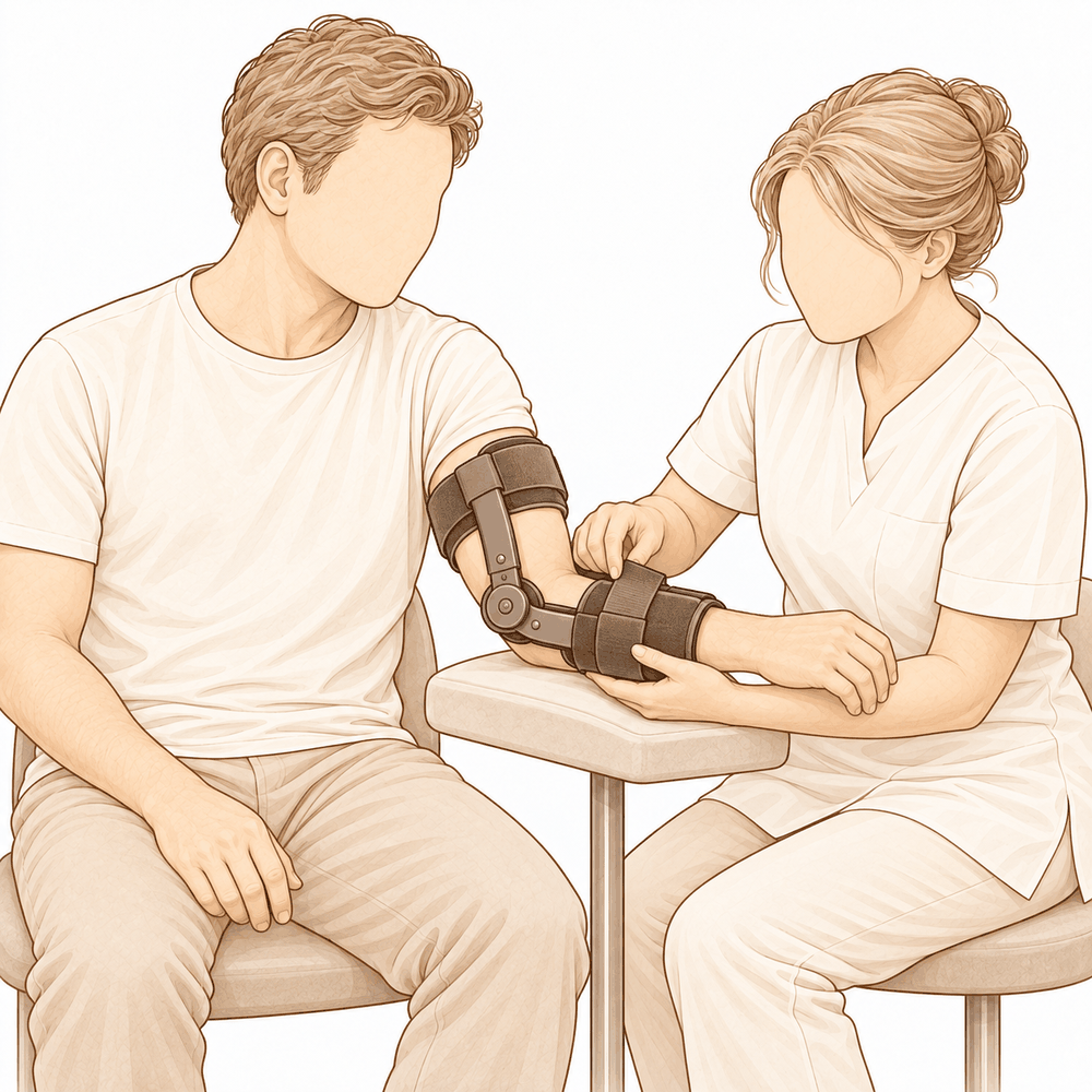
உங்கள் கை கைத்தாங்கியில் இருக்க, நீங்கள் வீட்டுக்குச் செல்வீர்கள். உடனடியாக நடமாடவும் இலகுவான வேலைகளைச் செய்யவும் முடியும்; ஆனால் எழுந்து நடமாடும்போது முழங்கையைக் கைத்தாங்கியில் ஓய்வாக வைத்திருங்கள். உங்கள் கையையும் மணிக்கட்டையும் ஆரம்பத்திலேயே அசைக்கலாம்; அவற்றுக்கான மென்மையான பயிற்சிகள் பொதுவாகச் சில நாட்களுக்குள் தொடங்கும். அறுவை சிகிச்சைக்குப் பிந்தைய கை சிகிச்சை, Extend Rehabilitation-இல் உள்ள Ruby Doolan-இடம் நடைபெறும். Ruby ஒரு கை சிகிச்சையாளர் (hand therapist): அவர் உங்கள் பயிற்சிகளுக்கு வழிகாட்டி, உங்களுக்குத் தேவையான சிம்பைச் (splint) செய்து தருவார். சிகிச்சையின் நோக்கம், சீரான, ஆரம்பகால அசைவே; ஏனெனில் முழங்கையை நீண்ட காலம் அசைக்காமல் வைத்திருப்பது விறைப்பை ஏற்படுத்திவிடும்.
வீக்கம் தணியத் தணிய, உங்கள் கையை அதிகமாகப் பயன்படுத்துவீர்கள்: உண்பது, எழுதுவது, இலகுவான வீட்டு வேலைகள். சரிசெய்யப்பட்ட பகுதி உறுதியாக இருக்கிறது என்று உங்கள் அறுவை சிகிச்சை நிபுணர் திருப்தி அடைந்ததும், கைத்தாங்கி அகற்றப்பட்டு, முழங்கையை மடக்கவும் நீட்டவும் தொடங்குவீர்கள். அசைவு பெரும்பாலும் முதலில் விரைவாகவும், பின்னர் மெதுவாகவும் முன்னேறும். வலியின்றிப் பொருட்களைப் பிடிக்கவும் தாங்கவும் முடியும்போது, அன்றாட வேலைகள் எளிதாக உணரப்படும். உங்கள் கை கைத்தாங்கியில் இருக்கும்போது வாகனம் ஓட்டுவது பாதுகாப்பானது அல்ல; மேலும், வலுவான வலி நிவாரண மருந்துகளை நிறுத்திய நிலையில், இரு கைகளாலும் ஸ்டியரிங்கைப் பிடிக்கவும் அவசரகால நிறுத்தத்தில் உடனடியாகச் செயல்படவும் உங்களால் முடிய வேண்டும். எப்போது நீங்கள் மீண்டும் ஓட்டத் தொடங்கலாம் என்பதை எங்களுடைய மேல் அவயவ அறுவை சிகிச்சைக்குப் பிறகு வாகனம் ஓட்டுதல் வழிகாட்டி விளக்குகிறது.
ஒவ்வொருவரும் அவரவர் வேகத்தில் குணமடைகிறார்கள். உங்கள் கால அளவு மாறுபடலாம்; உங்கள் அறுவை சிகிச்சை நிபுணரும் சிகிச்சையாளரும் வழி நெடுகிலும் உங்களுக்கு வழிகாட்டுவார்கள்.
என்ன தவறு நேரலாம்
பெரும்பாலான நோயாளிகள் நன்றாக இருக்கிறார்கள், ஆனால் எப்போதாவது பிரச்சினைகள் ஏற்படலாம். எந்தப் பிரச்சினையையும் முன்கூட்டியே கண்டறிய உங்கள் அறுவை சிகிச்சை நிபுணரும் குழுவும் உங்களை நெருக்கமாகக் கண்காணிக்கிறார்கள்.
உங்கள் முழங்கையின் உட்புறமாகச் செல்லும் நரம்பு, அறுவை சிகிச்சைக்குப் பிறகு எரிச்சலடையலாம். உங்கள் மோதிர விரலிலும் சுண்டு விரலிலும் கூச்சம், ஊசி குத்துவது போன்ற உணர்வு, அல்லது மரத்துப்போதல் ஏற்படுவதை நீங்கள் கவனிக்கலாம். பெரும்பாலும் இது தானாகவே சரியாகிவிடும்; ஆனால் அது குறையவில்லை என்றாலோ, விரல்கள் பலவீனமாக உணரப்பட்டாலோ எங்களிடம் சொல்லுங்கள்.
முழங்கை சில சமயம் தளர்ச்சியுடனேயே இருக்கலாம் அல்லது மீண்டும் விலகிச் செல்வது போல் உணரப்படலாம். உங்கள் முழங்கை உறுதியற்றதாக உணரத் தொடங்கினாலோ, அதன் இடத்திலிருந்து நழுவினாலோ, உடனடியாகக் கிளினிக்கைத் தொடர்பு கொள்ளுங்கள்.
முழங்கை விறைப்பாகவும் ஆகலாம். கையை முழுமையாக நீட்டுவதோ மடக்குவதோ கடினமாக இருக்கலாம்; அசைவு வலியாக அல்ல, தடைபட்டது போல் உணரப்படலாம். கூடுதல் சிகிச்சையோ மேலதிக சிகிச்சையோ உதவும் என்பதால், உங்கள் மறுபரிசோதனையின்போது இதைச் சொல்லுங்கள்.
தொற்று (infection) அரிதானது, ஆனால் தீவிரமானது. எளிய வலி நிவாரணிகளால் குறையாத ஆழமான, துடிக்கும் வலி, காயத்திலிருந்து வெளிப்புறமாகப் பரவும் சிவப்பு, அல்லது அதிலிருந்து திரவம் கசிவது ஆகியவற்றைக் கவனியுங்கள். இவற்றை நீங்கள் கவனித்தால், அன்றைய தினமே கிளினிக்கை அழையுங்கள்; காய்ச்சல் போலவோ உடல்நலம் சரியில்லாமலோ உணர்ந்தால் அவசர சிகிச்சைப் பிரிவுக்குச் செல்லுங்கள்.
சரிசெய்யப்பட்ட பகுதியைப் பிடித்து வைக்கப் பயன்படுத்தப்படும் சிறிய உலோக நங்கூரங்களோ திருகுகளோ சில சமயம் எரிச்சலை ஏற்படுத்தலாம்; அல்லது தற்காலிகக் கீல் அமைப்பு தளர்ந்துவிடலாம். புதிதாக ஒரு 'கிளிக்' சத்தம், எதோ சிக்கிக்கொள்வது போன்ற உணர்வு, அல்லது தோலுக்கு அடியில் ஒரு கட்டி ஆகியவற்றை நீங்கள் உணரலாம். உங்கள் அடுத்த மறுபரிசோதனையின்போது அதைச் சொல்லுங்கள்.
தசைநாரை மீண்டும் உருவாக்க உங்கள் சொந்த தசைநாணின் ஒரு துண்டு எடுக்கப்பட்ட இடத்தில், சிறிது காலத்துக்கு வலியோ ஒரு தொடர் வலியோ இருக்கலாம். அது குறைவதற்குப் பதிலாக மோசமானால் எங்களிடம் தெரிவியுங்கள்.
சில சமயம் எலும்பு இருக்கக் கூடாத இடங்களில் — மூட்டைச் சுற்றியோ அதன் உள்ளேயோ — உருவாகலாம். இது அசைவைக் கட்டுப்படுத்தலாம், அல்லது எதோ சிக்கிக்கொள்வது போலவும் உரசுவது போலவும் உணர்த்தலாம். உங்கள் முழங்கையின் முன்னேற்றம் நின்றுவிட்டால், மறுபரிசோதனையின்போது அதைச் சொல்லுங்கள்.
தழும்பே எப்போதாவது பிரிந்துவிடலாம் அல்லது கசியலாம். காயம் விரிந்தாலோ, மேலும் சிவந்தாலோ, சீழோ திரவமோ வரத் தொடங்கினாலோ, காத்திராமல் எங்களைத் தொடர்பு கொள்ளுங்கள்.
விவரங்கள் வேண்டுமென்றால், இந்தப் பக்கத்தில் உள்ள சிக்கல்கள் அட்டவணை வழக்கமான விகிதங்களைப் பட்டியலிடுகிறது.
எப்போது எங்களை அழைக்க வேண்டும்
பெரும்பாலான பிரச்சினைகள் ஆரம்பத்திலேயே வெளிப்படுகின்றன; அவற்றைப் பிற்பாடு அறிவதைவிட விரைவில் அறிவதையே நாங்கள் விரும்புகிறோம். உங்களுக்குக் காய்ச்சல் இருந்தால், காயம் மேலும் சிவந்தாலோ திரவம் கசியத் தொடங்கினாலோ, அல்லது வலி குறைவதற்குப் பதிலாகத் தொடர்ந்து அதிகரித்தால், எங்களை அழையுங்கள். உங்கள் கெண்டைக்காலில் வீக்கமோ வலியோ இருந்தாலோ, திடீரென மூச்சுத் திணறல் ஏற்பட்டாலோ, அவசர சிகிச்சைப் பிரிவுக்குச் செல்லுங்கள். உங்கள் மோதிர விரலும் சுண்டு விரலும் மரத்துப்போனாலோ, உங்கள் கை பலவீனமாக உணரப்பட்டாலோ, அல்லது உங்கள் கையை அறவே அசைக்க முடியாவிட்டாலோ, உடனடியாக எங்களை அழையுங்கள். உங்கள் முழங்கை இடம் விட்டு நழுவினாலோ, மீண்டும் விலகிச் செல்வது போல் உணரப்பட்டாலோ, அன்றைய தினமே கிளினிக்கைத் தொடர்பு கொள்ளுங்கள்.
Evidence & references
This is the clinical evidence summary written for health professionals. It is technical, and it lists the research this page was built from. You do not need to read it to understand your treatment or to make a decision about it.
Overview
- Surgical ligament reconstruction or repair is the most favorable treatment option for restoration of normal elbow function in posterolateral rotatory instability [4].
- Repair and reconstruction of the lateral ulnar collateral ligament offers practical and often successful solutions for patients with posterolateral rotatory instability of the elbow [10].
- Improving arthroscopic techniques provide a satisfactory alternative in stabilizing the elbow [7].
- The gravity varus test has biomechanical plausibility and confirms that pronation stabilizes the lateral elbow [1].
- An all-arthroscopic reconstruction technique for the lateral ulnar collateral ligament is reproducible and avoids residual instability [2].
- Open posterolateral ligament plication and lateral ulnar collateral ligament repair using an all-suture construct allows for complete posterolateral stabilization of the elbow with a single implant and bone preservation [3].
- Suture-augmented lateral ulnar collateral ligament and radial collateral ligament reconstruction provides a reproducible, anatomically based construct that restores posterolateral elbow stability and addresses the complex spectrum of lateral-sided injuries observed in posterolateral rotatory instability [5].
- Further follow-up and wider experience of the Surgilig/LockDown technique is required to assess its long-term functional outcome in comparison with more established surgical techniques for stabilization of posterolateral elbow instability [6].
- Arthroscopic lateral ulnar collateral ligament plication/reconstruction with augmented lateral collateral ligament imbrication allows effective management of elbow instability while promoting quicker patient recovery and long-term functional restoration [8].
- Acute repair of the lateral ulnar collateral ligament with a cortical button resulted in satisfactory clinical outcomes and successful restoration of elbow stability [9].
- The use of suture button fixation for repair of the lateral ulnar collateral ligament in terrible triad injuries has not been previously described [11].
- Corrective open-wedge osteotomy with structural bone allograft restored radial head alignment, eliminated dynamic subluxation, and achieved stable elbow function without the need for ligament reconstruction in complex posterolateral rotatory instability due to malunited radius fracture [12].
- Reconstruction of the lateral ulnar collateral ligament with a tendon graft offers an alternative that restores stability through a dynamic “sling effect” rather than rigid constraint [13].
Anatomy & Pathophysiology
Bony Anatomy
- The elbow is a trocho-ginglymoid joint consisting of medial and lateral articulations that provide bony stability [22].
- The ulnohumeral joint is formed by the articulation of the trochlea with the ulna within the greater sigmoid notch [22].
- The ulnohumeral joint provides highly congruent anatomy through almost 180° of articular contact, except for a bare area of the greater sigmoid notch devoid of cartilage [22].
- The radiocapitellar joint is formed by the articulation of the capitellum and the radial head [22].
- The radial head is a concave elliptical structure covered with articular cartilage along the radiocapitellar joint and approximately 270° of the articular margin [22].
- The radius is held in close approximation to the ulna at the proximal radioulnar joint by the annular ligament [22].
- The distal humeral articulation is angled 30° from the longitudinal axis of the humerus [22].
- The axis of rotation is angulated 5° to 7° in the coronal plane relative to the epicondylar axis, with the medial side more distal than the lateral side [22].
- The articular surface of the distal humerus is angled 30 degrees anterior to the humeral shaft axis [28].
- In full extension, 60% of axial load is transmitted through the radiocapitellar joint [28].
- The medial column of the distal humerus diverges from the humeral shaft at a 45-degree angle in the coronal plane [30].
- The lateral column of the distal humerus diverges from the humeral shaft at a 20-degree angle in the coronal plane [30].
- The trochlea is covered by articular cartilage anteriorly, inferiorly, and posteriorly, creating an arc of almost 270 degrees [33].
- The olecranon fossa and coronoid fossa are separated by a thin bony septum which is occasionally absent [33].
- The ulna medially bends approximately 8° at 8 cm from the tip of the olecranon [22].
- The articulation to the tip of the coronoid is approximately 30° from the long axis of the ulna in the sagittal plane [22].
Ligamentous Anatomy
- The lateral collateral ligament complex consists of the radial collateral ligament, the lateral ulnar collateral ligament (LUCL), and the annular ligament [33].
- The radial collateral ligament originates from an isometric point on the lateral epicondyle and fans out to attach to the annular ligament [33].
- The LUCL arises from an isometric point on the lateral epicondyle and attaches to the crista supinatoris of the proximal ulna [33].
- The annular ligament attaches to the anterior and posterior margins of the lesser sigmoid notch [33].
- The LUCL origin center is located 10.7 mm from the lateral epicondyle [21].
- The LUCL insertion is located 3.3 mm from the apex of the supinator crest [21].
- The LUCL acts as a posterior buttress for the radial head and the ulna to prevent subluxation [17].
- The LUCL is considered the principal constraint of the elbow joint against posterolateral rotatory instability (PLRI) [17].
- The lateral collateral ligament contributes 14% of varus stability of the elbow with the joint in full extension [37].
- The lateral collateral ligament contributes 9% of varus stability of the elbow with the joint in 90 degrees of flexion [37].
- The medial ulnar collateral ligament (MUCL) is the primary restraint to valgus stress within functional elbow range of motion [28].
- The MUCL originates on the posterior medial epicondyle and inserts on the sublime tubercle of the medial coronoid process [28].
- The anterior bundle of the MUCL is the primary restraint to valgus stress within functional elbow range of motion [28].
- The posterior bundle of the MUCL is the primary restraint to valgus stress with the elbow in maximal flexion [28].
- The common extensor tendon is a secondary stabilizer of the lateral elbow [16].
- The joint capsule is a secondary stabilizer of the elbow [19].
- The radiocapitellar articulation is a secondary stabilizer of the elbow [19].
Biomechanics & Pathophysiology
- Elbow stability is determined by primary and secondary stabilizers, with injury to these structures causing instability [19].
- The three primary stabilizers of the elbow are the ulnohumeral articulation, the MUCL, and the LUCL complex [19].
- Secondary stabilizers of the elbow include the radiocapitellar articulation, common flexor tendon, common extensor tendon, and joint capsule [19].
- Tensile forces are present at the medial elbow while compressive forces are present at the lateral elbow [23].
- The capsule allows maximum distension at approximately 70 to 80 degrees of flexion [23].
- The anterior capsule attaches at a point approximately 6 mm distal to the tip of the coronoid [23].
- Pronation stabilizes the lateral elbow [1].
- The LUCL is taut in both flexion and extension [37].
- Disruption of the LUCL results in posterolateral rotatory instability [37].
- Failure of primary stabilizers results in increased stress on secondary stabilizers, leading to capsular contractures, chondromalacia, osteophytes, and loose bodies [37].
- Ulnar nerve symptoms may develop from traction, scarring, or osteophyte impingement following primary stabilizer failure [37].
- The typical injury pattern for traumatic elbow dislocation involves a valgus internal rotation force of the forearm on a slightly flexed extremity [37].
- In traumatic elbow dislocation, structures are disrupted on the lateral side first, progressing medially as more force is applied [37].
- Posterolateral structures are most commonly affected when recurrence or persistence of instability results from traumatic dislocation [37].
- A coronoid fracture in association with disruption of the posterior band of the ulnar collateral ligament can result in symptomatic posteromedial instability [37].
- The normal elbow has a range of motion from 0° to 140° from extension to flexion [19].
- The normal elbow has a range of motion of 75° and 85° in pronation and supination respectively [19].
- A functional arc for elbow flexion and extension is 100° [19].
- A functional arc for forearm rotation is 100° [19].
- The normal valgus carrying angle of the elbow is 5 to 10 degrees for men and 10 to 15 degrees for women [28].
- The overall valgus angle of the elbow in extension, including the ulna, is 10 to 17 degrees [33].
- The distal humerus articular surface is internally rotated 3 to 8 degrees axially [33].
- As the elbow flexes, it internally rotates resulting in slight varus alignment [33].
Classification
- The lateral ulnar collateral ligament (LUCL) acts as a posterior buttress for the radial head and the ulna to prevent subluxation [17].
- Pronation stabilizes the lateral elbow in a cadaveric model of posterolateral rotatory instability [1].
Clinical Presentation
- The gravity varus test has biomechanical plausibility for evaluating posterolateral rotatory instability of the elbow [1].
- Complex posterolateral rotatory instability can be caused by a malunited radius fracture [12].
- Four-dimensional computed tomography scan can be used to diagnose complex posterolateral rotatory instability due to malunited radius fracture [12].
- Dynamic subluxation is a feature of complex posterolateral rotatory instability associated with malunited radius fracture [12].
Investigations
Physical Examination
- Pronation stabilizes the lateral elbow during the gravity varus test [1].
- Elbow stability is determined by primary stabilizers, which include the ulnohumeral articulation, the medial ulnar collateral ligament (MUCL), and the lateral ulnar collateral ligament (LUCL) complex [19].
- Secondary stabilizers of the elbow include the radiocapitellar articulation, the common flexor tendon, the common extensor tendon, and the joint capsule [19].
- The physical examination for elbow pathology is directed by the history and the location of patient pain in the anterior, posterior, medial, or lateral aspect of the elbow [19].
- The LUCL is considered the principal constraint of the elbow joint against posterolateral rotatory instability [17].
- The lateral ulnar collateral ligament injury is the cause of recurrent instability following elbow dislocation [41].
Imaging
- Plain radiographs remain the hallmark and best screening test for elbow evaluation [19].
- Standard radiographic views for elbow evaluation include AP, lateral, and oblique views [36].
- CT is helpful when assessing for malunion architecture and the location and pattern of osteophytes or loose bodies [36].
- Three-dimensional CT is used to check for heterotopic ossification [36].
- CT is not necessary when elbow stiffness is entirely soft-tissue related [36].
- CT is beneficial if any joint incongruity or abnormal bony anatomy is present [36].
- MRI can be used to evaluate ligaments and tendons, but it is rarely indicated for elbow stiffness [36].
- Magnetic resonance evaluation of the elbow includes imaging of ligament complexes [20].
- MRI findings in acute elbow dislocation provide insight into the mechanism of injury [20].
- MR evaluation of instability patterns emphasizes the soft-tissue lesions that result from dislocation [20].
- Four-dimensional computed tomography scan can be used for diagnosis in complex posterolateral rotatory instability due to malunited radius fracture [12].
Treatment
Operative Techniques and Constructs
- Surgical ligament reconstruction or repair is the most favorable treatment option for restoration of normal elbow function [4].
- Repair and reconstruction of the lateral ulnar collateral ligament (LUCL) offers practical and often successful solutions for patients with posterolateral rotatory instability (PLRI) of the elbow [10].
- An all-arthroscopic reconstruction technique for the LUCL is reproducible and achieves reconstruction while avoiding residual instability [2].
- Open posterolateral ligament plication and LUCL repair using an all-suture construct allows for complete posterolateral stabilization of the elbow with a single implant and bone preservation [3].
- A suture-augmented LUCL and radial collateral ligament reconstruction method provides a reproducible, anatomically based construct that restores posterolateral elbow stability and addresses the complex spectrum of lateral-sided injuries observed in PLRI [5].
- Acute repair of the LUCL with a cortical button resulted in satisfactory clinical outcomes and successful restoration of elbow stability [9].
- Arthroscopic LUCL plication/reconstruction with augmented lateral collateral ligament imbrication is a minimally invasive method that allows effective management of elbow instability while promoting quicker patient recovery and long-term functional restoration [8].
- Arthroscopic-assisted LUCL reconstruction provides less insult and dissection to the soft tissue at the lateral side of the elbow while being an excellent tool to diagnose any concomitant intraarticular pathologies [15].
- A knotless, onlay technique for LUCL reconstruction performs reconstruction with a tendon graft without violation of the extensor origin and soft tissue envelop [16].
- The use of knotless anchors and an onlay technique shortens operative time, reduces the required surgical exposure, and removes the risk of tunnel osteolysis or fracture and resultant graft failure while maintaining a broad bone surface for graft incorporation [16].
- A minimally-invasive dissection prevents iatrogenic injury to the common extensor origin, an important secondary stabilizer of the lateral elbow, and the articular capsule [16].
- Remaining extracapsular with a minimally-invasive technique avoids plication of the capsular structures or risk of formation of intra-articular adhesions, theoretically reducing the risk of any postoperative loss of range of motion [16].
Outcomes and Considerations
- The docking technique originally described by Jones et al. in 2012 is the most common method in use in contemporary practice for LUCL reconstruction [16].
- The docking technique has shown recurrent instability rates as high as 25% [16].
- A minimally-invasive approach allows for earlier rehabilitation and return of range of motion, reduced postoperative pain, and reduced operative time as a repair of the common extensor origin is not required following LUCL reconstruction [16].
- Fluoroscopic guidance can help to reduce injuries to unintended structures that could be foreseen due to a limited exposure [16].
- Postoperative stiffness is a known complication of LUCL reconstruction and occurs not uncommonly [16].
Non-Ligamentous Interventions
- Corrective open-wedge osteotomy with structural bone allograft restored radial head alignment, eliminated dynamic subluxation, and achieved stable elbow function without the need for ligament reconstruction in cases of complex PLRI due to malunited radius fracture [12].
Instability and Recurrence
- The docking technique for LUCL reconstruction has shown recurrent instability rates as high as 25% [16].
- The knotless, onlay technique for LUCL reconstruction removes the risk of tunnel osteolysis or fracture and resultant graft failure [16].
- The arthroscopic technique for LUCL reconstruction achieves reconstruction and avoids residual instability [2].
- The all-suture construct for posterolateral ligament plication and LUCL repair allows for complete posterolateral stabilization of the elbow [3].
- The suture-augmented LUCL and radial collateral ligament reconstruction method restores posterolateral elbow stability [5].
- Acute repair of the LUCL with a cortical button resulted in successful restoration of elbow stability [9].
- Repair and reconstruction of the LUCL offers practical and often successful solutions for patients with PLRI of the elbow [10].
- Reconstruction of the lateral ulnar collateral ligament with a tendon graft restores stability through a dynamic “sling effect” rather than rigid constraint [13].
Stiffness and Range of Motion
- The minimally-invasive, extracapsular technique for LUCL reconstruction theoretically reduces the risk of postoperative loss of range of motion by avoiding plication of capsular structures or risk of formation of intra-articular adhesions [16].
- The minimally-invasive method for managing elbow instability promotes quicker patient recovery and long-term functional restoration [8].
Iatrogenic Injury and Soft Tissue
- The minimally-invasive dissection for LUCL reconstruction prevents iatrogenic injury to the common extensor origin, an important secondary stabilizer of the lateral elbow [16].
- The minimally-invasive dissection for LUCL reconstruction prevents iatrogenic injury to the articular capsule [16].
- The arthroscopic-assisted LUCL reconstruction provides less insult and dissection to the soft tissue at the lateral side of the elbow [15].
Long-Term Outcomes
Recovery
- The minimally invasive arthroscopic method allows effective management of elbow instability while promoting quicker patient recovery and long-term functional restoration [8].
- The all-arthroscopic reconstruction technique is reproducible and achieves the reconstruction of the lateral ulnar collateral ligament as well as avoids residual instability [2].
- The open posterolateral ligament plication and lateral ulnar collateral ligament repair technique allows for complete posterolateral stabilization of the elbow with a single implant and bone preservation [3].
- The suture-augmented lateral ulnar collateral ligament and radial collateral ligament reconstruction method provides a reproducible, anatomically based construct that restores posterolateral elbow stability and addresses the complex spectrum of lateral-sided injuries observed in posterolateral rotatory instability [5].
- Reconstruction of the lateral ulnar collateral ligament with a tendon graft offers an alternative, restoring stability through a dynamic “sling effect” rather than rigid constraint [13].
- Clinical practice supports formal reconstruction of the lateral ulnar collateral ligament for chronic late presenting injury with posterolateral rotatory instability in the patient with open physes, without concern for causing significant growth arrest [14].
Key Evidence
- [L5] These findings support the biomechanical plausibility of the test and confirm that pronation stabilizes the lateral elbow. [1] (10.1016/j.jseint.2026.101792)
- [L5] The presented arthroscopic technique is reproducible and achieves the reconstruction of the LUCL of the elbow as well as avoids residual instability. [2] (10.1016/j.eats.2024.103096)
- [L5] The technique allows for complete posterolateral stabilization of the elbow with a single implant and bone preservation. [3] (10.1016/j.eats.2024.103172)
- [L4] Surgical ligament reconstruction or repair is the most favorable treatment option for restoration of normal elbow function. [4] (10.1016/s1058-2746(03)00091-0)
- [L5] The described method provides a reproducible, anatomically based construct that restores posterolateral elbow stability and addresses the complex spectrum of lateral-sided injuries observed in PLRI. [5] (10.1016/j.eats.2025.103797)
- [L4] Further follow-up and wider experience of the technique is required to assess its long-term functional outcome in comparison with more established surgical techniques for stabilization of posterolateral elbow instability. [6] (10.1097/bte.0000000000000025)
- [L5] Improving arthroscopic techniques provide a satisfactory alternative in stabilizing the elbow. [7] (10.1016/s0278-5919(05)70246-5)
- [L5] This minimally invasive method allows effective management of elbow instability while promoting quicker patient recovery and long-term functional restoration. [8] (10.1016/j.eats.2025.103529)
- [L4] The study found that acute repair of the LUCL with a cortical button resulted in satisfactory clinical outcomes and successful restoration of elbow stability. [9] (10.1097/bte.0000000000000178)
- [L5] Repair and reconstruction of the LUCL now offers practical and often successful solutions for patients with PLRI of the elbow. [10] (10.1016/j.csm.2004.06.010)
- [L4] The use of suture button fixation for repair of lateral ulnar collateral ligament has not been previously described. [11] (10.1016/j.eats.2023.10.004)
- [L5] Corrective open-wedge osteotomy with structural bone allograft restored radial head alignment, eliminated dynamic subluxation, and achieved stable elbow function without the need for ligament reconstruction. [12] (10.1016/j.xrrt.2026.100840)
- [L5] Reconstruction of the lateral ulnar collateral ligament with a tendon graft offers an alternative, restoring stability through a dynamic “sling effect” rather than rigid constraint. [13] (10.1002/atn2.70037)
- [L5] Clinical practice supports formal reconstruction of the LUCL for chronic late presenting injury with PLRI in the patient with open physes, without concern for causing significant growth arrest. [14] (10.1097/bth.0b013e3181dd88f3)
- [L5] It provides less insult and dissection to the soft tissue at the lateral side of the elbow while being an excellent tool to diagnose any concomitant intraarticular pathologies. [15] (10.1016/j.eats.2024.103101)
- [L5] [16] (10.1002/atn2.70135)
- [L5] On the basis of these morphological features, the LUCL might act a posterior buttress for the radial head and the ulna to prevent its subluxation, and therefore it is considered to be the principal constraint of the elbow joint against PLRI. [17] (10.1177/1558944716660555mm)
References
[1] Biomechanical Evaluation of the Gravity Varus Test in a Cadaveric Model of Posterolateral Rotatory Instability of the Elbow. JSES International. 2026. DOI: 10.1016/j.jseint.2026.101792
[2] Posterolateral Elbow Dislocation: An All‐Arthroscopic Reconstruction of the Lateral Ulnar Collateral Ligament. Arthroscopy Techniques. 2024. DOI: 10.1016/j.eats.2024.103096
[3] Open Posterolateral Ligament Plication and Lateral Ulnar Collateral Ligament Repair in Posterolateral Rotatory Instability of the Elbow Using an All‐Suture Construct. Arthroscopy Techniques. 2024. DOI: 10.1016/j.eats.2024.103172
[4] Surgical reconstruction for posterolateral rotatory instability of the elbow. Journal of Shoulder and Elbow Surgery. 2003. DOI: 10.1016/s1058-2746(03)00091-0
[5] Suture‐Augmented Lateral Ulnar Collateral Ligament and Radial Collateral Ligament Reconstruction for Subacute and Chronic Posterolateral Rotatory Instability. Arthroscopy Techniques. 2025. DOI: 10.1016/j.eats.2025.103797
[6] Surgical Repair of Posterolateral Rotatory Instability of the Elbow Using Surgilig/LockDown. Techniques in Shoulder & Elbow Surgery. 2014. DOI: 10.1097/bte.0000000000000025
[7] Posterolateral Rotatory Instability of the Elbow. Clinics in Sports Medicine. 2001. DOI: 10.1016/s0278-5919(05)70246-5
[8] Arthroscopic Lateral Ulnar Collateral Ligament Plication/Reconstruction With Augmented Lateral Collateral Ligament Imbrication. Arthroscopy Techniques. 2025. DOI: 10.1016/j.eats.2025.103529
[9] Clinical Outcomes of Lateral Ulnar Collateral Ligament Repair of the Elbow Using a Cortical Button. Techniques in Shoulder & Elbow Surgery. 2019. DOI: 10.1097/bte.0000000000000178
[10] PLRI: posterolateral rotatory instability of the elbow. Clinics in Sports Medicine. 2004. DOI: 10.1016/j.csm.2004.06.010
[11] Suture Button Repair for Lateral Ulnar Collateral Ligament in Terrible Triad Injuries: Surgical Technique. Arthroscopy Techniques. 2024. DOI: 10.1016/j.eats.2023.10.004
[12] Complex posterolateral rotatory instability due to malunited radius fracture: diagnosis with four-dimensional computed tomography scan and corrective osteotomy. JSES Reviews, Reports, and Techniques. 2026. DOI: 10.1016/j.xrrt.2026.100840
[13] Arthroscopic Lateral Collateral Ligament Reconstruction With Tendon Graft in High‐Grade Atraumatic Posterolateral Rotatory Instability in Elbows. Arthroscopy Techniques. 2026. DOI: 10.1002/atn2.70037
[14] Surgical Treatment of Posterolateral Rotatory Instability of the Elbow in Children. Techniques in Hand & Upper Extremity Surgery. 2010. DOI: 10.1097/bth.0b013e3181dd88f3
[15] Arthroscopic‐Assisted Lateral Ulnar Collateral Ligament Reconstruction for Posterolateral Rotatory Instability of the Elbow: A Technical Note. Arthroscopy Techniques. 2024. DOI: 10.1016/j.eats.2024.103101
[16] Lateral Ulnar Collateral Ligament Reconstruction Through a Mini‐Invasive Approach. Arthroscopy Techniques. 2026. DOI: 10.1002/atn2.70135
[17] Morphology of the Lateral Ulnar Collateral Ligament of the Elbow. HAND. 2016. DOI: 10.1177/1558944716660555mm
[19] Orthopaedic Knowledge Update 13 Ebook Without Multimedia. Anatomy, Biomechanics, Physical Examination, and Imaging of the Elbow > Summary and Conclusions.
[20] Orthopaedic Knowledge Update Sports Medicine 6. Magnetic Resonance Imaging of the Elbow > Annotated References.
[21] Orthopaedic Knowledge Update 13 Ebook Without Multimedia. Anatomy, Biomechanics, Physical Examination, and Imaging of the Elbow > Annotated References.
[22] Orthopaedic Knowledge Update 13 Ebook Without Multimedia. Anatomy, Biomechanics, Physical Examination, and Imaging of the Elbow > Anatomy > Bony Anatomy.
[23] Miller S Review Of Orthopaedics. SECTION 16 PATELLAR TRACKING IN TOTAL KNEE ARTHROPLASTY > TABLE 2.3 Shoulder Spaces.
[28] Miller S Review Of Orthopaedics. ELBOW.
[30] A Lange Medical Book Current Diagnosis Treatment In Orthopedics Fifth Edition. 2Musculoskeletal Trauma Surgery > INJURIES AROUND THE ELBOW.
[33] Rockwood And Green S Fractures In Adults. 29: Principles of Nonunion and Bone Defect Treatment > Pathoanatomy and Applied Anatomy Related to Distal Humerus Fractures.
[36] Aaos Comprehensive Orthopaedic Review 3. Elbow Stiffness* > IV. Evaluation.
[37] Campbell S Operative Orthopaedics 4 Volume Set. POSTERIOR SURGICAL APPROACH FOR QUADRILATERAL SPACE SYNDROME > MCLAUGHLIN PROCEDURE > ARTHROSCOPIC SURGERY.
[41] A Lange Medical Book Current Diagnosis Treatment In Orthopedics Fifth Edition. 2Musculoskeletal Trauma Surgery > ELBOW DISLOCATION.