நீங்கள் உணர்வது
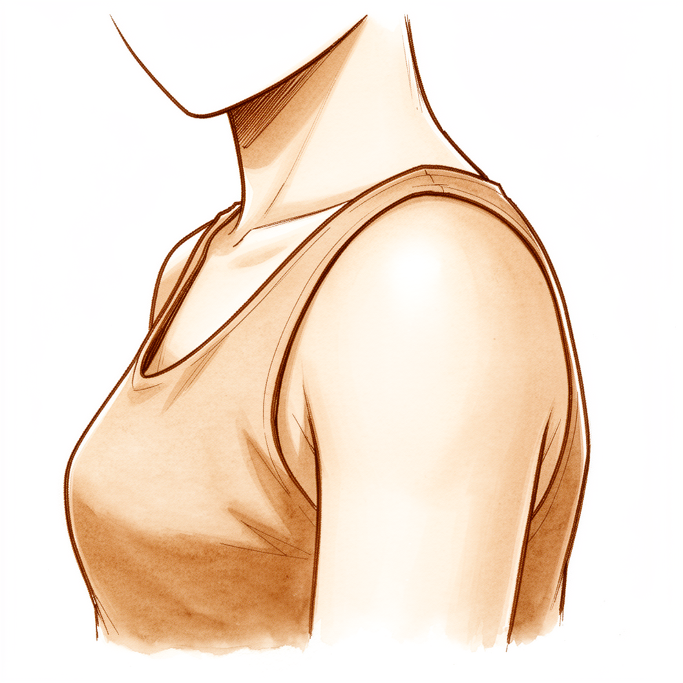
ஒலியெழுப்பும் தோள்பட்டை எலும்பு (snapping scapula) என்பது, உங்கள் தோள்பட்டை எலும்பு (shoulder blade) மார்புச் சுவரின் மேல் நகரும்போது, அதிலிருந்து ஓர் உரசும் ஒலி, 'கிளிக்' என்ற ஒலி அல்லது 'படக்' என்ற ஒலி வருவதை நீங்கள் கேட்பதையோ உணர்வதையோ குறிக்கிறது. இந்த ஒலி, துணி துவைக்கும் அலைப் பலகையின் (washboard) மேல் ஒரு சக்கரம் ஓடுவதைப் போல உணரப்படலாம் என்பதால், மருத்துவர்கள் சில சமயங்களில் இதை "வாஷ்போர்டு நோய்க்குறி" ("washboard syndrome") என்று அழைக்கிறார்கள். இந்த ஒலி தானே பொதுவானது. எந்த அறிகுறியும் இல்லாதவர்களில் சுமார் மூன்றில் ஒருவருக்குத் தோள்பட்டை எலும்பிலிருந்து ஓரளவு உரசல் ஒலி இருக்கிறது. வலி இல்லாமல் அது மட்டும் இருந்தால், அதற்குச் சிகிச்சை தேவையில்லை.
பொதுவாக மக்களை மருத்துவரிடம் கொண்டு வருவது வலிதான். அது தோள்பட்டை எலும்பைச் சுற்றியோ அதன் கீழோ இருக்கும்; பெரும்பாலும் உங்கள் முதுகெலும்புக்கு மிக அருகிலுள்ள அதன் உள் ஓரத்துக்கு அருகில் இருக்கும். தலைக்கு மேலான செயல்பாடுகள், மேலே எட்டுவது, அல்லது உடலின் குறுக்கே கையை நீட்டுவது ஆகியவற்றால் அது மோசமாகும் போக்குடையது; விளையாட்டில் உங்கள் திறன் குறைவதாகவும் அது வெளிப்படலாம். இந்த வலி, அழற்சியடைந்த பர்சாவிலிருந்து (bursa) — தோள்பட்டை எலும்பு விலா எலும்புகளின் மேல் சீராக வழுக்கிச் செல்ல உதவும், திரவம் நிரம்பிய மெல்லிய மெத்தை அது — அல்லது எலும்பின் வடிவத்திலிருந்தே வருகிறது. மார்புச் சுவர் ஓர் ஒலிப் பெட்டியைப் போலச் செயல்படுவதால், நீங்கள் கேட்கும் ஒலி, அதை உண்டாக்கும் அசைவைவிடச் சத்தமாக இருக்கும்.
வலி அன்றாட வேலைகளைக் கடினமாக்கலாம். கனமான வாணலியை உயரமான அலமாரியில் தூக்கி வைப்பது, கொடியில் துணிகளைக் காயப்போடுவது, இருக்கைப் பட்டையை (seatbelt) எட்டிப் பிடிப்பது ஆகிய அனைத்தும் அதைத் தூண்டலாம். சிலர் செயல்பாட்டின்போது அல்லாமல், அதற்குப் பிறகே அதை அதிகமாகக் கவனிக்கிறார்கள். தூக்கத்தைக் கெடுக்கும் அளவுக்கோ, உங்கள் கையை இயல்பாகப் பயன்படுத்த விடாமல் தடுக்கும் அளவுக்கோ அது கடுமையாக இருக்கலாம்.
உங்கள் அறிகுறிகள் தணியாமல் இருந்தால், வாரக்கணக்கில் மோசமாகிக்கொண்டிருந்தால், இரவில் உங்களை எழுப்பினால், அல்லது வேலை செய்யவோ கையைப் பயன்படுத்தவோ விடாமல் தடுத்தால், உங்கள் GP-யைப் பாருங்கள் அல்லது சிறப்பு மருத்துவரின் மதிப்பாய்வைக் கேளுங்கள்.
உண்மையில் என்ன நடக்கிறது
உங்கள் தோள்பட்டை எலும்பு வழுக்கிச் செல்லும்படி வடிவமைக்கப்பட்டுள்ளது. அது உங்கள் மார்புச் சுவரின் மேல் அமர்ந்திருக்கிறது; வழுவழுப்பான, கொழுப்புள்ள ஒரு மெல்லிய திசு அடுக்கு அதை மார்புச் சுவரிலிருந்து பிரிக்கிறது. நகரும் இரண்டு பரப்புகளுக்கு இடையிலான ஒரு கேஸ்கெட் (gasket) போல அந்த அடுக்கு செயல்படுகிறது. உங்கள் தோள்பட்டையை அசைக்க நான்கு மூட்டுகள் சேர்ந்து செயல்படுகின்றன; அசைவில் சுமார் மூன்றில் இரண்டு பங்கு பந்து-கிண்ண மூட்டிலிருந்தே (ball-and-socket joint) வருகிறது; மீதமுள்ள மூன்றில் ஒரு பங்கு, தோள்பட்டை எலும்பு விலா எலும்புகளின் மேல் நழுவிச் செல்வதிலிருந்து வருகிறது. அந்த வழுக்கல் சீராக இருக்கும்போது, அதை நீங்கள் ஒருபோதும் கவனிப்பதில்லை.
ஒலியெழுப்பும் தோள்பட்டை எலும்பில் உள்ள பிரச்சினை என்னவென்றால், ஏதோ ஒன்று இந்த வழுக்கலைக் குலைக்கிறது. சில சமயங்களில் அது எலும்பின் வடிவம்: தோள்பட்டை எலும்பின் உள் ஓரம் வழக்கத்தைவிட அதிகமாக விலா எலும்புகளை நோக்கிச் சாய்ந்தோ வளைந்தோ இருக்கலாம்; அதனால் அது இருக்க வேண்டியதைவிட மார்புச் சுவருக்கு நெருக்கமாக அமர்கிறது. சில சமயங்களில் அது தோள்பட்டை எலும்பின் மேல் உள்ள கூடுதல் எலும்பின் ஒரு சிறிய புடைப்பு. பெரும்பாலும், தோள்பட்டை எலும்பைச் சுற்றியுள்ள தசைகள் சமமற்ற முறையில் செயல்படுவதே காரணம்; அதனால் நீங்கள் அசையும்போது தோள்பட்டை எலும்பு சீரான பாதையில் நகர்வதில்லை. இவற்றில் எதுவும், தோள்பட்டை எலும்பு வழுக்கிச் செல்வதற்குப் பதிலாக உரசவோ சிக்கவோ காரணமாகலாம்.
இந்த உரசல், தோள்பட்டை எலும்புக்கும் விலா எலும்புகளுக்கும் இடையிலான மெத்தையை எரிச்சலடையச் செய்கிறது. பர்சா (bursa) என்று அழைக்கப்படும் அந்த மெத்தை, எலும்புக்கும் மென்திசுவுக்கும் இடையே அதிர்வு தாங்கியைப் (shock absorber) போலச் செயல்படும், திரவம் நிரம்பிய மெல்லிய பை. அது அழற்சியடையும்போது தடித்து, தொட்டால் வலிக்கும் தன்மையுடையதாகிறது; தோள்பட்டை எலும்பின் கீழோ அதைச் சுற்றியோ நீங்கள் உணரும் வலி அதுவே. இந்த உராய்வு, திசுவிலேயே ஓர் எதிர்வினைத் தடிப்பையும் ஏற்படுத்தலாம் — மீண்டும் மீண்டும் நிகழும் உரசலுக்கு உடல் தரும் பதில் அது.
மேலே விவரிக்கப்பட்ட அறிகுறிகளை இது விளக்குகிறது. சொரசொரப்பான பரப்புகள் ஒன்றின் மேல் ஒன்று நகர்வதிலிருந்து ஒலி வருகிறது; மார்புச் சுவர் ஓர் ஒலிப் பெட்டியைப் போல அதைப் பெருக்குகிறது. வலி, ஒலியிலிருந்து அல்ல, அழற்சியடைந்த மெத்தையிலிருந்து வருகிறது. அதனால்தான் இந்த இரண்டு பிரச்சினைகளுக்கும் வெவ்வேறு விதமாகச் சிகிச்சையளிக்கப்படுகிறது; அதனால்தான் சிகிச்சை, அழற்சியைத் தணிப்பதிலும், தோள்பட்டை எலும்பைக் கட்டுப்படுத்தும் தசைகளுக்கு மீண்டும் பயிற்சி அளிப்பதிலும் கவனம் செலுத்துகிறது.
இதற்கு நாங்கள் என்ன செய்ய முடியும்
Mater Private Hospital Rockhampton-இல் மேல் அவயவ (upper-limb) அறுவை சிகிச்சை நிபுணரான டாக்டர் கியரன் ஹிர்பரா, உங்கள் நிலைக்குப் பொருத்தமான, உடலுக்குள் மிகக் குறைவாகத் தலையிடும் (least invasive) தேர்வுகளுடன் தொடங்குகிறார். நோயாளிகள் பொதுவாக அவர்களுடைய GP (பொது மருத்துவர்) மூலம் எங்கள் கிளினிக்கிற்குப் பரிந்துரைக்கப்படுகின்றனர்; ஒரு இயன்முறை சிகிச்சையாளர் (physiotherapist) எங்களைப் பார்க்கும்படி பரிந்துரைத்திருந்தாலும், Medicare தள்ளுபடியைப் (rebate) பெறத் தகுதி பெற உங்கள் GP-யிடமிருந்து பரிந்துரைக் கடிதம் (referral) இன்னும் தேவைப்படும். கிளினிக்கில், ஒலிக்கும் வலிக்கும் என்ன காரணம் என்பதைக் கண்டறிய, உங்கள் மருத்துவ வரலாற்றைக் கேட்டறிந்து, உங்கள் தோள்பட்டையைப் பரிசோதித்து, தேவைப்படும் இடங்களில் ஸ்கேன்களுக்கு ஏற்பாடு செய்கிறோம்.
பெரும்பாலானோருக்கு அறுவை சிகிச்சை இல்லாமலேயே இது தணிகிறது; எனவே சிகிச்சை அறுவை சிகிச்சை இல்லாமல் தொடங்குகிறது. முதல் படி, உங்கள் தோள்பட்டை எலும்பைக் கட்டுப்படுத்தும் தசைகளை வலுப்படுத்தும் ஓர் இயன்முறை சிகிச்சை (physiotherapy) திட்டம்; அதனுடன், இறுக்கமாகிவிட்ட தசைகளை நீட்டும் பயிற்சிகளும் இருக்கும். அறிகுறிகளைத் தூண்டும் செயல்பாடுகளை மாற்றுவதும் உதவுகிறது. வேறு எதையும் பற்றி யோசிப்பதற்கு முன், இதற்கு 3 முதல் 6 மாதங்கள் முறையான வாய்ப்பு கொடுங்கள்.
வலி நீடித்தால், அழற்சியைத் தணிக்க மருந்து உதவலாம். பலர் தசை வலிகளுக்கு எடுத்துக்கொள்ளும் அதே வகையான அழற்சி எதிர்ப்பு மாத்திரைகள் (anti-inflammatory tablets) இந்தக் கட்டத்தின் ஒரு பகுதி. உங்கள் தோள்பட்டை எலும்பின் கீழ் உள்ள பர்சாவுக்குள், ஸ்கேன் வழிகாட்டுதலுடன் (image-guided) ஸ்டீராய்டும் உள்ளூர் மயக்க மருந்தும் (local anaesthetic) கலந்த ஊசியையும் நாங்கள் வழங்க முடியும். இது இரண்டு வேலைகளைச் செய்கிறது: அழற்சியடைந்த மெத்தையைத் தணிக்கிறது; உங்கள் வலி வருவது இங்கிருந்துதான் என்பதையும் உறுதிப்படுத்துகிறது.
இந்தச் சிகிச்சைகளைப் பல மாதங்கள் மேற்கொண்ட பிறகும் வலி தொடர்ந்து, அவை உங்களுக்குப் போதுமான முன்னேற்றத்தைத் தராதபோது மட்டுமே அறுவை சிகிச்சை பரிசீலிக்கப்படுகிறது. இந்த அறுவை சிகிச்சை, சிறிய வெட்டுகள் வழியாக ஒரு கேமராவுடன் செய்யப்படும் சாவித்துளை (keyhole) அறுவை சிகிச்சை. இது உங்கள் தோள்பட்டை எலும்புக்கும் விலா எலும்புகளுக்கும் இடையில் உள்ள அழற்சியடைந்த பர்சாவை அகற்றுகிறது. அறுவை சிகிச்சையின்போது உங்கள் தோள்பட்டை எலும்பின் மேல் உள் மூலை (superomedial angle) துருத்திக்கொண்டிருப்பது கண்டறியப்பட்டால் மட்டுமே அது சீவப்படுகிறது. அறுவை சிகிச்சைக்குப் பிறகு பெரும்பாலானோரின் வலி கணிசமாக மேம்படுகிறது; ஆனால் சிறிதளவு 'கிளிக்' ஒலி அடிக்கடி அப்படியே இருக்கும். அறுவை சிகிச்சை உங்களுக்கு என்ன அர்த்தம் தரக்கூடும் என்பதைப் பற்றிப் பேசி, அது சரியான அடுத்த படியா என்பதை நாம் சேர்ந்து முடிவு செய்வோம்.
என்ன எதிர்பார்க்கலாம்
பெரும்பாலானோருக்கு அறுவை சிகிச்சை இல்லாமலேயே இது தணிகிறது. உங்கள் தோள்பட்டை எலும்பைக் கட்டுப்படுத்தும் தசைகளை வலுப்படுத்தும் இயன்முறை சிகிச்சை, அழற்சி எதிர்ப்பு மருந்து, அறிகுறிகளைத் தூண்டும் செயல்பாடுகளை மாற்றுதல் ஆகியவற்றுடன் சிகிச்சை தொடங்குகிறது. இதற்கு 3 முதல் 6 மாதங்கள் முறையான வாய்ப்பு கொடுங்கள். இந்தக் காலத்தில் தங்கள் வலி தணிவதைப் பலர் காண்கிறார்கள்; அவர்களுக்கு ஒருபோதும் அறுவை சிகிச்சை தேவைப்படுவதில்லை.
இந்தச் சிகிச்சைகளைப் பல மாதங்கள் மேற்கொண்ட பிறகும் வலி தொடர்ந்தால், அறுவை சிகிச்சை பரிசீலிக்கப்படலாம். சாவித்துளை அறுவை சிகிச்சை, உங்கள் தோள்பட்டை எலும்புக்கும் விலா எலும்புகளுக்கும் இடையில் உள்ள அழற்சியடைந்த பர்சாவை அகற்றுகிறது; தோள்பட்டை எலும்பின் மேல் உள் மூலை துருத்திக்கொண்டிருப்பது கண்டறியப்பட்டால் மட்டுமே அதைச் சீவுகிறது. அறுவை சிகிச்சைக்குப் பிறகு பெரும்பாலானோரின் வலி கணிசமாக மேம்படுகிறது. சிறிதளவு 'கிளிக்' ஒலி அடிக்கடி அப்படியே இருக்கும்; ஏனெனில் வலிக்கு உதவும் சிகிச்சைகள் பெரும்பாலும் வலியைத்தான் சரிசெய்கின்றன, ஒலியை அல்ல. ஒவ்வோர் அறிகுறியும் முழுமையாக நீங்குவது வழக்கமான விளைவு அல்ல.
சிகிச்சை இல்லாவிட்டால், அறிகுறிகள் நீடிக்கலாம்; தூக்கத்தைக் கெடுக்கும் அளவுக்கோ, உங்கள் கையை இயல்பாகப் பயன்படுத்த விடாமல் தடுக்கும் அளவுக்கோ கடுமையாகவும் இருக்கலாம். இந்த நிலையை அடையாளம் காண்பதும் அதற்குச் சிகிச்சையளிப்பதும் கடினமாக இருக்கலாம்; எனவே அது தானாகத் தணியும் வரை காத்திருப்பதைவிட, தொடக்கத்திலேயே தெளிவான நோயறிதலைப் பெறுவது பயனுள்ளது.
உங்கள் அறிகுறிகள் தணியாமல் இருந்தால், வாரக்கணக்கில் மோசமாகிக்கொண்டிருந்தால், இரவில் உங்களை எழுப்பினால், அல்லது வேலை செய்யவோ கையைப் பயன்படுத்தவோ விடாமல் தடுத்தால், உங்கள் GP-யைப் பாருங்கள் அல்லது சிறப்பு மருத்துவரின் மதிப்பாய்வைக் கேளுங்கள்.
எப்போது மருத்துவரைப் பார்க்க வேண்டும்
ஒலி மட்டுமே கவலைப்படுவதற்கான காரணம் அல்ல. எந்த அறிகுறியும் இல்லாதவர்களில் சுமார் மூன்றில் ஒருவருக்குத் தோள்பட்டை எலும்பிலிருந்து ஓரளவு உரசல் ஒலி இருக்கிறது; அது மட்டும் இருந்தால் அதற்குச் சிகிச்சை தேவையில்லை. முக்கியமானது வலிதான். தோள்பட்டை எலும்பைச் சுற்றியோ அதன் கீழோ உள்ள வலி தணியாமல் இருந்தால், வாரக்கணக்கில் மோசமாகிக்கொண்டிருந்தால், இரவில் உங்களை எழுப்பினால், அல்லது வேலை செய்யவோ கையைப் பயன்படுத்தவோ விடாமல் தடுத்தால், உங்கள் GP-யைப் பாருங்கள் அல்லது சிறப்பு மருத்துவரின் மதிப்பாய்வைக் கேளுங்கள். வலி தூக்கத்தைக் கெடுக்கும் அளவுக்குக் கடுமையாக இருந்தாலோ, விளையாட்டில் உங்கள் திறன் குறைந்திருந்தாலோ, அல்லது மேலே எட்டுவது அல்லது உடலின் குறுக்கே கையை நீட்டுவது போன்ற தலைக்கு மேலான செயல்பாடுகள் வலியைத் தூண்டினாலோ, மதிப்பாய்வைக் கேளுங்கள். ஒலியெழுப்பும் தோள்பட்டை எலும்பை அடையாளம் காண்பது கடினமாக இருக்கலாம்; எனவே தொடக்கத்திலேயே தெளிவான நோயறிதலைப் பெறுவது பயனுள்ளது.
ஆழமான விளக்கம்
Advanced reading: the deeper science (optional)
உங்கள் சொந்த சிகிச்சை முடிவுகளுக்குத் தேவையானதைவிட இந்தப் பகுதி மேலும் ஆழமாகச் செல்கிறது. ஒலியெழுப்பும் தோள்பட்டை எலும்பு இந்தக் கூடுதல் வாசிப்புக்குத் தகுதியானது; ஏனெனில் ஒலியும் வலியும் இரண்டு வெவ்வேறு பிரச்சினைகள்; உதவும் சிகிச்சைகள் பெரும்பாலும் இரண்டாவதைத்தான் சரிசெய்கின்றன, முதலாவதை அல்ல.
ஒலி பொதுவானது; வலிதான் பிரச்சினை
தோள்பட்டை எலும்பிலிருந்து வரும் உரசல் ஒலி, அது மட்டுமே இருந்தால், நோயின் அறிகுறி அல்ல. இந்த நிலை பற்றிய ஒரு முக்கியமான மதிப்பாய்வு, எந்த அறிகுறியும் இல்லாத 100 பேரில் 31% பேருக்குத் தோள்பட்டை எலும்பு உரசல் ஒலி (scapular crepitus) இருப்பதைக் கண்டறிந்த ஒரு தொடக்கக் கால ஆய்வை மேற்கோள் காட்டுகிறது [1]. ஒலியை ஒரு நோய்நிலையாக மாற்றுவது வலி; அது பொதுவாக அழற்சியடைந்த பர்சாவிலிருந்து வருகிறது: தோள்பட்டை எலும்பு விலா எலும்புகளின் மேல் வழுக்கிச் செல்ல உதவும், உள்ளே திரவம் படிந்த மெல்லிய திசு அடுக்கு அது.
வடிவமும் ஒரு பங்கு வகிக்கிறது. ஒரு ஸ்கேன் ஆய்வில், உள் ஓரத்தில் முன்னோக்கி வளைந்திருக்கும் தோள்பட்டை எலும்பு உள்ளவர்களுக்கு (நேர்மறை "medial scapula corpus angle" கொண்ட, உட்குழிவான வடிவம்) ஒலியெழுப்பும் தோள்பட்டை எலும்புக்கான அபாயம் 12 மடங்கு அதிகமாக இருந்தது [2]. அந்த அளவீடு முதலில் தோன்றியதைவிடக் குறைவான பயனுடையதாக மாறியது: அதே கோணம், அது பார்க்கப்படும் திசையைப் பொறுத்துக் கணிசமாக மாறுபடுகிறது என்று பின்னர் வந்த ஓர் ஆய்வு கண்டறிந்தது; எனவே அதை மட்டும் வைத்து இந்த நிலையை நோயறிவதற்கு அது போதுமான நம்பகத்தன்மை உடையதல்ல [3]. இறந்த உடல் (cadaver) மாதிரி ஒன்றில், மிக முக்கியமான எலும்பு அம்சம், தோள்பட்டை எலும்பின் உள் பகுதி விலா எலும்புகளை நோக்கி எவ்வளவு முன்னோக்கி அமர்ந்திருந்தது என்பதே [4].
பெரும்பாலானோருக்கு அறுவை சிகிச்சை இல்லாமலேயே தணிகிறது
ஒரு குறிப்பிட்ட தசை வலுப்படுத்தும் திட்டத்தின் மூலம் சிகிச்சையளிக்கப்பட்ட 28 நோயாளிகளின் 30 தோள்பட்டை எலும்புகள் கொண்ட ஒரு தொடர் ஆய்வில், 14 (50%) பேர் பயிற்சிகளால் மட்டுமே நன்றாக இருந்தனர்; 6 (21%) பேருக்குக் கூடுதலாக அழற்சி எதிர்ப்பு மருந்து மட்டுமே தேவைப்பட்டது; ஆக, ஆய்வின் சொந்த மதிப்பீட்டு முறைப்படி 71% பேர் அறுவை சிகிச்சை இல்லாமல் நல்ல அல்லது அதைவிடச் சிறந்த விளைவை அடைந்தனர் [5].
மிக நீண்ட காலப் பின்தொடர்தல் பின்லாந்திலிருந்து வருகிறது. திறந்த அறுவை சிகிச்சை செய்துகொண்ட பன்னிரண்டு பேரும், அறுவை சிகிச்சை இல்லாமல் சிகிச்சையளிக்கப்பட்ட 9 பேரும், சராசரியாக 22 ஆண்டுகள் கழித்து மீண்டும் மதிப்பிடப்பட்டனர். உடல் உழைப்பின்போது ஏற்படும் வலி இரு குழுக்களிலும் குறைவாக இருந்தது: அறுவை சிகிச்சைக்குப் பிறகு 10-க்கு 0.8, அது இல்லாமல் 10-க்கு 1.5; இந்த வேறுபாடு உண்மையானது என்று உறுதியாகச் சொல்ல முடியாத அளவுக்குச் சிறியது (p = 0.357) [6]. 12 பேரில் 7 பேருக்கு அறுவை சிகிச்சைக்குப் பிறகு வலி உடனடியாக மேம்பட்டது; ஆனால் நீண்ட காலத்தில் இரண்டு வழிகளும் ஏறக்குறைய ஒரே இடத்தில் முடிந்தன.
பர்சாவுக்குள் செலுத்தப்படும், உள்ளூர் மயக்க மருந்தும் ஸ்டீராய்டும் கலந்த ஊசி இரண்டு வேலைகளைச் செய்கிறது: அழற்சிக்குச் சிகிச்சையளிக்கிறது; வலிக்கான மூலம் உண்மையிலேயே பர்சாதானா என்பதையும் சோதிக்கிறது. ஐக்கிய இராச்சியத்தின் (UK) ஒரு சிகிச்சை வழிமுறையில், 94 நோயாளிகளில் 74 பேர் இயன்முறை சிகிச்சைக்குப் பிறகும் தொந்தரவுக்கு உள்ளாகியிருந்தனர்; அவர்களுக்கு அல்ட்ராசவுண்ட் வழிகாட்டுதலுடன் (ultrasound-guided) ஊசி போடப்பட்டது; 55 பேருக்குத் தொடக்கத்தில் நல்ல பலன் கிடைத்தது; அந்தப் பலன் 17 பேரிடம் நீடித்தது [7]. பின்தொடரப்பட்ட, அறுவை சிகிச்சை செய்யப்பட்ட 42 நோயாளிகளில், ஊசிக்கு நன்றாகப் பதிலளித்தவர்கள், பதிலளிக்காதவர்களைவிட அறுவை சிகிச்சைக்குப் பிறகு அதிகமாக மேம்பட்டனர் [7]. குறைந்தது ஐந்து ஆண்டுகள் பின்தொடரப்பட்ட, பெரிய அமெரிக்கத் தொடர் ஆய்வு ஒன்று இதே தொடர்பைக் கண்டறியவில்லை [8]; எனவே ஊசிக்குப் பலன் குறைவாக இருந்தது என்பதற்காக அறுவை சிகிச்சை சாத்தியமில்லை என்று பொருள் அல்ல.
அறுவை சிகிச்சை எதைச் சாதிக்கிறது, எதைச் சாதிப்பதில்லை
மிகப் பெரிய நீண்ட காலத் தொடர் ஆய்வு, சாவித்துளை முறையில் பர்சாவை அகற்றி, தோள்பட்டை எலும்பின் மூலையைச் சீவிய பிறகு 66 பேரை 5 முதல் 15 ஆண்டுகள் பின்தொடர்ந்தது. சராசரி தோள்பட்டை மதிப்பெண் 100-க்கு 56.7-இலிருந்து 87.2 ஆக உயர்ந்தது; வலி 10-க்கு 5.2-இலிருந்து 1.4 ஆகக் குறைந்தது; 77.6% பேர் கவனிக்கத்தக்க அளவுக்குப் பெரிய முன்னேற்றத்தை அடைந்தனர். விளையாட்டில் ஈடுபட்டிருந்த 42 பேரில், 38 (90.5%) பேர் மீண்டும் அதற்குத் திரும்பினர்; 31 (73.8%) பேர் தங்கள் முந்தைய நிலைக்கோ அதற்கு நெருக்கமான நிலைக்கோ திரும்பினர். எட்டு (12.1%) பேருக்கு மேலும் ஓர் அறுவை சிகிச்சை தேவைப்பட்டது [8]. 74 தோள்பட்டை எலும்புகள் கொண்ட மற்றொரு தொடர் ஆய்வில், மேலும் அறுவை சிகிச்சைக்கான விகிதம் ஏறக்குறைய இதே அளவில் இருந்தது (74-இல் 8); வயதான நோயாளிகள், நீண்ட காலமாக அறிகுறிகள் இருந்தவர்கள், அறுவை சிகிச்சைக்கு முன் மன நலத்துக்கான மதிப்பெண்கள் குறைவாக இருந்தவர்கள் ஆகியோர் குறைவாகவே மேம்படும் போக்கு இருந்ததையும் அது கண்டறிந்தது [9].
முழுமையாகக் குணமாவது விதிவிலக்குதான். அறுவை சிகிச்சை செய்யப்பட்ட 203 தோள்பட்டைகள் பற்றிய ஒரு முறையான மதிப்பாய்வு (systematic review), 42 (20.7%) முழுமையாகக் குணமடைந்ததையும், 139 (68.5%) மேம்பட்டிருந்தாலும் இன்னும் சில அறிகுறிகளுடன் இருந்ததையும், 22 (10.8%) மோசமான விளைவைக் கொண்டிருந்ததையும் கண்டறிந்தது [10]. 464 நோயாளிகள் பற்றிய 2026-ஆம் ஆண்டு மதிப்பாய்வு ஒன்றும் அதே ஒட்டுமொத்த முடிவுக்கு வந்தது: அறுவை சிகிச்சை அல்லாத சிகிச்சையும் அறுவை சிகிச்சையும் வலியைக் குறைக்கின்றன; ஆனால் இயந்திரவியல் அறிகுறிகள் (mechanical symptoms) — 'கிளிக்' ஒலியும் உரசல் ஒலியும் — பெரும்பாலும் முழுமையாக நீங்குவதில்லை [11].
எலும்பை அகற்றுவது பயனுள்ளதா?
தோள்பட்டை எலும்பின் மேல் உள் மூலையைச் சீவுவது இந்த அறுவை சிகிச்சையின் பாரம்பரியப் பகுதி; எனவே அது சிறிதளவே கூடுதல் பலனைத் தரக்கூடும் என்பது ஆச்சரியமானது. ஒரு மையம், பர்சாவை மட்டும் அகற்றிக்கொண்ட 24 பேரை, மூலையையும் சீவிக்கொண்ட 32 பேருடன் ஒப்பிட்டது. இறுதி வலி மதிப்பெண்களும் (10-க்கு 3.4 மற்றும் 2.8, p = 0.351) தோள்பட்டை மதிப்பெண்களும் (75.8 மற்றும் 76.5, p = 0.895) ஒரே அளவில் இருந்தன; 'கிளிக்' ஒலி 58% மற்றும் 59% பேரிடம் இன்னும் இருந்தது [12]. எலும்பு வேலை இல்லாதபோது அறுவை சிகிச்சை குறைவான நேரம் எடுத்தது: 37.5 நிமிடங்கள், எதிராக 61.2 நிமிடங்கள் [12]. இந்தக் குழுக்கள் வெவ்வேறு கால அளவுகளுக்குப் பின்தொடரப்பட்டன; சீரற்ற முறையில் (randomised) பிரிக்கப்படவும் இல்லை; எனவே இதுவே இறுதியான முடிவு அல்ல; ஆனால் எலும்பைச் சீவுவதற்கான முடிவு முன்கூட்டியே அல்லாமல் அறுவை சிகிச்சையின்போது எடுக்கப்படுவதற்கு இதுவே காரணம்.
மேற்கோள்கள்
[1] Kuhn JE, Plancher KD, Hawkins RJ. Symptomatic Scapulothoracic Crepitus and Bursitis. J Am Acad Orthop Surg. 1998;6(5):267-73. https://doi.org/10.5435/00124635-199809000-00001 [2] Spiegl UJ, Petri M, Smith SW, Ho CP, Millett PJ. Association between scapula bony morphology and snapping scapula syndrome. J Shoulder Elbow Surg. 2015;24(8):1289-95. https://doi.org/10.1016/j.jse.2014.12.034 [3] Percin B, Featherall J, Tashjian RZ, Chalmers PN, Joyce CD, Mortensen AJ, et al. The influence of scapular orientation on the medial scapula corpus angle in snapping scapula syndrome. JSES Int. 2025;9(1):70-8. https://doi.org/10.1016/j.jseint.2024.08.179 [4] Tahal DS, Katthagen JC, Marchetti DC, Mikula JD, Montgomery SR, Brady A, et al. A Cadaveric Model Evaluating the Influence of Bony Anatomy and the Effectiveness of Partial Scapulectomy on Decompression of the Scapulothoracic Space in Snapping Scapula Syndrome. Am J Sports Med. 2017;45(6):1276-82. https://doi.org/10.1177/0363546516687755 [5] Groh G, Simoni M, Allen T, Dwyer T, Heckman M, Rockwood C. Treatment of snapping scapula with a periscapular muscle strengthening program. J Shoulder Elbow Surg. 1996;5(2):S6. https://doi.org/10.1016/s1058-2746(96)80089-9 [6] Vastamäki M, Vastamäki H. Open Surgical Treatment for Snapping Scapula Provides Durable Pain Relief, but so Does Nonsurgical Treatment. Clin Orthop Relat Res. 2016;474(3):799-805. https://doi.org/10.1007/s11999-015-4614-1 [7] Tytherleigh-Strong G, Gill J, Griffiths E, Al-Hadithy N. Prognosis After Arthroscopic Superior Medial Scapuloplasty for Snapping Scapula Syndrome Improves After a Transient Beneficial Response With an Ultrasound-Guided Subscapular Cortisone Injection. Arthroscopy. 2020;36(12):2965-72. https://doi.org/10.1016/j.arthro.2020.07.024 [8] Rupp M, Geissbuhler AR, Rutledge JC, Amendola R, Hanson JA, Doan KC, et al. Minimum 5-Year Clinical and Return-to-Sport Outcomes After Primary Arthroscopic Scapulothoracic Bursectomy and Partial Scapulectomy for Snapping Scapula Syndrome. Am J Sports Med. 2024;52(6):1449-56. https://doi.org/10.1177/03635465241243072 [9] Menge TJ, Horan MP, Tahal DS, Mitchell JJ, Katthagen JC, Millett PJ. Arthroscopic Treatment of Snapping Scapula Syndrome: Outcomes at Minimum of 2 Years. Arthroscopy. 2016;33(4):726-32. https://doi.org/10.1016/j.arthro.2016.08.029 [10] Memon M, Kay J, Simunovic N, Ayeni OR. Arthroscopic management of snapping scapula syndrome improves pain and functional outcomes, although a high rate of residual symptoms has been reported. Knee Surg Sports Traumatol Arthrosc. 2017;26(1):221-39. https://doi.org/10.1007/s00167-017-4693-5 [11] Wallace K, Mitchell JK, Lauck BJ, Reed J, Conlon S, Kamath GV. Snapping Scapula Syndrome: A Systematic Review of Treatment Options and Outcomes. J Shoulder Elbow Surg. 2026. https://doi.org/10.1016/j.jse.2026.05.023 [12] Featherall J, Christensen GV, Mortensen AJ, Wheelwright JC, Chalmers PN, Tashjian RZ. Arthroscopic scapulothoracic bursectomy with and without superomedial angle scapuloplasty: a comparison of patient-reported outcomes. J Shoulder Elbow Surg. 2023;32(9):1945-52. https://doi.org/10.1016/j.jse.2023.03.020
Evidence & references
This is the clinical evidence summary written for health professionals. It is technical, and it lists the research this page was built from. You do not need to read it to understand your treatment or to make a decision about it.
Overview
- Snapping scapula syndrome is caused by either osseous lesions or scapulothoracic bursitis [3].
- The condition is also referred to as “washboard syndrome” and is attributed to bony and soft tissue abnormalities [9].
- Snapping scapula represents the clinical expression of scapulothoracic impingement due to mechanical factors and/or muscle imbalance [7].
- Osteochondroma of the scapula is a rare entity that may present with snapping scapula syndrome when it alters scapulothoracic mechanics [6].
- Snapping scapula syndrome can be a debilitating disorder [2].
- Diagnosis and management of snapping scapula syndrome remains challenging despite recent advances [1].
- The condition can be difficult to recognize and treat [3].
- Four-dimensional computed tomography scans facilitate preoperative planning by defining pathology and improving assessment of the amount and location of scapular bone and soft tissue causing symptoms [18].
Treatment
- Treatment of patients with snapping scapula syndrome begins with nonoperative methods [5].
- When nonoperative treatment fails, several surgical options exist [5].
- Arthroscopic management of snapping scapula syndrome yields improvement in pain, crepitus, and range of motion in a majority of patients [4].
- Most patients experience residual symptoms following arthroscopic management [4].
- Arthroscopic scapulothoracic treatment provides significant pain relief and functional improvement for painful snapping scapula symptoms not responding to non-surgical treatment modalities [8].
- Arthroscopic treatment for snapping scapula syndrome resulted in significant improvement in pain and function with high patient satisfaction at a mean follow-up of 3.4 years [14].
- Arthroscopic scapuloplasty leads to significant improvement in pain and function in patients with snapping scapula syndrome refractory to conservative treatment [15].
- Arthroscopic surgery is an effective treatment for snapping scapula syndrome in both primary and revision cases, showing significant improvements in all postoperative outcome scores at a mean of 3.4 years [17].
- Arthroscopic resection of the superomedial corner of the scapula is a useful technique in patients whose pain and clicking can be clinically localized and who have failed to respond to conservative measures [19].
- Arthroscopic resection of the medial superior hook formation in combination with partial bursectomy provided good results with significant gains with respect to the WORC score in 90 % of the patients troubled by painful snapping scapula for a long time and without relief by exercise-based rehabilitation [35].
- At the end of 1 year, all patients in a case series had a constant score above 90 and were completely relieved of snapping around the scapula having full pain-free movements at the shoulder and scapulothoracic joints [11].
- A patient remained asymptomatic with full range of motion and no further complaints of pain or snapping at twenty-six months postoperatively [12].
- At 2 years 6 months after the operation, snapping and pain were completely gone and the patient was satisfied with the results [13].
Anatomy & Pathophysiology
Etiology and Mechanism
- Snapping scapula is the clinical expression of scapulothoracic impingement due to mechanical factors and/or muscle imbalance [7].
- The condition is attributed to bony and soft tissue abnormalities [9].
- Osteochondroma of the scapula may present with snapping scapula syndrome when it alters scapulothoracic mechanics [6].
- Subscapular elastofibroma is a reactive pseudotumor resulting from mechanical friction of the scapula on the chest wall [86].
Bony Anatomy and Morphology
- The scapula is separated from the chest wall by thin gliding fibro-fatty tissue, allowing its smooth excursion over the chest wall [53].
- Anterior angulation of the medial scapula in the axial plane was associated with snapping scapula syndrome [34].
- Patients with a concave-shaped scapula and a positive medial scapula corpus angle may be at risk for snapping scapula syndrome [47].
- The anterior offset of the scapula appeared to be the most important bony parameter to consider during preoperative planning and the evaluation of scapulothoracic space decompression with partial scapulectomy [46].
- The weakest area of the circumference of the biomechanical body of the scapula is the connection of the scapular spine and the medial border of the scapula, known as the spinomedial angle [53].
- The superior angle and the adjacent part of the supraspinous fossa form an appendage that serves as a surface of insertion or origin of muscles but does not transmit compressive forces from the glenoid [53].
Soft Tissue and Neurovascular Anatomy
- Neurologic structures around the scapula vary significantly between the neutral arm position and the chicken-wing position commonly used in the arthroscopic treatment of snapping scapula syndrome [85].
- The scapula is attached to the axial skeleton by the clavicle, specifically by the acromioclavicular and sternoclavicular joints [53].
- The scapula serves for attachment of a number of muscles originating from the axial skeleton that control its motion [53].
Classification
- Mauclaire classified scapulothoracic crepitus into three types: froissement (a gentle friction sound thought to be physiologic), frottement (a louder sound with grating, which is usually pathologic), and craquement (a loud snapping sound, which is always pathologic) [26].
- Frottement may suggest a soft-tissue lesion or bursitis, while craquement may suggest an osseous lesion as the source of symptomatic scapulothoracic crepitus [26].
- The snapping scapula syndrome is caused by either osseous lesions or scapulothoracic bursitis [3].
- Snapping scapula is the clinical expression of the scapulothoracic impingement due to mechanical factors and/or muscle imbalance [7].
- The snapping scapula, also called “washboard syndrome,” is a controversial condition attributed to bony and soft tissue abnormalities [9].
- Rib fractures at the posterior angle could cause snapping scapula syndrome [20].
Clinical Presentation
- Snapping scapula syndrome is an uncommon condition that is likely underdiagnosed [42].
- The condition can produce severe shoulder pain and dysfunction [24].
- Patients often present with decreased athletic performance and increased pain with overhead activities [42].
- Crepitus is a reported symptom in patients with snapping scapula syndrome [42].
- High clinical suspicion is necessary in patients presenting with medial scapular border tenderness, palpable crepitus, and audible snapping [40].
- Potential factors causing snapping scapula syndrome include bony prominence at the superomedial scapular angle, imbalance of the periscapular muscles, and scapulothoracic bursitis [42].
- Scapulothoracic crepitus is not necessarily a pathologic condition, as it was found in 31% of 100 normal asymptomatic persons [26].
- Scapulothoracic crepitus is considered pathologic if it is associated with pain, winging, or other disorders of the scapulothoracic articulation [26].
- The condition has been referred to by various names, including snapping scapula, washboard syndrome, scapulothoracic syndrome, rolling scapula, grating scapula, and scapulocostal syndrome [26].
- Mauclaire classified scapulothoracic crepitus into three types: froissement (gentle friction sound), frottement (louder sound with grating), and craquement (loud snapping sound) [26].
- Scapular noises arise from anatomic changes in the tissue interposed between the scapula and the chest wall and/or an incongruent scapulothoracic articulation [26].
- The air-filled thoracic cavity acts as a resonating chamber that amplifies scapulothoracic crepitus [26].
- A mass composed of alternating strands of fatty and fibrous tissue located at the inferior pole of the scapula is pathognomonic for elastofibroma dorsi [36].
- Scapular osteochondroma is a rarely reported cause of scapular pseudo-winging and should be considered in the differential diagnosis when winging and a mass are present [43].
- The differential diagnosis for causes of snapping scapula includes idiopathic causes, scapulothoracic bursitis, osteochondromas, and elastofibromas [42].
- Four-dimensional computed tomography scans facilitate preoperative planning by defining pathology and assessing the amount and location of scapular bone and soft tissue causing symptoms [18].
- Plain radiographs and CT scans provide detailed information about osseous abnormalities in snapping scapula syndrome [42].
- MRI characterizes bursal and other soft-tissue pathologies in snapping scapula syndrome [42].
Investigations
- Symptomatic scapulothoracic crepitus is considered pathologic when associated with pain, winging, or other disorders of the scapulothoracic articulation [26].
- Scapulothoracic crepitus is attributed to anatomic changes in the tissue interposed between the scapula and the chest wall or an incongruent scapulothoracic articulation [26].
- The air-filled thoracic cavity acts as a resonating chamber that amplifies scapulothoracic crepitus noises [26].
- Frottement (a louder sound with grating) usually suggests a soft-tissue lesion or bursitis, while craquement (a loud snapping sound) always suggests an osseous lesion [26].
- Four-dimensional computed tomography (4D CT) scans define pathology well and improve the assessment of the amount and location of scapular bone and soft tissue causing symptoms in snapping scapula syndrome [18].
- Fluoroscopy provides a real-time method for evaluating and documenting abnormal osseous mechanics of the scapulothoracic bursal region [31].
- Anterior angulation of the medial scapula in the axial plane is associated with snapping scapula syndrome [34].
- The anterior offset of the scapula is the most important bony parameter to consider during preoperative planning and evaluation of scapulothoracic space decompression with partial scapulectomy [46].
- Imaging-guided injections provide an alternative means of diagnosis in cases where direct imaging is usually not successful [48].
- Rib fractures at the posterior angle can cause snapping scapula syndrome [20].
Treatment
Non-Operative Management
- In most cases, nonoperative treatment is curative and includes physical therapy for scapular muscle strengthening and nonsteroidal anti-inflammatory medications [29].
- Nonsurgical management remains the first treatment option, with surgery recommended after 6 months of nonsurgical treatment with no improvement [42].
- Nonoperative management of snapping scapula syndrome in the form of analgesia, physiotherapy, local corticosteroid injection, and/or extracorporeal shockwave therapy should be initiated for 3 to 6 months before considering surgical management [40].
- Strengthening weak periscapular muscle groups combined with simultaneous stretching of contracted muscles and training of antagonistic muscle groups can yield good clinical results [42].
- Physical therapy, injections with local anesthetics and/or steroids, and NSAIDs can be useful in nonsurgical management [42].
- Patients treated nonsurgically achieved results that were not distinguishable from the surgically treated group in a small series with a minimum of 16 years follow-up [24].
- At a minimum of 16 years follow-up, patients treated nonsurgically reported little pain, some crepitus (usually painless), and a consistent ability to return to work [24].
Operative Management
- When nonoperative treatment fails, several surgical options exist for snapping scapula syndrome [5].
- Most patients experience residual symptoms after arthroscopic management of snapping scapula syndrome [4].
- For patients who fail conservative management, arthroscopic bursectomy and partial scapulectomy is a reasonable option [23].
- Arthroscopic bursectomy and partial scapulectomy is technically demanding and requires an in-depth understanding of the complex anatomy of the scapulothoracic region [23].
- Improvement in functional outcomes and pain relief are reliably achieved after surgical treatment of snapping scapula syndrome [28].
- Poorer outcomes can be seen in workers' compensation patients as well as those with residual symptomatic shoulder pathology following surgical treatment [28].
- Operative time is shorter without scapuloplasty when performing arthroscopic scapulothoracic bursectomy [30].
- Open surgical treatment for snapping scapula provides durable pain relief [24].
- Patients treated surgically for snapping scapula report little pain, some crepitus (usually painless), and a consistent ability to return to work at a minimum of 16 years follow-up [24].
- At the end of 1 year, all patients in a case series of arthroscopic management for recalcitrant scapulothoracic bursitis had a constant score above 90 and were completely relieved of snapping around the scapula [11].
- Patients in a case series of arthroscopic management for recalcitrant scapulothoracic bursitis had full pain-free movements at the shoulder and scapulothoracic joints at 1 year [11].
- A patient with snapping scapula syndrome remained asymptomatic with full range of motion and no further complaints of pain or snapping at twenty-six months postoperatively [12].
- Snapping and pain were completely gone and the patient was satisfied with the results at 2 years 6 months after operation for abnormal angulation of the superior angle of the scapula [13].
- Arthroscopic access to the scapulothoracic space is simple and reproducible, with bursal tissue clearance optimizing visualization [25].
- Arthroscopic treatment of snapping scapula follows 8 key steps to ensure a safe and effective procedure [10].
- A technique for arthroscopic management of snapping scapula syndrome includes scapulothoracic bursectomy and superomedial partial scapulectomy, which has been refined over nearly two decades of experience [16].
- Most authors have recommended partial scapulectomy or resection of the superomedial angle of the scapula in patients with scapular crepitus and pain caused by bony incongruity [81].
- The decision for and timing of operative management should be individualized for each patient [81].
- Operative options include partial scapulectomy and open versus arthroscopic bursectomy [81].
Complications
- Arthroscopic management of snapping scapula syndrome results in most patients experiencing residual symptoms [4].
- Snapping scapula syndrome is difficult to recognize and treat [3].
Recovery
Non-Operative
- Nonoperative treatment for snapping scapula syndrome includes physical therapy for scapular muscle strengthening and nonsteroidal anti-inflammatory medications [29].
- In most cases, nonoperative treatment is curative [29].
Operative Outcomes
- Improvement in functional outcomes and pain relief are reliably achieved after surgical treatment [28].
- Poorer outcomes can be seen in workers' compensation patients as well as those with residual symptomatic shoulder pathology [28].
- At the end of 1 year, all the patients had a constant score above 90 and were completely relieved of snapping around the scapula having full pain-free movements at the shoulder and scapulothoracic joints [11].
- The patient remained asymptomatic with full range of motion and no further complaints of pain or snapping at twenty-six months postoperatively [12].
- A year after excision, the patient has not suffered recurrent pain in the left shoulder [90].
Surgical Technique and Planning
- Operative time is shorter without scapuloplasty [30].
- The 4D CT scan images defined pathology well in patients with snapping scapula syndrome and improved assessment of the amount and location of the scapular bone and soft tissue causing symptoms [18].
Key Evidence
- [L5] Diagnosis and management of snapping scapula syndrome remains challenging despite recent advances. [1] (10.5435/jaaos-21-04-214)
- [L4] Snapping scapula syndrome can be a debilitating disorder. [2] (10.1016/j.arthro.2012.05.889)
- [L5] The snapping scapula syndrome is caused by either osseous lesions or scapulothoracic bursitis and can be difficult to recognize and treat. [3] (10.1155/2013/635628)
- [L1] Arthroscopic management of snapping scapula syndrome yields improvement in pain, crepitus, and range of motion in a majority of patients; however, most patients experience residual symptoms. [4] (10.1007/s00167-017-4693-5)
- [L5] Treatment of patients with snapping scapula syndrome begins with nonoperative methods; when nonoperative treatment fails, several surgical options exist. [5] (10.1177/0363546504268790)
- [Case_report] Osteochondroma of the scapula is a rare entity that may present with snapping scapula syndrome when it alters scapulothoracic mechanics. [6] (10.1016/j.otsr.2017.01.019)
- [L4] Snapping scapula is the clinical expression of the scapulothoracic impingement due to mechanical factors and/or muscle imbalance. [7] (10.1016/s1058-2746(96)80466-6)
- [L4] Arthroscopic scapulothoracic treatment provides significant pain relief and functional improvement for painful snapping scapula symptoms not responding to non-surgical treatment modalities. [8] (10.2174/1874325001711010785)
- [L5] The snapping scapula, also called “washboard syndrome” is a controversial condition attributed to bony and soft tissue abnormalities. [9] (10.11138/mltj/2013.3.2.080)
- [L5] Arthroscopic treatment of snapping scapula follows 8 key steps to ensure a safe and effective procedure. [10] (10.1016/j.eats.2025.103671)
- [L4] At the end of 1 year, all the patients had a constant score above 90 and were completely relieved of snapping around the scapula having full pain-free movements at the shoulder and scapulothoracic joints. [11] (10.13107/jocr.2025.v15.i02.5290)
- [Case_report] The patient remained asymptomatic with full range of motion and no further complaints of pain or snapping at twenty-six months postoperatively. [12] (10.2106/00004623-198264060-00020)
- [L5] At 2 years 6 months after the operation, snapping and pain were completely gone and the patient was satisfied with the results. [13] (10.1016/s1058-2746(03)00178-2)
- [L3] Arthroscopic treatment for snapping scapula syndrome resulted in significant improvement in pain and function with high patient satisfaction at a mean follow-up of 3.4 years. [14] (10.1177/2325967116s00097)
- [L3] Arthroscopic scapuloplasty leads to significant improvement in pain and function in patients with snapping scapula syndrome refractory to conservative treatment. [15] (10.1016/j.arthro.2020.07.024)
- [L5] This Technical Note presents a technique for arthroscopic management of snapping scapula syndrome that includes scapulothoracic bursectomy and superomedial partial scapulectomy, which has been refined over nearly two decades of experience. [16] (10.1016/j.eats.2022.02.028)
- [L5] The editorial commentary notes that the referenced study by Menge et al. concludes that arthroscopic surgery is an effective treatment for snapping scapula syndrome in both primary and revision cases, showing significant improvements in all postoperative outcome scores at a mean of 3.4 years. [17] (10.1016/j.arthro.2016.11.017)
- [L5] The 4D CT scan images defined pathology well in patients with snapping scapula syndrome and improved assessment of the amount and location of the scapular bone and soft tissue causing symptoms. [18] (10.1016/j.jse.2014.09.020)
- [L4] Arthroscopic resection of the superomedial corner of the scapula is a useful technique in patients whose pain and clicking can be clinically localized and who have failed to respond to conservative measures. [19] (10.1016/s1058-2746(99)90056-3)
- [L5] It should be noted that rib fractures at the posterior angle could cause snapping scapula syndrome. [20] (10.1016/s1058-2746(03)00055-7)
- [Paper] For those patients who fail conservative management, arthroscopic bursectomy and partial scapulectomy is a reasonable option, but is technically demanding and requires an in-depth understanding of the complex anatomy of the scapulothoracic region. [23] (10.1016/j.csm.2014.06.003)
- [L3] [24] (10.1007/s11999-015-4614-1)
- [Paper] Arthroscopic access to the scapulothoracic space is simple and reproducible, with bursal tissue clearance optimizing visualization. [25] (10.1016/j.eats.2015.07.002)
- [L4] [26] (10.5435/00124635-199809000-00001)
- [L4] Improvement in functional outcomes and pain relief are reliably achieved after surgical treatment, although poorer outcomes can be seen in workers' compensation patients as well as those with residual symptomatic shoulder pathology. [28] (10.1177/2325967113505739)
- [L5] In most cases, nonoperative treatment is curative and includes physical therapy for scapular muscle strengthening and nonsteroidal anti-inflammatory medications. [29] (10.1016/j.arthro.2008.12.022)
- [L3] Operative time is shorter without scapuloplasty. [30] (10.1016/j.jse.2023.03.020)
- [L5] Fluoroscopy provides a realtime method of evaluating and documenting abnormal osseous mechanics of the scapulothoracic bursal region. [31] (10.1007/s11420-007-9047-2)
- [L3] Anterior angulation of the medial scapula in the axial plane was associated with SSS. [34] (10.1016/j.jse.2014.12.034)
- [L4] In this study, it was found that arthroscopic resection of the medial superior hook formation in combination with partial bursectomy provided good results with significant gains with respect to the WORC score in 90 % of the patients troubled by painful snapping scapula for a long time and without relief by exercise-based rehabilitation. [35] (10.1007/s00590-012-1154-1)
- [L4] A mass composed of alternating strands of fatty and fibrous tissue is pathognomonic if it is located at the inferior pole of the scapula. [36] (10.1016/j.jbspin.2004.04.006)
- [L4] [40] (10.1177/19417381211029211)
- [Case_report] Scapular osteochondroma is a rarely reported cause of scapular pseudo-winging, and should be considered in the differential diagnosis when winging and a mass are present. [43] (10.1007/s11552-014-9659-1)
- [L5] The anterior offset of the scapula appeared to be the most important bony parameter to consider during preoperative planning and the evaluation of scapulothoracic space decompression with partial scapulectomy. [46] (10.1177/0363546516687755)
- [L3] Those patients with a concave-shaped scapula and a positive MSCA may be at risk for SSS. [47] (10.1177/2325967114s00063)
- [L4] [48] (10.2214/ajr.181.5.1811232)
- [L5] [81] (10.1177/1941738109338359)
- [L5] Neurologic structures around the scapula vary significantly between the neutral arm position and the chicken-wing position commonly used in the arthroscopic treatment of SSS. [85] (10.1016/j.jse.2022.03.029)
- [L4] Subscapular elastofibroma is a reactive pseudotumor resulting from mechanical friction of the scapula on the chest wall. [86] (10.1016/s1058-2746(05)80008-4)
- [L5] A year after excision, the patient has not suffered recurrent pain in the left shoulder. [90] (10.1016/1058-2746(93)90078-u)
References
[1] Snapping Scapula Syndrome: Diagnosis and Management. Journal of the American Academy of Orthopaedic Surgeons. 2013. DOI: 10.5435/jaaos-21-04-214
[2] Technique and Outcomes of Arthroscopic Scapulothoracic Bursectomy and Partial Scapulectomy. Arthroscopy. 2012. DOI: 10.1016/j.arthro.2012.05.889
[3] Scapulothoracic Anatomy and Snapping Scapula Syndrome. Anatomy Research International. 2013. DOI: 10.1155/2013/635628
[4] Arthroscopic management of snapping scapula syndrome improves pain and functional outcomes, although a high rate of residual symptoms has been reported. Knee Surgery, Sports Traumatology, Arthroscopy. 2017. DOI: 10.1007/s00167-017-4693-5
[5] Nonoperative and Operative Management of Snapping Scapula. The American Journal of Sports Medicine. 2004. DOI: 10.1177/0363546504268790
[6] The unusual osteochondroma: A case of snapping scapula syndrome and review of the literature. Orthopaedics & Traumatology: Surgery & Research. 2017. DOI: 10.1016/j.otsr.2017.01.019
[7] Surgical management of snapping scapula. Journal of Shoulder and Elbow Surgery. 1996. DOI: 10.1016/s1058-2746(96)80466-6
[8] Outcome of Scapulothoracic Arthroscopy for Painful Snapping Scapula. The Open Orthopaedics Journal. 2017. DOI: 10.2174/1874325001711010785
[9] Snapping scapula syndrome: current concepts review in conservative and surgical treatment. Muscles, Ligaments and Tendons Journal. 2013. DOI: 10.11138/mltj/2013.3.2.080
[10] Full‐House Arthroscopic Treatment of Snapping Scapula Syndrome: Bursectomy, Levator Scapulae Release, and Partial Scapulectomy. Arthroscopy Techniques. 2025. DOI: 10.1016/j.eats.2025.103671
[11] Arthroscopic Management of Snapping Scapular Syndrome due to Recalcitrant Scapulothoracic Bursitis – A Case Series. Journal of Orthopaedic Case Reports. 2025. DOI: 10.13107/jocr.2025.v15.i02.5290
[12] The snapping scapula syndrome. A case report.. The Journal of Bone & Joint Surgery. 1982. DOI: 10.2106/00004623-198264060-00020
[13] Snapping scapula caused by abnormal angulation of the superior angle of the scapula. Journal of Shoulder and Elbow Surgery. 2004. DOI: 10.1016/s1058-2746(03)00178-2
[14] Two-Year Outcomes following Arthroscopic Treatment for Snapping Scapula Syndrome. Orthopaedic Journal of Sports Medicine. 2016. DOI: 10.1177/2325967116s00097
[15] Prognosis After Arthroscopic Superior Medial Scapuloplasty for Snapping Scapula Syndrome Improves After a Transient Beneficial Response With an Ultrasound‐Guided Subscapular Cortisone Injection. Arthroscopy. 2020. DOI: 10.1016/j.arthro.2020.07.024
[16] Arthroscopic Treatment of Snapping Scapula Syndrome With Scapulothoracic Bursectomy and Partial Scapulectomy. Arthroscopy Techniques. 2022. DOI: 10.1016/j.eats.2022.02.028
[17] Editorial Commentary: Arthroscopy of the Scapulothoracic Joint for Treatment of Snapping Scapula Syndrome—Minimum 2‐Year Results in a Large Series, a Long‐Awaited Publication. Arthroscopy. 2017. DOI: 10.1016/j.arthro.2016.11.017
[18] Four-dimensional computed tomography scans facilitate preoperative planning in snapping scapula syndrome. Journal of Shoulder and Elbow Surgery. 2015. DOI: 10.1016/j.jse.2014.09.020
[19] Arthroscopic partial resection of the scapula for snapping scapula: A new technique. Journal of Shoulder and Elbow Surgery. 1999. DOI: 10.1016/s1058-2746(99)90056-3
[20] Snapping scapula syndrome due to malunion of rib fractures: a case report. Journal of Shoulder and Elbow Surgery. 2004. DOI: 10.1016/s1058-2746(03)00055-7
[23] Snapping Scapula Syndrome in the Military. Clinics in Sports Medicine. 2014. DOI: 10.1016/j.csm.2014.06.003
[24] Open Surgical Treatment for Snapping Scapula Provides Durable Pain Relief, but so Does Nonsurgical Treatment. Clinical Orthopaedics & Related Research. 2016. DOI: 10.1007/s11999-015-4614-1
[25] Arthroscopic Scapulothoracic Decompression for Snapping Scapula Syndrome. Arthroscopy Techniques. 2015. DOI: 10.1016/j.eats.2015.07.002
[26] Symptomatic Scapulothoracic Crepitus and Bursitis. Journal of the American Academy of Orthopaedic Surgeons. 1998. DOI: 10.5435/00124635-199809000-00001
[28] Functional Outcomes After Arthroscopic Scapulothoracic Bursectomy and Partial Superomedial Angle Scapulectomy. Orthopaedic Journal of Sports Medicine. 2013. DOI: 10.1177/2325967113505739
[29] The Snapping Scapula: Diagnosis and Treatment. Arthroscopy. 2009. DOI: 10.1016/j.arthro.2008.12.022
[30] Arthroscopic scapulothoracic bursectomy with and without superomedial angle scapuloplasty: a comparison of patient-reported outcomes. Journal of Shoulder and Elbow Surgery. 2023. DOI: 10.1016/j.jse.2023.03.020
[31] Imaging-Guided Treatment of Scapulothoracic Bursitis. HSS Journal®: The Musculoskeletal Journal of Hospital for Special Surgery. 2007. DOI: 10.1007/s11420-007-9047-2
[34] Association between scapula bony morphology and snapping scapula syndrome. Journal of Shoulder and Elbow Surgery. 2015. DOI: 10.1016/j.jse.2014.12.034
[35] Arthroscopic treatment for snapping scapula: a prospective case series. European Journal of Orthopaedic Surgery & Traumatology. 2013. DOI: 10.1007/s00590-012-1154-1
[36] Imaging study findings in elastofibroma dorsi. Joint Bone Spine. 2004. DOI: 10.1016/j.jbspin.2004.04.006
[40] Diagnosis and Treatment of Snapping Scapula Syndrome: A Scoping Review. Sports Health: A Multidisciplinary Approach. 2021. DOI: 10.1177/19417381211029211
[42] Chapter 5 Adhesive Capsulitis, Cartilage Lesions, Nerve Compression Disorders, and Snapping Scapula. 2019.
[43] Pseudo-Winging of the Scapula Caused by Scapular Osteochondroma: Review of Literature and Case Report. HAND. 2014. DOI: 10.1007/s11552-014-9659-1
[46] A Cadaveric Model Evaluating the Influence of Bony Anatomy and the Effectiveness of Partial Scapulectomy on Decompression of the Scapulothoracic Space in Snapping Scapula Syndrome. The American Journal of Sports Medicine. 2017. DOI: 10.1177/0363546516687755
[47] Significant Association Between Snapping Scapula Syndrome and Anterior Angulation of the Superomedial Scapular Angle. Orthopaedic Journal of Sports Medicine. 2014. DOI: 10.1177/2325967114s00063
[48] Fluoroscopically Guided Scapulothoracic Injections. American Journal of Roentgenology. 2003. DOI: 10.2214/ajr.181.5.1811232
[53] Rockwood And Green S Fractures In Adults. 29: Principles of Nonunion and Bone Defect Treatment > Applied Anatomy Related to Scapular Fractures.
[81] Clinical Management of Scapulothoracic Bursitis and the Snapping Scapula. Sports Health: A Multidisciplinary Approach. 2010. DOI: 10.1177/1941738109338359
[85] Anatomic safe zones for arthroscopic snapping scapula surgery: quantitative anatomy of the superomedial scapula and associated neurovascular structures and the effects of arm positioning on safety. Journal of Shoulder and Elbow Surgery. 2022. DOI: 10.1016/j.jse.2022.03.029
[86] Subscapular elastofibroma: A reactive pseudotumor. Journal of Shoulder and Elbow Surgery. 1996. DOI: 10.1016/s1058-2746(05)80008-4
[90] Snapping scapula: Formation of an erosive, subscapular bursal cyst after partial scapulectomy. Journal of Shoulder and Elbow Surgery. 1993. DOI: 10.1016/1058-2746(93)90078-u